26 June 2013

(Not)Drugging the Undruggable

Well after spending last week at a Structure-based Drug Discovery conference that ended up being horribly academic and computational (think force fields and not the cool Star Trek kind), I come back to my blogging pile to find this paper

In this paper, the authors are looking at HIV Integrase.  HIV integrase has long been considered an "undruggable" target, although I would think that the presence of a marketed drug would kind of kill that perception.  But as we all know, preconceived notions die hard and slowly.  Anyhow, viral evolution necessitates the discovery of next generation integrase inhibitors.  To that end, the authors decided to use GLIDE to dock quinolines to the HIV Integrase-IN−LEDGF/p75 interface because some previous work had shown these molecules to have "been previously explored" for anti-integrase activity.  They quickly found out that 8-hydroxyquinoline made far more favorable contacts (one more hydrogen bond) than quinoline.  

They immediately started testing 8-OH-quinoline fragments for potency (via an Alpha-screen) and found molecules with very low micromolar potency but they do not report LEANs.  [It absolutely should be a requirement for any paper citing itself as "fragment" to include some sort of ligand efficiency metric with its data.] QA, QB, and QC have LEANS of 0.45, 0.41, and 0.37.  Yet, these compounds were cytotoxic.  

To expedite their search for non-cytotoxic, they generated a pharmacophore model.  This was used to screen ~7000 compounds generated from a query of 8-OH-quinoline and found that 5- (typically phenyl) and 7-substituted (typically phenyl) 8-OH-quinolines were the best output.
 In a perfect case of fitting the results to your preconceived notions the authors note: 
Although the pharmacophore identified the neutral form of compounds from the database, we have used the ionized form for the purpose of pharmacophore mapping, as we consider these compounds to be ionized in the context of receptor binding.
Despite adding a tremendous amount of heavy atoms, none of the compounds had activity much better than the original 12 HAC quinoline and were still cytotoxic.  They eventually found that only the 5-substituted-8-OH-quinolines did not have cytotoxicity. However, and please note, the potency is still NOT better than the original fragment hits.  Changing the C5-phenyl for piperazine or piperidine did increase potency (to 0.4uM, 0.35 LEAN) and reduced cytotoxicity.  What about the 8-OH moiety you ask?  Well, they found out that it could be substituted and/or be a thioether.  Of course, these weren't any more potent than the lead fragment and cytotoxicity remained an issue.

Well, to continue in a vein started by Dan, these authors seem to have wasted valuable NIH/NIAID and Campbell Foundation money.  Did they discover inhibitors of HIV Integrase, sure.  Are these useful frameworks?  Proabably not.  Did they establish a robust SAR from which they can move forward?  No.  Their activities floated right around 2 uM, with one or two getting below the uM line.  As I said, your computation is only as good as your experimental follow up.  In this case, it doesn't pass muster.


24 June 2013

GIGO: pollution of the literature continues

Practical Fragments has repeatedly warned about the dangers of what Jonathan Baell dubbed pan-assay interference compounds, or PAINS (see for example here, here, and here). These are compounds that hit numerous unrelated targets through mechanisms that can charitably be described as “non-druglike”. Regrettably, many people still do not recognize these nuisance compounds for the artifacts they are, and PAINS continue to show up in high-profile fragment libraries. An unintentional illustration of why this is a problem was recently published in J. Med. Chem.

The researchers were interested in STAT3, a popular oncology target. They used a computational approach to extract fragments from reported inhibitors and then recombined them into new molecules, a few of which were made and tested. Unfortunately, some of the previously reported inhibitors were PAINS, and, like HeLa cells contaminating cell cultures, the resulting pathological fragments contaminated this research. The most active molecule out of this exercise, compound 8, is a para-quinone:


Quinones are troublemakers for two main reasons. First, they can nonspecifically react with thiols (see figure), and STAT3 does indeed have several free cysteine residues. Second, quinones are well-known redox-cyclers: they can be reduced and then re-oxidize in air, generating reactive hydrogen peroxide in the process.

The researchers showed that compound 8 is active in several cell assays and a mouse xenograft tumor model, but of course any generic alkylator could also show these effects (mustard gas, anyone?) and hydrogen peroxide is itself an important second messenger. It is impossible to say whether the activity of compound 8 is due to interaction with STAT3 on the basis of the experiments reported in the paper. The only evidence that compound 8 interacts with STAT3 at all comes from a fluorescence-based assay which appears to show 70% inhibition at >100 micromolar compound 8, a concentration far higher than the cell experiments.

In other words, what this paper shows is that a quinone has modest but ill-characterized biological activity. Of course, just because a compound can be a bad actor doesn’t necessarily mean it is behaving as one, but in the case of PAINS it is best to assume guilty until proven innocent. Indeed, a figure in the Supporting Information shows that the compound also inhibits STAT5 phosphorylation, supporting the notion that it acts through multiple mechanisms.

We can do better than this.

I hesitated before writing this post – I don't want to come across as a mean-spirited vigilante – but one of the strengths of science is its self-correcting nature. Researchers should learn to recognize PAINS when they inevitably show up as screening hits. If and when they don’t, editors and reviewers evaluating manuscripts and grants have an obligation to hold them to account.

It is easy to ignore or shrug off sloppy science, perhaps with a cynical chuckle, but papers like this fill me with a mixture of sadness and outrage. This research consumed the time and efforts of four scientists, not to mention scarce funding from the NIH and Alex’s Lemonade Stand Foundation, a charity founded by a young girl who subsequently died of her cancer at the age of eight.

We owe it to society to stop wasting resources chasing artifacts.

19 June 2013

Fragments vs DAAO

Fragment-based approaches are often applied to tough targets, such as protein-protein interactions or BACE1, that have stymied more conventional approaches. Although kinases have certainly been the focus of many fragment efforts, other more “traditional” enzymes are sometimes ignored. In a recent paper in J. Med. Chem., Takeshi Hondo, Tatsuya Niimi, and colleagues at Astellas Pharma show that fragments can play a valuable role here too.

The researchers were interested in D-amino acid oxidase (DAAO), a potential schizophrenia target. This enzyme catalyzes the deamination of amino acids such as D-serine, so it is not surprising that very small, fragment-sized molecules can bind to it quite tightly (as indeed we noted here). Recognizing this, the researchers conducted a high-concentration screen of 3500 fragments. One of the more interesting hits was compound 8, which is actually a fragment of a previously reported molecule. With low micromolar potency and just 8 atoms, the fragment has a ligand efficiency of just over 1 kcal mol-1 atom-1, one of the highest values I’ve ever seen.

 In addition to its impressive affinity and ligand efficiency, compound 8 induced a conformational change in the protein to open a nearby subpocket, and growing into this pocket led to dramatic improvements in potency. Additional optimization for permeability and brain penetration eventually led to compound 30, with low nanomolar activity in both biochemical and cell-based assays. This compound proved to be selective against a panel of 57 potential off-targets, and was found to be active when dosed orally in a mouse model of schizophrenia.

This is a lovely example of structure-based fragment growing. Although it’s rare to find such a small, potent fragment, examples such as this do support the inclusion of very small fragments in screening libraries.

12 June 2013

What you are Missing.

The nice part about blogging is you can post anything you want.  Mostly, we post on interesting (to us at least) papers, news about fragments, conferences, and so on.  We have assiduously avoided being a commercial outfit; what we post is what we believe in and we don't post for cash or barter.  So, we tend to not promote people/companies.  I am so totally breaking that rule right now.  

Many of you probably know Chris Swain, the principal of Cambridge MedChem Consulting.  [Full Disclosure: Chris and I have worked and published together.  I hope that does not diminish your feelings about him.  :-)] If not, this post will introduce you to him and the many wonderful resources he curates, particularly in the FBHG world.  There is so much there, sometimes I forget what he has; in fact, Chris's site goes by the rule, "Why make them buy the milk, give it to them, and the cow too!"  

One of the great resources Chris has is a graphical snapshots of a metric pile of fragment collections.  On Monday, Chris added nPMI (Principal Moment of Inertia) to these snapshots.  As has been discussed some, I (and Justin Bower from the Beatson) think this is the best way to evaluate "3D-arity".  It is interesting to just browse through the snapshots.  Some of the collections that are VERY large do seem to have a good to excellent amount of 3D-arity.  Does this correlate with increased hit rates for certain target classes?  

Well, Chris has thought of that.  He has been collecting the compounds from the literature that are reported as fragment hits.  He has just updated that snapshot with nPMI also.  What do the reported fragment hits tell us?  I would say that the vast majority of the reported fragments are Voldemort Rule compliant.  No surprising.  What I would like to see is a breakdown of fragment property against target type.  This may the part of the cow Chris isn't giving away.  There may be other slices/dices too.  

Trust me, go and spend some time on Chris site.   I won't call it a time waster, but it will suck you in.

10 June 2013

Multiple methods find fragments on MEK1, but fluorimetry shines

The tendency of fragments found in one assay to reproduce – or not – in another assay is a frequent topic at Practical Fragments. In a recent paper in Bioorg. Med. Chem. Lett., researchers at Sanofi describe their experience screening the oncology-associated kinase MEK1.

The researchers were interested in the ATP-binding site of MEK1, and they started with a virtual screen (using Glide-SP) of a 10,000 compound library. The top 196 hits were then tested experimentally by differential scanning fluorimetry (DSF) and surface plasmon resonance (SPR), leading to 30 and 44 hits, respectively, with 12 in common. A subsequent biochemical assay of the same 10,000 compound library yielded 106 hits, only 13 of which were in common with the virtual screen. 158 different fragments were identified by one or more of the three experimental methods.

Of 13 hits selected for follow-up experiments, crystallography ultimately yielded structures for 7 of them, of which 5 had been identified in the virtual screen. Interestingly, SPR had only confirmed 2 of these molecules, while DSF had confirmed all of them. Thus, in contrast to some reports, the Sanofi folks are quite sanguine about DSF and advocate using it widely and early in a project (as indeed many people do seem to be doing). The technique is fast and easy, and in this case the researchers were able to run the DSF screen before they had finished developing their biochemical assay.

The paper includes detailed comparisons between virtual screening, DSF, SPR, biochemical, and X-ray approaches, and is well worth examining if you are putting together a screening cascade.

The researchers conclude:

There is no gold-standard method for screening fragments. The general approach is to conduct a primary screen and then follow this up with at least another method to confirm hits, which are subsequently prioritised for structure determination. Different groups adopt different methods based on availability of materials, in-house expertise and prior experiences screening fragments.

In other words, multiple methods can find fragments. Ultimately, you’ll probably find real hits whatever methods you use, as long as you’re careful.

05 June 2013

Inhibition in Solution Assay (ISA) – on a surface

As illustrated by our poll, surface plasmon resonance (SPR) is one of the most widely used techniques for finding fragments. However, as commonly practiced, SPR – like all techniques – has drawbacks. For one thing, despite impressive recent advances, it is still not particularly high throughput. Also, the protein is typically immobilized on a sensor chip, and the detection of binders depends on the mass ratio of the ligand to the protein. With larger proteins and smaller fragments, this can quickly push the signal below the noise.

A seemingly simple solution is to reverse the experiment: immobilize the small molecule and add the (comparatively large) protein to get a whopping signal. Indeed, this is the approach that Graffinity (now part of NovAliX) takes, and is conceptually similar to work done at RIKEN. However, both these techniques require dedicated surfaces functionalized with fragments.

In a recent paper in J. Med. Chem., Stefan Geschwindner, Jeffrey Albert, and colleagues at AstraZeneca sought to simplify matters. Their idea is to immobilize a single high-affinity molecule to a chip. Protein in solution should give a good signal when the protein binds to the surface, and adding competitor to the solution should decrease protein binding to the immobilized target compound, thereby reducing the signal. They call this the “inhibition in solution assay”, or ISA.

The researchers used the protein PDE10A as a test case and attached a previously characterized small molecule to the surface; this modified small molecule has an IC50 of 991 nM for the target. They then used two different approaches for detecting interactions, SPR (GE/Biacore) and a 384-well plate-based optical waveguide grating (OWG) from SRU Biosystems. Both formats work and give comparable results for a set of molecules ranging in affinities from 40 nM to 0.5 mM.

One nice feature of this approach is that, as a competition assay, it should only identify molecules that are competitive with a known binder. On the flip side, ISA does require a reasonably potent binder for your protein, and you must be able to modify this molecule such that it can be immobilized to the surface. And of course, there are still problems at high concentrations; the researchers mention that high loading of immobilized small molecule can cause other molecules to stick to the surface. Still, this is an interesting approach that should be easily applied to many systems. I’d be curious to know whether you’ve tried it or a variant, and how it compares to more conventional SPR methods.

03 June 2013

Poll results: how small are your fragments?


The results of our most recent poll are in - here are the smallest fragments readers would allow in their library:
 
It looks like the smallest fragment most people would include in their library has a median of 7 or 8 non-hydrogen atoms, just slightly smaller than azaindole. More than 85% of respondents set a minimum size of 5 to 10 heavy atoms, so if we take the Pfizer rule of thumb that each heavy atom averages 13.3 Da, we’re talking 67 to 133 Da for the smallest fragments.

These sound like reasonable limits; slightly smaller molecules start becoming too volatile to handle reliably. Also, as Teddy pointed out, you’ll probably need either very sensitive methods to detect the smallest fragments, or very impressive ligand efficiencies.

Our poll last year asked about the largest fragments, so together these polls set a range of 5 to 20 heavy atoms for typical fragment libraries.

Thanks to the 75 of you who voted in this most recent poll.

28 May 2013

A slew of sites for fragments in HIV Reverse Transcriptase

The protein HIV-1 reverse transcriptase (RT) has been something of an Achilles heel for HIV; 13 approved drugs inhibit this enzyme! However, HIV is more adaptable than Achilles, and can develop resistance to drugs, creating a need for new molecules. With this in mind, Eddy Arnold and colleagues at Rutgers University performed an extensive fragment campaign against this target; their work was recently published in J. Med. Chem.

The researchers assembled a library of 775 fragments, 500 from Maybridge and most of the rest from Sigma-Aldrich and Acros. These were combined into 143 pools of 4 to 8 fragments, each at 100 mM in DMSO. Crystals of RT grown with the drug rilpivirine were soaked with each of the pools; rilpivirine stabilizes the protein and yields crystals that diffract to high resolution. The researchers also added 80 mM arginine and 6% trimethylamine N-oxide (TMAO) to the soaking solutions; arginine helped solublize some of the more hydrophopic fragments and improved electron density, while TMAO improved diffraction.

Overall, the researchers found 34 fragments that bound to HIV RT, a hit rate just over 4%. Interestingly, halogenated fragments seemed to give a much higher hit rate: 7 of 29 fluorine-containing fragments produced structures, as did 4 of the 17 brominated fragments and one of the two chlorinated fragments. I don’t recall seeing halogens previously over-represented among fragment hits, though last year we did write about halogen-enriched fragment libraries. The sample sizes reported here are small, but if the findings hold up in other studies, fluorine fetishism may be further justified.

But just as interesting as the composition of the fragment hits is the number of binding sites in the protein: 16, with names ranging from the descriptive (“NNRTI Adjacent” and “Incoming Nucleotide Binding”) to the concise (“399”) to the downright thuggish (“Knuckles”). In the case of three of these sites, some of the fragments also inhibited enzymatic activity.

There is a lot of nice information here, and eight co-crystal structures have been deposited in the protein data bank. Still, I am left a bit dizzy at the sheer number of sites. In fact, one fragment (4-bromopyrazole) bound to all of the 16 sites! What are we to make of this – is this a privileged fragment or a promiscuous binder? And as for the sites with no known functional activity, are these useful? What do you think?

22 May 2013

Fragment Events in 2013 and 2014

If you missed Fragments 2013 and the Eighth Annual FBDD there are still a few more fragment events this year, and although we're not quite into summer it's not too early to start marking your calendar for 2014!

2013

June 19-21: CHI’s Thirteenth Annual Structure-Based Drug Design will be held in Boston, with several talks on FBLD.

September 3-5: LibPubMedia Conferences is organizing DrugDesign2013 in Oxford, UK, with a focus on fragment- and ligand-based drug design.

September 23: Teddy and I will be teaching a three-hour short course on FBLD in Boston as part of CHI’s 11th Annual Discovery on Target

2014

April 23-25: CHI’s Ninth Annual Fragment-Based Drug Discovery will be held in San Diego. You can read impressions of this year's meeting here and here, last year's meeting here, the 2011 meeting here, and 2010 here.

September 21-24: Finally, FBLD 2014 will be held in Basel, Switzerland. This marks the fifth in an illustrious series of conferences, the last of which was in San Francisco in 2012. I believe this will also be the first major dedicated fragment conference in continental Europe. You can read impressions of FBLD 2010 and FBLD 2009.

Know of anything else? Add it to the comments or let us know!


20 May 2013

Fragment Mixes for NMR

The number of fragments in a mixture for NMR screening has been the subject of a poll.  Some people have stated that they go much higher than 10 fragments (of course for 19F it is totally different).  What many people who are interested in doing ligand-observed NMR screening, it is daunting to figure out how to compute the mixes.  This paper addresses the issue.  Unlike many current approaches which use the spectra and then deconvolute them, this approach encodes the spectra into "fingerprints" and uses a Monte Carlo algorithm to minimize signal overlap.  The paper itself delves deeply into a lot of computer-ese gobbledygook (e.g. "the knapsack problem, one of the typical, non-deterministic polynomial time (NP-complete) problems") that I don't find interesting at all.  What I do find interesting is that they are targeting mixtures of 5 fragments. Other than that, they go into serious detail about their algorithm and what version was best.  They worked with 342 fragments from their in-house library.

However, after doing the initial POC on these they did not have a library big enough to test for scalability so they generated some virtual libraries: 500, 1000, 3000, and 5000 fragments.  Similar to discussion going on elsewhere, they clustered their fragments as strongly aromatic, strongly aliphatic, or balanced shown here.  As would be expected, library size and peak distribution did not affect the algorithm, but number of fragments per mixture did. As shown here, for the optimized libraries there is less overlap as you increase the number of fragments per mix (for 5 fragments it was ~0% to about 10-20% for 8-10 fragments).  This is a potentially huge increase in efficiency, simply increasing the number of compounds per mix from 5 (our poll found 5-7 to be the median number in mixes) to 10 would half the number of spectra that need to be acquired; hence lowering the potential cost to companies (especially if they are outsourcing (shameless self-promotion)). 

I have spoken to the authors and while, unlike the Beatson, their tool will not be available online, it is being incorporated into an upcoming release of Mnova's software. [Full disclosure: I have a business relationship with Mestrelab.]  Of the other software available, I believe only AMIX (Bruker) has built in screening tools, but I am not sure entirely as I have never used AMIX.  NMRpipe would be the one to be most likely to also have such tools, but their availability would be based upon the kindness of strangers.  Frankie D (Mr. NMRPipe) is at Agilent (nee Varian) now, so maybe vNMRJ will become more utile.  That last major software package from ACDLabs is not geared to this kind of work AFAIK. Additionally, this approach of course is just as "easily" applied to 19F, which could mean a mean increase of compounds from 10-15 to 25-30 routinely. 

I of course will update this if information on other software becomes available in the comments or via email.

[UPDATE #1: Ben Davis (Vernalis) pointed out CCPN has tools for this.  
Anna Vulpetti (Novartis) points out that python scripts for 19F have been published.
Arvin Moser (ACD) points out that ACD does offer screening tools.]

15 May 2013

30% of all Posts...

NOTE: Blogger blew up my post when I published it.  I have fixed what I can.  Blogger keeps on blowing up this post after I edit it.   I have removed what I think may have been causing some of the problems.  This post should be considered in "Wiki-ese" as a fragment.   Thankfully, the summary was unaffected. 

As I recently said, GPCRs are a theme around here, so this post will talk about work published last year by the folks at ZoBio and Heptares.  [In terms of full disclosure, I had a business relationship with ZoBio until recently.]  This work is also on STaRs, the stabilized GPCRs developed by Heptares.  I noted my concerns with this approach here.  These two papers focus on A2A GPCRs, while previous posts here were on A1A, A3A, and B1A GPCRs.

In the ACS Chemical Biology paper, the authors are using TINS to screen an antagonist-stabilized A2AR StAR.   The immobilized protein showed a ~50% greater retention in activity after 5 days at 4C compared to the native protein in membranes (60% vs. 30% binding competency).  So, immobilized stabilized protein is more stable than non-immobilized, non-stabilized protein.  They then took a subset (531 compounds) of the ZoBio fragment library picked for maximal chemical and shape diversity and screened using OmpA as the reference protein. As shown in the bucketing below the vast majority of the compounds cluster around a T/R of 1.  This indicates that they have a slight preference for the target or the reference. The used a T/R cutoff of less than 0 e.g
. aromatic and aliphatics.  Additionally, the use of the logarithmic plot for the bucketing obscures the spread around T/R=1.  I have never seen a discussion from the creators of TINS discussing the relative error of the method and how to best evaluate the screening data.  In this case, they chose a 0.7 cutoff because there appears to be a discontinuity in the data there.
They followed up on these (see Table 1 in the paper) as orthosteric hits by observing if they can inhibit binding of an inverse orthosteric agonist in a radiolabeled assay using WT A2AR in HEK membranes; five fragments inhibited binding by >30% at 500uM (see below). 


This data in conjunction with the TINS data shows the compounds bind reversibly with a 1:1 stoichiometry.  These fragments also inhibited A1AR, which would not be unexpected for such small molecules.  However, 3 of these compounds have poor LEAN values >0.3.  This is particularly poor for GPCR targeting compounds.

Four additional fragments either one or the other of the inverse agonist or agonist used.  The two most potent AM appear to have some subtype specificity (A2AR over A1AR).  When they tried to test these compounds in a cell-based assay, toxicity was observed so no data could be collected.

In summary, the authors show that TINS is productive in finding fragments that bind to GPCRs.  However, they have to rely on standard biochemical assays for follow up.  It would have been nice to see at least one other method used to verify the active fragments, like SPR.  What I really like is that they did the biochemical assays against WT, which does not necessarily alleviate my concerns about screening against a mutant.  I would have really liked to see a comparison of the biochemical data in the STaR and WT.

So, while people say 30% of marketed drugs target GPCRs, I can assure you 30% of all of our posts are not about GPCRs.


13 May 2013

Reversibly covalent fragments vs kinases

A big problem with small fragments is that they usually have low affinities for their targets; there is only so much binding energy you can pack into a dozen or so heavy atoms. Indeed, it wasn’t until the rise of sensitive biophysical methods such as NMR that fragment-based lead discovery really took off. But what if you could increase the affinity of fragments themselves?

One way to increase affinity is by introducing a covalent bond between the fragment and the protein: an irreversible covalent bond will, by definition, keep a fragment from ever dissociating from the protein. However, with this type of interaction, it may be difficult to distinguish between fragments with different inherent binding energies: iodoacetamide will alkylate every exposed cysteine residue, even though acetamide itself may have no inherent binding affinity. What you really need is a reversible covalent bond: something just strong enough to improve the affinity for the target, but still allow you to define structure-activity relationships among different fragments. This is the principle behind Tethering, which relies on (reversible) disulfide bonds between fragments and the amino acid cysteine. In a recent communication in J. Am. Chem. Soc., Jack Taunton and coworkers at UCSF apply a different chemistry to discover potent and selective kinase inhibitors.

Among the 518 human protein kinases, there are many non-conserved cysteine residues. Indeed, several advanced clinical candidates target a cysteine found just outside the ATP-binding site of certain kinases. These candidates are potent molecules in their own right, with irreversible covalent “warheads” attached to permanently knock out the kinases.

The UCSF researchers instead started with simple fragments (molecular weights between 96 and 250 Da) found in non-covalent kinase inhibitors. Each fragment was derivatized with a cyanoacrylamide moiety that could form a reversible covalent bond with cysteine residues, and these were screened against three of the eleven kinases that contain a cysteine residue at a certain spot within the active site. Remarkably, all showed activity against at least one of the kinases, though there were very different selectivities. Mutation of the targeted cysteine residue dramatically reduced affinity in all but one case, as did removal of the cyanoacrylamide.

Crystal structures of two fragments bound to the C-terminal domain of the kinase RSK2 were determined. In the case of compound 1, the indazole made the expected interactions to the so-called hinge region of the kinase. Interestingly, though, in the case of fragment 8, the azaindole moiety does not bind to the hinge. Instead, the ketone moiety serves as a hydrogen bond acceptor. Overlaying the two fragments suggested that adding an aromatic substituent to the indazole could improve affinity, a hypothesis that was nicely validated by compound 11. Addition of another small moiety gave compound 12, with improved selectivity over the kinases NEK2 and PLK1.


Compound 12 was tested against a panel of 26 kinases, 12 of which have active-site cysteine residues, and was found to be selective for RSK2 against all but NEK2 and PLK1 (and even then, the compound was more than 40-fold selective for RSK2). Crystallography confirmed the binding mode, complete with covalent bond to the cysteine residue, and mass-spectrometry of the denatured protein confirmed that the covalent bond is reversible.

One of the attractive features of the cyanoacrylamides is that they are quite stable, and in fact compound 12 showed respectable cell-based activity against RSK2 as well as the closely related C-terminal domain of the kinase MSK1, for which no inhibitors had previously been reported.

All in all this is a nice approach that should be broadly applicable not just to kinases but to a wide variety of targets. At least some of this technology has been licensed to Principia Biopharma, so it will be fun to watch this story progress.

08 May 2013

Fragments in Living Cells

GPCRs seem to be popular around here lately.  In this paper, a group of researchers in the UK developed a report-fluorescent assay to be used on living cells to screen fragments.  Recent advances have been made in structural studies of fragments (see Receptos and Heptares) and with this new assay, the entire suite of experiments for prosecuting FBHG exist for GPCRs.  As the authors point out, and I mentioned here, mucking around with GPCRs with things like detergent stabilization removes ancillary proteins which can provide allosteric interactions. As the authors state:
It is now acknowledged that GPCRs can adopt multiple active conformations as a consequence of protein-protein interactions that can lead to the activation or attenuation of different signaling pathways.  Furthermore, different agonists appear able to bias signaling in favor of a particular downstream pathway, including those that do not involve heterotrimeric G proteins . It is also clear that the binding affinity of antagonists can vary depending on the signaling pathway and agonist that is being studied. These data suggest that intracellular signaling proteins can elicit marked allosteric influences on the binding of both agonists and antagonists to a particular GPCR and as a consequence the cellular context in which binding affinities are measured will have a major impact on drug screening strategies. [Emphasis mine].
To build their assay, they used an existing fluorescent existing amine congener that was commercially available and ensured that it had competitive, antagonistic properties against A3AR (Adenosine-A3 receptor).  It exhibited many desirable characteristics for a high-content screening: including high affinity and slow off-rate.  It was able to quantify agonist displacement from A3AR.  

To test for the ability to detect weak binders, they deconstructed the high affinity A3AR antagonist (1) into its component fragments (2-7) [the affinity values are pKi].  They then acquired a 248 subset of the Maybridge fragment library that was Voldemort Rule compliant.
  They found 38 confirmed actives, with the compounds shown below as the top 6:

To me, the interesting part of this paper is NOT the subsequent SAR and the novel compounds that resulted, but instead the fact that there is now an assay for A3-and A1AR that can reliably detect fragments and support SAR studies.  Of course, one major caveat here is something that I learned from a venerable GPCR chemist at Lilly: the natural ligands of GPCRs are fragments, so of course fragment screening works.  I think most people who work in fragments would be very happy with 1 microM actives from a screen; I think most GPCR chemists would not.  I would also imagine that ligand efficiency is even more important when working in this class of compounds. 


06 May 2013

Fragment-based chemogenomics

An article with an intriguing title appeared recently in Drug Discovery Today: Small and colorful stones make beautiful mosaics: fragment-based chemogenomics. Iwan de Esch and colleagues at VU University Amsterdam and IOTA Pharmaceuticals define chemogenomics as:

The discovery of new connections between chemical and biological space leading to the discovery of new targets and biologically active molecules.

Thus, “fragment-based chemogenomics” is:

An approach to accurately characterize protein-ligand binding sites by interrogating protein families with libraries of small fragment-like molecules.

Like the “small, colorful stones” (or tesserae, though presumably not quantum) in a mosaic, fragments can be used to build up an understanding of protein-ligand interactions.

The authors start by constructing a fragment library consisting of 1010 compounds, most of which follow the rule of 3 (or, as Teddy would have it, the Voldemort Rule). An upper limit of 22 non-hydrogen atoms was used, and it looks like the lower limit was 7 atoms (vote on your own lower limit in the box at the right!), with a mean molecular weight of 211 Da. Most of the fragments were originally made as synthetic intermediates, but 117 were purchased specifically to add diversity to the library.

Having assembled the library, the authors then screened it against six targets: 4 GPCRs, 1 ion channel, and 1 kinase. Most of the assays involved displacement of a radioligand; hits were found against all of the targets, with hit rates ranging between 1 and 10%. Perhaps not surprisingly, different proteins preferred fragments with different physicochemical properties: histamine receptors selected polar, positively charged fragments (like histamine itself), while the kinase preferred rigid, hydrophobic, neutral fragments.

Although none of the fragments hit all six targets, a good proportion bound several (up to four). These tended to be larger than average, though no more hydrophobic, in contrast to results from other studies (see here and here).

Some of the most interesting results describe activity “cliffs,” ways to classify SAR observations for the GPCRs. An affinity cliff consists of two closely related fragments, one of which is active, the other of which is not, while a selectivity cliff consists of two fragments, one of which is active for a set of proteins, while the other is selective for one or more. The researchers show several examples where small changes – the introduction of a single heavy atom, the contraction of a ring, or the reversal of an amide bond – ablates activity.

Although crystallographically-enabled fragment optimization is now possible for GPCRs, the activity-guided SAR described here should be accessible to more researchers working on a wider range of proteins, and should prove powerful for tackling targets where structures are still elusive.

Nature recently declared that “‘omics bashing is in fashion,” but I do think there is something here. Whether or not it deserves its own ‘ome is open to debate, so feel free to weigh in!

01 May 2013

Fragments vs GPCRs

G protein-coupled receptors, or GPCRs, have been one of the most fruitful areas of drug discovery. Humans have 390 of them throughout the body (plus many more in the nose, where they are essential for smelling), and almost a quarter of new drugs approved in the past decade target GPCRs. Despite these successes, there are plenty of “difficult” GPCRs that have resisted drug-discovery efforts. Since GPCRs are membrane-bound proteins, crystallography has until very recently been all but impossible, making structure-based design and fragment approaches correspondingly more difficult. In a recent issue of J. Med. Chem., John Christopher and colleagues at Heptares Therapeutics describe their success against one member of this target class (see also here for In The Pipeline’s coverage).

Fragments have been screened against other types of membrane proteins using surface plasmon resonance (SPR) and TINS, but one of the particular challenges of GPCRs is that they are generally quite unstable and conformationally flexible. Heptares solves this problem by making a small number of targeted mutations to increase receptor stability and lock the conformation. In this case, the researchers targeted the human β1-adrenergic receptor (β1AR); both agonists and antagonists of β-adrenergic receptors are approved drugs.

Approximately 650 fragments were screened using SPR against the stabilized human β1AR as well as another GPCR, the adenosine A2A receptor. Selective hits were identified against both targets; among the β1AR-selective hits were compounds 7 and 8, both with impressive affinities and ligand efficiencies.


Co-crystal structures of various ligands bound to turkey β1AR (which is identical to its human counterpart in the ligand binding domain) had previously been solved, and molecular modeling of the fragment hits led to the purchase of a set of analogs, which were then tested in a radioligand membrane binding assay. Happily, compounds 19 and 20 both bound with improved affinity over the parent fragments. Crystal structures of these new molecules in complex with turkey β1AR were also determined, revealing that they do not completely fill the ligand-binding pocket, and suggesting additional modifications to further improve potency and alter their pharmacology.

There are still many unanswered questions. Phenylpiperazines such as these are unusual ligands for β-adrenergic receptors, but they are known to bind other GPCRs, so selectivity will need to be investigated thoroughly. Also, the researchers don’t say whether their molecules are agonists or antagonists, though they suggest the later. Some of this work was publicly presented as early as 2010, so presumably there is plenty more data beyond what’s reported here.

All that said, this is a nice milestone in fragment-based ligand discovery, and it will be fun to see how crystal structures play a role in understanding (and drugging!) this important class of targets.

29 April 2013

Fragment merging for renin

Renin, an aspartic protease involved in regulating blood pressure, is one of those drug targets that has been around forever; it took decades before the first direct inhibitor was approved. In a recent paper in J. Med. Chem., Daniel Baeschlin and colleagues at Novartis (where the approved drug was discovered) describe how they’ve taken a fragment-merging approach to look for additional inhibitors of this target.

The researchers started by assembling a small (113 compound) fragment library designed to target aspartic proteases. This was screened against renin by NMR, resulting in hits such as compound 3. Although these were too weak to yield dissociation constants or IC50 values by NMR or biochemical screens, the researchers were able to obtain crystal structures of at least two of these fragments bound to renin, including compound 3. Interestingly, although the amino alcohol moiety of this compound was designed to target the catalytic aspartic acids, this turned out not to be the case. Instead the binding appears to be largely driven by hydrophobic contacts between the tricyclic moiety of the fragment and the so-called S3-S1 pocket of the protein.


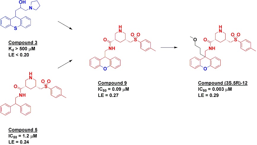
Along with the fragment effort, the researchers also undertook an HTS screen, resulting in the discovery of compound 5, which itself had come from a 950-compound library targeted towards aspartic proteases. Crystallography revealed that this molecule binds with the diphenylmethane moiety in a similar position as the tricycle of fragment 3, and indeed when a rigid tricyclic framework was grafted onto compound 5 the resulting compound 9 showed a satisfying boost in potency. Further optimization led to a pure enantiomer of compound 12 with low nanomolar potency, good selectivity, moderate oral bioavailability and efficacy in rats, though it did also show some time-dependent CYP3A4 inhibition.

This is really a structure-based design paper, and there is obviously much more detail than would be appropriate here. What caught my eye is that it is a nice example of fragment-assisted drug discovery, in which fragment information is used as one aspect of an overall lead discovery program. In this case a cynic could argue that the only bit that came from the fragment was the tricylic motif. However, given the limitless number of analogs that could be made, such information can be both unexpected and valuable.

23 April 2013

Poll: how many atoms are too few?

Last year we asked how large a fragment you would include in your library, but the related question, how low will you go, is also interesting (see poll to right).

Azaindole, with 9 non-hydrogen atoms, has been the starting point for more than one clinical compound, and 5-atom acetohydroxamic acid also figures rather prominently in fragment history, but would you include something this small? How seriously should we take the Rule of 1?

22 April 2013

Poll Results - Number of Fragments in the Screen

The poll is closed!  So, the question was
I think it is fair to say that the average number of fragments per fragment mixture for NMR pooling is 6.  I typically say five is the goal and if you can squeeze in more good on ya!  I don't think the limitation is chemical shifts, I think it is solubility.  I would love to hear what others think is the limitation.

20 April 2013

Eight Annual Fragment-Based Drug Discovery Meeting (part 2)

The last major fragment event of 2013, CHI’s FBDD, wrapped up earlier this week in San Diego. As with last year this summary is not meant to be comprehensive (and you can also read Teddy’s impressions here.)

The FBDD track is just one of six within the CHI Drug Discovery Chemistry Conference. One indication of the success of the field is its appearance in several of the other tracks: attendees were likely to hear about fragments without going to the FBDD track at all.

In the GPCR track, Robert Cooke from Heptares discussed the application of fragments to the β1-adrenergic receptor (see also here). In the kinase track, Hongtao Zhao presented in silico fragment work (see also here). And in the protein-protein interaction track, David Fry from Roche described a deconstruction of the p53-HDM2 inhibitor RG7112 into its component fragments to see whether the molecule could have been identified from FBLD. RG7112 consists of a central core with four appendages, and although the mono-substituted core was too weak to detect, some of the cores with two substituents could be identified and bound to the protein in the same manner as the parent compound. However, these did tend to be super-sized fragments, with molecular weights in the 300-350 Da range.

Protein-protein interactions were also a theme of Richard Taylor, from the company UCB. They built a sizable fragment library of about 23,000 (mostly commercial) compounds designed to cover molecular frameworks found in known drugs. UCB has invested heavily in SPR technology, and with a stable of four Biacore 3000 instruments could rapidly screen this entire library against a dozen protein-protein interaction targets. Not surprisingly, given the difficulty of this target class, the hit rate was much lower than in conventional fragment screens, averaging just about 1%. What was interesting is that only 964 fragments hit any target – at less than 5%, this is much lower than the roughly 33% hit rate seen in other fragment libraries. Most of these fragments were reasonably specific, though; 908 hit ≤ 8 targets. It will be interesting to see whether anything can be learned about “privileged” protein-protein interaction fragments from this set.

Of course, extracting general trends from collections of fragments is not necessarily straightforward. Teddy has already brought up the difficulties of describing molecular shape; as he pointed out in his presentation, Fsp3 is not the best measure of “three-dimensionality” for several reasons. For example, even toluene has an Fsp3 = 0.14, and while Pete Kenny correctly points out that aromatic molecules do have volume, most chemists would think of this as a very “flat” compound. Principal moment of inertia (PMI) is better, but is harder to calculate. Happily, as Justin Bower described in his presentation, the Beatson Institute is allowing other researchers to use their 3DFIT software to calculate PMI and other properties.

One of the criticisms of 3D fragments is that, as Rod Hubbard pointed out, they can be a “pain in the neck” for chemistry. One solution that researchers at Vernalis took was to do analog work on a simpler molecule, then scaffold-hop back to the original fragment once the SAR was sufficiently understood to justify investment in more challenging chemistry.

Finally, the question of what to do with fragment hits that don’t reproduce in different assays was the topic of at least one breakout discussion and was also extensively discussed by Peter Kutchukian, who presented an analysis of 134 fragment screens using a variety of techniques against 34 different targets at Novartis. Some of this was presented at FBLD 2012, but one interesting finding was that hits from biophysical screens (such as SPR, NMR, or DSF) tended to cluster separately from hits in biochemical assays. Given the number of ways to find fragments, pursuing hits that confirm in both a biochemical and a biophysical method may help to weed out artifacts, though at the risk of increasing false negatives.

Feel free to chime in with your thoughts and impressions, whether or not you were there. And if you are kicking yourself for not attending, next year’s meeting is scheduled to return to San Diego from April 22-25.

17 April 2013

What's the Fire behind the Smoke?

Dan and I are here at the CHI FBDD conference, with of course other luminaries.  I am not going to get into details about all the talks, Dan does that much better than I anyways.  I wanted to set up some future blog posts by sharing what I think are some trends.

1.  3D fragments are real and very useful.  However, fSP3 is a horrible way to measure 3D-arity.  Principal Moment of Inertia (PMI) is a much better way to determine this.  This was mentioned in a brilliant talk (watered down from full-fledged rant) by me.  But much better explained by Justin Bower of the Beatson Institute. 

2.  Solubility, Solubility, and Solubility.  Experimentally determined solubility is a much better indicator of fragment quality (for a library) than cLogP, for example. 

3.  The Voldemort Rule (or the rule that shall not be named).  Arbitrary "rules" created as a marketing tool have no place in a discipline that has over a decade of results and empirical evidence.  While Dan is not ready to put a stake in its heart, I am.  Rod Hubbard is.  Who else wants to join Zartler-Dore's Army?
Look for more updates, thoughts, comments, and general frivolity as follow up to the conference.