Showing posts with label mass spectrometry. Show all posts

14 September 2026

Covalent allosteric inhibitors of NLRP3

NLRP3 has received considerable attention as an inflammatory disease target. The protein normally exists in an auto-inhibited conformation, but in response to various stimuli it changes shape to help form a multimeric complex called the inflammasome. This in turn activates various inflammatory pathways, including cell death. Several drugs targeting NLRP3 have gone into the clinic, but none have been approved. In a new open-access Br. J. Pharmacol. paper, Brian Cook, Gabriel Simon, and colleagues at Vividion describe a covalent inhibitor that binds to a previously unknown site.
 
As we wrote nearly a decade ago, Vividion pioneered the use of chemoproteomics to identify covalent binders in cells and cell lysates. While exploring the mechanism of the previously reported NLRP3 inhibitor MCC950, they found that cysteine 280 became resistant to modification by electrophiles in the presence of the ligand. MCC950 does not contain an electrophile, arguing against direct interaction with Cys280 and suggesting instead that binding causes a conformational change to the protein which makes Cys280 less accessible. In other words, protein inhibition correlated with Cys280 being less exposed.
 
A screen of Vividion’s library yielded no hits against Cys280 but more than 300 against cysteine 463. Interestingly, modification of C463 also caused a decrease in reactivity of Cys280, suggesting a conformational change similar to the one caused by MCC950. Thus, the researchers pursued Cys463 binders.
 
Two parameters were used to guide compound optimization, target engagement and promiscuity. The first number, TE50, measures the extent of covalent engagement of a given cysteine after 1 hour in cell lysates. The second, P500, measures the percentage of all measured cysteines in the sample labeled by at least 40% after incubation with 500 µM compound for 1 hour. One of the initial hits, VVD-124476, gave a low micromolar value for TE50 but labeled some 20% of the measured proteome. Indeed, it was previously reported as a covalent ligand for a completely different protein. To be useful as a NLRP3 inhibitor, the researchers needed to reduce promiscuity, and the most straightforward approach is to reduce general reactivity.
 

To decrease reactivity, the researchers swapped the acrylamide warhead with a butynamide (VVD-142397). Butynamides are reported to be less reactive than acrylamides, a result corroborated at Vividion with chemoproteomic data on 400 matched molecular pairs. The acrylamide to butynamide swap improved selectivity some 20-fold, though it also decreased potency against NLRP3. Increasing the core ring size regained activity, and rigidifying the molecule led to further improvements. Further optimization ultimately led to VVD-338213 and several related molecules.
 
Although the researchers do not report kinact/KI values, a back of the envelope calculation puts it around 1200 M-1s-1 for VVD-338213, a potency where cell activity might be expected. Happily this turned out to be the case: the molecule blocked IL-1β secretion from human cells at lower concentrations than MCC950. Importantly, in cells where Cys463 had been mutated to alanine, MCC950 was still active while the covalent molecules were inactive.
 
In addition to promising cell activity, many of the covalent molecules had reasonable stability in whole blood, good permeability, and low efflux. (I do wish the ligand efficiency reactivities of the molecules were reported; after all Vividion, introduced this metric.) Some of the molecules also showed good brain penetration. In vivo pharmacodynamic studies are complicated by the fact that Cys463 is not conserved in mice, but fortunately the researchers could run studies on transgenic mice in which human NLRP3 had been introduced. In these animals, IL-1β was decreased by both VVD-338213 and by MCC950.
 
The researchers were also able to obtain a cryo-EM structure of one of their molecules bound to NLRP3, which confirmed covalent binding to Cys463. More importantly, it revealed that the binding site is in a cryptic pocket some 20 Å from where MCC950 binds. (One wonders if the computational technique we discussed last week would have been able to predict this pocket.) Similarly to the WRN inhibitor Vividion has taken into the clinic, the molecule appears to make only hydrophobic contacts with the protein, with no polar interactions. Mechanistically, the “Cys463 ligands act as molecular doorstops, stabilizing the inactive conformation and preventing the structural rearrangements necessary for transition to the active inflammasome disc.”
 
This is a lovely paper and a case study in transforming a highly reactive molecule into a selective molecule with in vivo activity. It is interesting that the starting molecule, with 28 non-hydrogen atoms, flagrantly violates the rule of three. Last year we asked whether covalent fragments need to be larger than their non-covalent counterparts, and this paper provides another example where the answer is yes. Whether or not this chemical series ultimately advances, it is nice to have another chemical probe for this target.

17 August 2026

Site-specific HDX-MS for increased resolution

Most methods to characterize protein-ligand binding interactions have drawbacks. Crystallography, obviously, requires your protein to crystallize, while NMR and cryo-EM both have size limitations and are resource-intensive. Hydrogen-deuterium exchange mass spectrometry (HDX-MS), which we wrote about earlier this year, is theoretically faster and can apply to more proteins, but the technique provides limited resolution. Now a new open-access paper in J. Am. Chem. Soc. by Derek Wilson and collaborators at York University, the Structural Genomics Consortium, and SCIEX have improved the resolution down to single amino acids.
 
As we wrote in March, HDX-MS relies on the exchange between deuterium and protons on proteins; slower exchange is associated with more stable secondary structures. The exchange is detected by digesting the protein with proteases and determining the mass of the resulting peptide fragments using MS; incorporation of deuterons increases the mass of the peptides. If, for example, a peptide has three deuterons incorporated, the mass would increase by 3. However, in conventional HDX-MS, one can’t tell exactly where the deuterons have exchanged.
 
Last year the researchers described a method in J. Am. Soc. Mass Spectrom. to do just this using some clever mass spectrometry experiments. I won’t go into details as the paper is open access, but suffice it to say that it involves electron-activated dissociation (EAD) to fragment the peptides while preventing the scrambling of the protons and deuterons that normally limits resolution.
 
In the new paper, the approach was applied to the oncology target WDR5, which we previously discussed here. The researchers applied site-specific HDX-MS to three small molecule and two peptide ligands. All five of these ligands had previously been characterized by conventional HDX-MS as well as surface plasmon resonance (SPR), and four had also been studied crystallographically.
 
Reassuringly, the results from site-specific HDX-MS were mostly consistent with conventional HDX-MS, but there were some surprises. For example, the site-specific approach revealed protection from exchange for one amino acid (which makes a hydrogen bond to the ligand) and enhanced exchange for the adjacent amino acid on the same peptide. In conventional HDX-MS, the “peptide contains offsetting uptake increases and decreases, resulting in a ‘net-zero’ signal.”
 
The researchers also found that, for another residue, the change in HDX-MS signal correlates with the affinity of the ligand, as assessed by SPR. The mechanism is unclear since the concentrations of ligands were far higher than their affinities, but the results are intriguing.
 
The new paper describes just a single, well-behaved target, but if site-specific HDX-MS applies more broadly, it would offer a significant advance in obtaining structural information. The researchers note that the data analysis is currently “manual and prohibitively cumbersome,” a problem that should be solvable with automation. I look forward to seeing it applied prospectively to more targets, particularly if it works for weak binders such as fragments.

02 March 2026

Best practices for applying HDX-MS to FBLD

Among the many biophysical techniques presented at the recent Novalix meeting, hydrogen/deuterium exchange mass spectrometry (HDX-MS) was mentioned only a few times. Practical Fragments last covered it nearly four years ago in the context of a paper that described theoretical challenges and improvements to the method for assessing fragment binding modes. A new open-access Comm. Chem. paper from Tiago Bandeiras, Alessio Bortoluzzi, and collaborators at iBET-Instituto de Biologia Experimental e Tecnológica and Merck KGaA implements some of these suggestions.
 
The researchers find only two examples where HDX-MS had been applied to fragments, one of which we covered here. The new paper focuses mostly on Cyclophilin D (CypD), a mitochondrial protein implicated in several diseases. A screen we wrote about in 2020 described several fragment hits, some of which were crystallographically found to bind in three overlapping sites designated the proline pocket, the aniline pocket, and the pyrazolo pocket.  The researchers chose a binder from each pocket to study as well as two more fragments whose binding sites were not known. All five fragments are extremely weak hits, with at best 7 mM (yes, millimolar) affinity as assessed by SPR, making this a particularly challenging test case.
 
As a reminder, HDX-MS examines the exchange of deuterium from D2O to protein backbone amides. Nearby ligands can slow this exchange, and mapping the locations and magnitude of these changes reveals where the ligand binds. Optimization experiments were initially run on a compound with a KD of 44 mM for the aniline pocket. Three protein concentrations were tested. Since the highest (10 µM) yielded the highest number of detected peptides, it was used for subsequent experiments.
 
As suggested in the 2022 paper, compound was added both to the initial protein solution as well as to the deuterium exchange buffer. The fragment was tested at 2.5, 5, and 10 mM, and changes in deuterium uptake were found in all cases, which is remarkable given that the theoretical occupancies range from 5 to 18%. An experiment using substoichiometric concentrations of a high affinity ligand confirmed that 18% protein occupancy is sufficient to generate a reliable signal.
 
Still, given the low occupancy, the researchers used statistical methods to ensure that changes in signals were significant. Mapping those that were onto the structure of the protein confirmed that the fragment bound to the aniline pocket.
 
A second fragment was tested by HDX-MS, and the results confirmed that it binds in the pyrazolo pocket, as previously shown by crystallography. However, for a third fragment that had been shown to bind in the proline pocket, the results suggested instead that it binds in the aniline pocket. The proline pocket exhibited very low levels of deuterium exchange, but when the researchers increased the pH from 7.4 to 9 they did find evidence that the fragment binds here as well.
 
Next, the researchers turned to the two fragments with unknown binding sites. HDX-MS revealed that these bind to the aniline binding site, though with subtly different protection patterns suggesting that they bind in slightly different regions of the pocket.
 
Finally, the researchers used their optimized HDX-MS conditions on a previously identified fragment that binds the kinase FAK with low millimolar affinity. This showed that it binds in the so-called hinge region, in agreement with crystallography. This fragment was also used as a negative control for CypD, showing that it does not bind.
 
This paper is a nice resource for those hoping to apply HDX-MS to fragments. The fact that the binding sites of such weak binders can be determined is quite remarkable. That said, the resolution is not as good as crystallography or protein-detected NMR; for FAK in particular, the ligand reduces deuterium update across a large fraction of the protein surface. And the fact that a proline-pocket binder was initially mapped to the aniline pocket also gives one pause. Perhaps the binding mode in solution really is different from that found crystallographically. It would be interesting to see whether NMR can resolve the conundrum.

10 March 2025

Crude success against the SARS-CoV-2 main protease: From covalent fragment to noncovalent lead

With increased throughput and reliability of biophysical and other methods, finding fragments against most targets is now fast and easy. Advancing these fragments to leads, not so much. In a new open-access Angew. Chem. Int. Ed. paper, Jacob Bush and collaborators at GSK, University of Strathclyde, and the Francis Crick Institute provide a case study for how to accelerate the process.
 
Almost exactly five years ago we highlighted early efforts against the main protease (Mpro) from SARS-CoV-2. This target turned out to be a good choice, as demonstrated by the rapid discovery and approval of the drug nirmatrelvir. Mpro is a cysteine protease and thus ideally suited for covalent fragment screening.
 
In the new paper, the researchers screened a library of 219 chloroacetamide-containing fragments (each at 5 µM) individually against 0.5 µM protein for 16 hours at 4 ºC and then analyzed them by intact protein mass spectrometry. Six of these gave at least 75% modification, and further characterization found that the most potent, compound 2, had a kinact/KI = 170 M-1s-1. This (and the other hits) also inhibited the protein in an enzymatic assay, and additional chemoproteomic experiments revealed that compound 2 could bind to the active site cysteine of Mpro in living cells with surprising selectivity; just 11 targets were more strongly engaged than Mpro.
 
To optimize compound 2, the researchers turned to crude reaction screening, also known as direct-to-biology or D2B. As we described here and here, this entails running reactions at small scale and testing them directly, without purification. To validate the approach, the researchers synthesized a subset of the original 219 chloroacetamides in 384-well plates. HPLC studies confirmed the desired product as the major component for 43 of the 69 attempted syntheses; only four failed. Importantly, there was a good correlation in activity between the crude reaction mixtures and the pure molecules.
 
Next, the researchers synthesized a new D2B library of 193 molecules related to compound 2. HPLC analysis of the crude products showed a 77% success rate, with just nine outright failures. The library was screened against Mpro for 1 hour (as opposed to 16 hours in the first screen), resulting in 14 hits. The best of these, compound 7a, was such a rapid modifier that the a kinact/KI could not be easily calculated, but it showed nanomolar activity in the enzymatic assay. It was also more selective than compound 2 in cell-based experiments.
 

Chloroacetamides are not considered advanceable as drugs, so the researchers sought to remove the warhead, initially by replacing it with the simple acetamide in compound 12. Although this molecule showed almost no activity in the enzymatic assay, the researchers coupled a diverse set of 146 carboxylic acids to the amine building block and screened the crude reaction mixtures in a functional assay at 50 µM to identify seven molecules that gave nearly complete inhibition, with compound 13 being the most potent. A second D2B library of analogs around compound 13 was screened at 1 µM, leading to the mid-nanomolar compound 14.
 
This is a nice illustration of the power of crude reaction screening to rapidly identify new chemical matter. It is true that Mpro is quite ligandable; we wrote about other non-covalent fragment success stories here and here. However, as we discussed here, D2B can be applied to more challenging targets. The supporting information in the new paper should be particularly valuable for those hoping to try the approach themselves.
 
At FBLD 2024 Frank von Delft set a goal of taking a “100 µM binder to a 10 nM lead in less than a week for less than £1000.” We’re not there yet, but developments in D2B are moving us forward.

22 July 2024

Multiplexing (native) mass spectrometry

Native mass spectrometry (nMS) is one of the less commonly used fragment-finding methods. The approach entails mixing proteins and ligands and gently ionizing them under non-denaturing conditions to look for complexes. As with many other methods, multiple fragments can be screened in a single sample. In a new ACS Med. Chem. paper, Ray Norton and collaborators at Monash University and CSIRO report screening multiple proteins in a single sample.
 
The researchers were interested in fatty acid-binding proteins, or FABPs. As their name suggests, these transporter proteins shuttle lipophilic molecules such as fatty acids around cells. The ten human isoforms are expressed in different tissues and have different functions in metabolic signaling, but their similarity to one another has made finding selective chemical probes difficult. Enter nMS.
 
FABP isoforms 1-5 are the most heavily studied, and these were first assessed individually. They ionized well, though in some cases peaks corresponding to both the native protein and a complex with acetic acid was observed, not surprising given that the buffer contained 50 mM ammonium acetate.
 
Next, all five proteins were mixed together at 10 µM each. All the proteins could still be observed (with or without bound acetate), though some proteins did give stronger signals than others due to differences in ionization efficiency.
 
Adding small molecule WY14643, which the researchers had previously found to bind to FABPs in a fluorescence polarization (FP) assay, led to a more complex spectrum, with peaks corresponding to unbound proteins, proteins bound to WY14643, proteins bound to acetate, and proteins bound to both acetate and WY14643. When WY14643 was added at 10 µM, the selectivity profile was consistent with the FP data. Interestingly though, when ligand was added at the total concentration of all protein isoforms (50 µM), the selectivity profile changed. The researchers suggest this may be due to nonspecific binding at higher ligand concentrations, as has been seen previously for nMS.
 
To explore the generalizability of multiplexing nMS, the researchers turned to more potent (nanomolar) ligands. As with WY14643, these molecules showed good agreement with published selectivity rankings at lower ligand concentrations with some non-specific binding at higher concentrations.
 
When I first wrote about nMS back in 2010, I noted that “the stability of protein-small molecule complexes in native mass spectrometry assays does not necessarily correlate with the (more relevant) solution-phase affinity,” and this fact is investigated in the paper. Careful optimization of the experimental conditions, including ionization voltage and temperature, led to good relative selectivity rankings for a given ligand across the different FABP isoforms but differences in absolute values from those measured by ITC.
 
Another challenge is the fact that the five FABP isoforms tested have similar molecular weights; in one case a ligand complexed with FABP3 was difficult to distinguish from free FABP2. The researchers could solve this by using different protein constructs, such as a hexa-histidine-tagged version of FABP3.
 
Overall this is an interesting approach, and the paper does an excellent job describing the technical details and limitations. Along with protein-observed 19F NMR, mass spectrometry is a rare experimental technique suitable for screening mixtures of proteins in solution. Indeed, this becomes even easier when screening covalent binders, as seen in this paper from 2003, since there is no need to worry about ligand dissociation during ionization. And with the increasing interest in covalent drugs, the use of MS is only likely to increase.

13 May 2024

Fragments in cells, writ large

Earlier this year we highlighted work in which a dozen fragments were screened against cells to look for noncovalent binders across the proteome. A new paper in Science by Georg Winter and collaborators at the Austrian Academy of Sciences, Pfizer, and several other organizations ups the game by more than an order of magnitude, and uses machine learning to make predictions about fragments’ cellular destinations and binding partners. (See also Derek Lowe’s post here.)
 
The researchers started with 407 diverse fully functionalized fragments (FFFs), which as we previously discussed consist of a variable fragment coupled to a photoreactive group and an alkyne moiety that can be used to pull down any bound proteins using click chemistry. These were selected from a larger set of ~6000 FFFs available from Enamine. The FFFs were incubated at 50 µM with intact HEK293T cells, followed by ultraviolet crosslinking.
 
Next, cells were lysed and treated with a biotin-azide probe that reacts with the alkyne on the FFFs. Covalently modified proteins were captured on streptavidin resin and proteolytically digested. Tandem mass tag (TMT) proteomics, which we wrote about here, was used to identify captured proteins. Unlike earlier methods, the researchers did not pinpoint the specific fragment binding sites on proteins.
 
In total the researchers found 2667 proteins bound to one or more fragments, of which ~86% had no reported ligands. Both proteins and ligands varied considerably in promiscuity: some proteins bound to more than half of the FFFs, and some fragments bound to hundreds of proteins, while others bound only a few, or none. To look for specific interactions, the researchers focused on proteins bound by fewer than 10 different ligands.
 
Three protein-ligand interactions were analyzed in some detail: the kinase CDK2 (and other CDK family members), the adapter protein DDB1, and the solute carrier protein SLC29A1. In each case the researchers confirmed the results from their chemoproteomic screens. Follow-up studies with related molecules led to more potent derivatives, with a CDK2 inhibitor showing low micromolar activity in a biochemical assay and an SLC29A1 inhibitor showing micromolar activity in a cell-based assay.
 
The researchers also found patterns in their larger data set. Armed with 47,658 protein-ligand interactions, the researchers were able to use machine learning to start to predict which molecular features were associated with binding. They ranked fragments as promiscuous or nonpromiscuous and built a promiscuity model. Molecules with higher lipophilicity and a greater fraction of aromatic carbon atoms tended to be more promiscuous, but the model could correctly categorize compounds as promiscuous even if they had lower ClogP values, or nonpromiscuous even if they had higher ClogP values.
 
Beyond promiscuity, the researchers used machine learning to predict other behavior, such as subcellular localization. A relatively easy case was to predict which molecules would accumulate in lysosomes; these tended to be hydrophobic basic amines. More impressively, the researchers could predict fragments likely to bind to transmembrane transporters, RNA binding proteins, and even intrinsically disordered proteins. And this is just the start: they hope one day to predict “target proteins from an input chemical structure alone.”
 
Perhaps most exciting, all of the data and models are available for free at Ligand Discovery. You can explore the proteins bound across all 407 fragments, input one or more proteins and find ligands, predict whether any given FFF is likely to be promiscuous or not, and even “build a machine learning model on the fly to predict potential interactions.” 
 
Check it out and let us know your experience.

06 May 2024

Covalent fragments vs WRN

Last week Practical Fragments highlighted a covalent clinical compound from Vividion and Roche against the oncology target WRN. Another series of inhibitors against this protein are described in a recent Cancer Discov. paper by Gabriele Picco, Mathew Garnett, and collaborators at the Wellcome Sanger Institute, GSK, IDEAYA, and several academic institutes.
 
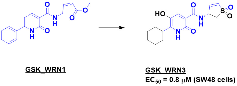
As we described in more detail last week, WRN is a synthetic lethal target for microsatellite instability (MSI) cancers. In contrast to the Vividion paper, which started by screening covalent fragments against cell lysates, here the researchers incubated purified WRN protein against each member of their covalent library (at 20 µM for 24 hours at 21 ºC) and analyzed the reactions by intact protein mass spectrometry. The fragment library was based around the methyl acrylate warhead, which, as we discussed a decade ago, has a narrower range of reactivities than more common acrylamides.
 
GSK_WRN1 was one of the prominent hits, with 81% modification. Tryptic digestion revealed that it modified C727, the same cysteine found by the Vividion researchers. Medicinal chemistry led to GSK_WRN3, with sub-micromolar activity in MSI SW48 cells. (Unfortunately no other details on the chemistry are provided; the paper states that these will be written up separately.)
 
GSK_WRN3 or a closely related compound were tested in a battery of assays and found to be inactive against three other helicases, which is not surprising given that C727 is unique to WRN. Chemoproteomic studies in cells also revealed the compound to be quite selective towards WRN vs other proteins. The compounds selectively inhibited MSI cancer cell lines and patient-derived organoids while sparing microsatellite stable (MSS) cell lines and organoids. One of the compounds showed activity in a mouse xenograft model.
 
In a useful public service, the researchers tested two previously reported WRN inhibitors, MIRA-1 and NSC617145, in the same set of several dozen cell lines and found that they were not only ineffective, they lacked selectivity for MSI cells over MSS cells. Although Dr. Saysno might object, I nominate these molecules to be added to the “Unsuitables” bestiary at the Chemical Probes Portal.
 
I do wish more details about the molecules were provided, especially the kinact/Ki values. It is interesting that GSK_WRN3 bears remarkable structural similarities to VVD-109063. IDEAYA recently announced that their collaboration with GSK has resulted in a development candidate targeting WRN, and it will be fun to see the full story emerge.

12 February 2024

Fragment screening across the proteome, noncovalently

Last week we discussed methodological improvements to industrialize covalent fragment screening across the proteome. While I’m a huge fan of covalent binders, their noncovalent counterparts are the vanilla ice cream of FBLD: also tasty and much more common. Back in 2017 we described how “fully functionalized fragments,” or FFFs, could be used to screen noncovalent fragments in cells. A new paper in Nat. Chem. Biol. by Christopher Parker and collaborators at Scripps and BMS further optimizes the approach.
 
FFFs contain, in addition to the variable fragment, a photoreactive group (often a diazirine) and an alkyne tag. When exposed to light the photoreactive group can react with nearby proteins and the alkyne tag can be used to fish out the proteins. In the new paper the researchers started with a dozen FFFs.
 
One challenge, which we discussed in 2021, is that the FFFs may react with many sites on a given protein. During analysis, a protein is typically digested into peptides for mass spectrometry. If a FFF reacts at several sites on a peptide the resulting spectra will be “chimeric” and more difficult to characterize.
 
The researchers developed methods to take these chimeric spectra into account when searching for sites of modification. The approach, called Dizco (for diazirine probe-labeled peptide discoverer) can identify three times as many peptides as standard approaches, as well as more detailed information on sites of modifications. 
 
Two pairs of FFF probes consisted of enantiomers, and these showed differential labeling across the proteome, consistent with specific molecular recognition. The researchers also confirmed binding of a few FFF probes to several proteins using a cellular thermal shift assay (CETSA).
 
In all, the probes modified 3603 peptides on 1669 proteins. The sites of modification were then mapped onto predicted or modeled three dimensional structures of the proteins. Importantly, and consistent with the 2017 work, most of the labeled sites were near predicted pockets. The researchers confirmed this for four proteins by showing that FFF probe binding could be competed by adding ligands known to bind to the pockets.
 
Next, the researchers docked (using AutoDock) their FFF probes onto 175 proteins (108 from structures in the Protein Data Bank and 67 from AlphaFold structures). They found that the docking experiments recapitulated the experimental data, and in fact often placed the diazirine tag near the protein residues found to react. Strikingly, and in another step forward for in silico approaches, docking against structures from AlphaFold was nearly as effective as those from the protein data bank.
 
As the researchers conclude, “we identified many binding pockets that have no reported ligands… these probes may serve as leads for further optimization.” It will be fun to see how far they go.

05 February 2024

Fragment screening across the proteome, industrialized

Last week we discussed covalent fragment screens against isolated enzymes, which can be very effective. But screening in cells or cell lysates preserves proteins in a more physiological environment and allows many proteins across the proteome to be screened simultaneously. In 2016 we wrote about covalent screens in human cell lysates which identified fragment hits for 758 cysteine residues in 637 proteins. Mass spectrometry techniques have improved since then in terms of both speed and sensitivity, as illustrated in a new Cell Chem. Biol. paper from Steve Gygi, Qing Yu, and collaborators at Harvard Medical School and Biogen. (Disclosure: Steve Gygi is on the Scientific Advisory Board of my current company, Frontier Medicines.)
 
The approach is called TMT-ABPP, or tandem mass tag activity-based protein profiling, and it involves multiple improvements to previous methods, some of which Steve discussed at the Discovery on Target meeting last year. Covalent fragments are added separately to cell lysate aliquots, after which a desthiobiotin iodacetamide (DBIA) probe is introduced. If a given site on a protein has reacted with a fragment, it will not be available to react with the DBIA probe.
 
Next, proteins are digested to peptides and labeled with TMT (tandem mass tag) reagents, which allow multiple samples (18 in this case, either individual fragments or DMSO-only controls) to be combined for simultaneous analysis. Peptides functionalized with the DBIA probe are captured on streptavidin resin while those that had previously reacted with a covalent fragment will not stick to the resin and be lost. Peptides eluted from the resin are then analyzed by mass spectrometry. The “competition ratio” between treated and untreated lysate gives a measure of how strongly a given site on a given protein is labeled by a fragment.
 
Multiple other tweaks, such as capturing proteins using magnetic beads and using a special type of mass-spectrometry (high-field asymmetric waveform ion mobility spectrometry, or FAIMS), further streamline the process to a 96-well plate format, with each well containing a mere 10-20 µg of cell lysate, as much as 100-fold less than earlier approaches.
 
The researchers benchmarked TMT-ABPP using three reactive “scout fragments,” including compound 1 from last week’s post. Collectively they identified 6813 cysteine residues hit by one or more of the scouts.
 
To demonstrate throughput, the researchers next screened 192 fragments, a third of which were acrylamides while the rest were chloroacetamides. Even with two controls for every 16 samples, this only required 12 injections on a mass spectrometer and resulted in hits against 38,450 cysteines, about 50-fold more than the 2016 paper. Proteins that were more highly expressed were better represented, as were proteins with known reactive cysteine residues, such as thioredoxins. Surprisingly though, surface-exposed cysteine residues were only slightly enriched over more buried cysteines.
 
The researchers also applied TMT-ABPP to five well-characterized covalent molecules, including the mutant KRASG12C inhibitor ARS-1620, which we wrote about here. In addition to the G12C site of KRAS, several other proteins were also liganded, including adenosine kinase (ADK). The researchers confirmed that ARS-1620 inhibited ADK in an enzymatic assay.
 
As the researchers note, “proteome-wide profiling of thousands of compounds remains a formidable challenge, both technically and financially.” This paper reveals how to significantly reduce the costs. By using such approaches, it is possible to build a catalog of fragment ligands for thousands of proteins. Doing so with a well-curated library could enable rapid fragment-to-lead campaigns.

29 January 2024

Covalent fragments vs a SARS-CoV-2 helicase

Last week we wrote about the difficulties of trying to understand even well-characterized covalent inhibitors of well-characterized targets. Most projects have far less information, as illustrated in a recent paper in J. Am. Chem. Soc. by Ekaterina Vinogradova, Tarun Kapoor, and collaborators at Rockefeller University and Sanders Tri-Institutional Therapeutics Discovery Institute, who report the first inhibitors of a particular SARS-CoV-2 enzyme.
 
The researchers were interested in helicases, enzymes that unwind DNA, RNA, or both. To do so, helicases cycle between “open” and “closed” forms, with conformational changes of as much as 15 Å. That dynamism complicates structure-based drug design, and many screens have yielded false positives. An irreversible covalent inhibitor that remained bound to the enzyme through its gyrations would potentially be easier to optimize.
 
The protein nsp13 from SARS-CoV-2 is essential for viral replication and thus an attractive drug target. The researchers started by testing previously reported and reactive “scout fragments” in a functional assay. Compound 1 inhibited the enzyme, and mass-spectrometry (MS) assays revealed that it modified three sites on the protein. Although multiple modifications are not desirable, the enzyme does contain 26 cysteine residues, so it could be worse. Peptide mapping and mutagenesis experiments revealed that modification of cysteine 556 (C556) is responsible for the inhibitory activity of compound 1.
 
A series of analogs culminated in compound 3b, which had low micromolar activity after a four hour incubation and also seemed more selective than compound 1, with less modification of other cysteine residues. The enantiomer of compound 3b was at least 6-fold less potent, suggesting molecular recognition rather than simple reactivity. In addition to nsp13, the researchers examined two mammalian helicases with disease relevance, WRN and BLM, and found that compound 3b was modestly selective for nsp13. (The researchers find different inhibitors for these two enzymes, though these are weaker and not as extensively characterized as those for nsp13.)

Cysteine 556 is not in the ATP-binding site and does not seem to be involved with RNA binding, and the researchers suggest that compound 3b may act allosterically. It seems to be highly conserved too, which might mean mutational resistance is less likely to evolve.
 
As the researchers acknowledge, compound 3b contains a chloroacetamide warhead, which is likely too reactive and unstable to move forward into in vivo studies, let alone the clinic. Also, had I reviewed the manuscript I would have requested the researchers to provide kinact/KI values rather than merely IC50 values; a rough calculation using the methodology in this paper suggests a modest 10 M-1s-1 for compound 3b. That said, the discovery that liganding C556 inhibits nsp13 means that working to develop more potent and selective molecules may be worth the effort.

19 June 2023

Nucleophilic covalent fragments against cysteine (!)

Covalent fragment-based drug discovery continues to gain momentum, as evidenced by the number of talks at the CHI Drug Discovery Chemistry meeting in April. All of those involved nucleophilic residues on proteins, especially cysteine, reacting with electrophilic fragments. However, as we noted last year, it is possible to do the reverse. This is the topic of a new paper in Nat. Chem. Biol. by Jing Yang (Beijing Institute of Lifeomics), Kate Carroll (UF Scripps), and collaborators.
 
The reason most covalent fragments are not nucleophiles is that none of the twenty standard amino acids are particularly electrophilic. The work we mentioned last year focused on post-translational modifications introducing aldehydes or ketones into proteins. But it turns out that the thiol group of cysteine, which is normally nucleophilic, can be oxidized to a sulfenic acid, which is electrophilic. The Carroll group has been studying this “cysteine redoxome” for years and found that S-sulfenation can serve a regulatory function akin to phosphorylation.
 
To assess the reactivity of sulfenic acids across the proteome, the researchers synthesized cyanoacetamide and nitroacetamide derivatives of 3,5-bis(trifluoromethyl)aniline. The chloroacetamide and acrylamide derivatives of this fragment have previously been used in chemoproteomics experiments to probe for reactive cysteines. Cell lysates were treated with one of these four fragments, followed by treatment with a generic probe for sulfenic acids or thiols. If a cysteine residue is modified with the fragment, it will be unavailable to react with the second probe, and this loss in signal can be quantifiably detected using mass spectrometry.
 
For the thiol-reactive chloroacetamide and acrylamide, the researchers found that 25.2% and 11.0% of quantifiable cysteines in the proteome could form adducts. But only 24 cysteine residues formed adducts with the nitroacetamide (1.3% of the total sulfenic acids quantified), and none formed adducts with the cyanoacetamide.
 
Despite this lower hit rate, the researchers constructed a library of 65 cyanoacetamide- and nitroacetamide-containing fragments, which was similarly screened in cell lysates. Adding hydrogen peroxide to cell lysates to mimic oxidative stress increased the number of sulfenic acid sites. In total the researchers found 524 liganded sites across 441 proteins. As expected from the earlier experiments, nitroacetamides tended to bind to more sites than cyanoacetamides.
 
The researchers studied the functional effect of covalent modification for several proteins. The enzymes GAPDH, GSTO1, and ACAT1 all have active-site cysteine residues that can be reversibly modified by oxidation to the sulfenic acid. Reaction of this form of the recombinant proteins with one of the covalent fragments led to irreversible inhibition.
 
Similarly, the researchers demonstrated that a fragment which hits the enzyme PRXL2A activated MAPK signaling in cells, as expected. Importantly, this effect was not seen in cells containing PRXL2A with a cysteine to serine mutation. The non-enzyme proteins HDGF and BCCIP could also be functionally inhibited in cells with covalent fragments.
 
This “umpolung” approach to covalent ligand discovery is scientifically interesting, but how useful will it be? In most cases sulfenic acid formation is already deactivating, so targeting this form of the protein will simply keep it in the off-state. However, the researchers do note that sulfenic acid formation can be activating.
 
A second challenge is that sulfenic acid formation tends to be substoichiometric, with only a small percentage of cysteines existing predominantly in the sulfenic acid form. Thus, it will be difficult to achieve the near quantitative level of modification often required for biological effects. That said, there are cases where you want to tweak a pathway rather than shut it down entirely. Protein activation or the development of new PROTACs could also benefit from limited target protein engagement.
 
As for the covalent fragments themselves, nitroacetamides may be too reactive, but the cyanoacetamide moiety is actually found in a few approved drugs, such as the anti-inflammatory tofacitinib. And if compelling sulfenic acid targets are identified, chemists will likely develop additional nucleophilic probes suitable for dosing in humans.

28 March 2022

Why HDX-MS is rare in FBLD – and practical tips to change this

Hydrogen/deuterium exchange mass spectrometry (HDX-MS) can help identify the binding site of a ligand, sometimes. Briefly, a protein-ligand complex is diluted into a solution of D2O; exchangeable hydrogens on the protein will be replaced by deuterium, and those that interact with the ligand will be protected. A comparison with the protein alone will thus reveal which region interacts with the ligand. Practical Fragments discussed the technique back in 2012 and 2014, but since then it has been mentioned only a handful of times. In a new paper in J. Am. Soc. Mass Spectrom. Yoshitomo Hamuro and Stephen Coales (ExSAR) provide insights into why it is rarely used in FBLD, and offer solutions.
 
The researchers argue that the main problem with using HDX-MS in FBLD is that fragments often have low affinities and low solubilities. A theoretical analysis reveals that “the concentration of a ligand, not the molar excess of a ligand over a protein, is the key to drive the equilibrium to complex formation.” A series of calculations with hypothetical ligands having dissociation constants of either 100 µM or 1000 µM reveals that changing the concentration of protein is unlikely to have much effect on the outcome of the experiment, whereas increasing the concentration of ligand will give cleaner data. The problem is that many fragments may not be soluble at sufficiently high concentrations.
 
To solve this challenge, the researchers provide two solutions. First, they suggest spiking ligand into the D2O exchange buffer; this will keep the ligand from being diluted.
 
A second fix is similar: rather than diluting a protein-ligand complex 1:9 into D2O, the researchers suggest a 1:1 dilution, so the ligand concentration drops only by half rather than by 10-fold.
 
High concentrations of ligand can potentially interfere with the mass spectrometry measurements, so the researchers also suggest using smaller volumes with higher concentrations of protein.
 
These all seem like simple, practical measures to make HDX-MS more applicable to FBLD, but unfortunately the paper does not actually provide any experimental proof of concept data, so I’ll put the question to you, dear readers: have you found HDX-MS useful in FBLD? If so, under what conditions?

21 March 2022

Nucleophilic fragments: the other kind of covalent inhibitors

Covalent fragment-based lead discovery is becoming increasingly popular, spurred on by the rapid discovery and approval of sotorasib. In general, covalent inhibitors contain cysteine-reactive electrophiles, though efforts are also targeting other amino acid residues such as serine and lysine. In all these cases though, the fragment contains an electrophile, while the protein contains the nucleophile. A new paper in J. Am. Chem. Soc. by Megan Matthews and collaborators at University of Pennsylvania and Oberlin College turns things around.
 
None of the twenty standard amino acids are electrophilic, but some proteins do use electrophilic cofactors, such as pyridoxal phosphate. Moreover, some proteins undergo post-translational modifications which introduce a pyruvoyl (Pyvl) or glyoxylyl (Glox) group onto the N-terminus; these contain, respectively, an electrophilic ketone or aldehyde. As we wrote about here, aldehydes and ketones can react covalently with hydrazines, and the new paper shows that the kinetics of this reaction vary – as expected – with the nucleophilicity of the hydrazine.
 
Next, the researchers assembled a library of 17 fragment probes containing both a nucleophile as well as an alkyne that could be used for click chemistry. These probes were screened against cells for 30 minutes at 37 °C, the cells were lysed, labeled proteins conjugated to a dye, and the whole gemish run on a denaturing gel; the results showed a wide range of reactivities for the different probes.
 
To assess which proteins were reacting with which probes, the researchers turned to isoTOP-ABPP, a chemoproteomic method we previously wrote about here in the context of electrophilic fragments. (Chemical biologists are fond of abbreviations, and they call this new approach with nucleophilic fragments “reverse-polarity activity-based protein profiling”, or RP-ABPP.) Three probes, P11, P12, and P13, were found to modify 98, 60, and 16 proteins, respectively. Remarkably, despite their small size and common hydrazine nucleophile, only a single protein was labeled by all three probes.
 

Two of the proteins labeled by P11 include secernin-2 and -3 (SCRN2 and SCRN3). The functions of these proteins are unknown, though genome-wide studies have associated SCRN3 with several diseases.
 
The requirement for the probes to contain both an alkyne handle and a nucleophile increases complexity, and the researchers recognized that they could use the probes in competition mode against fragments lacking the alkyne. They assembled a set of 45 nucleophile-containing fragments and treated cell lysates with these, followed by treatment with probe P11, click chemistry to introduce a fluorescent dye, and gel electrophoresis. Hydrazine-containing fragments that inhibited the binding of P11 were found for SCRN2, SCRN3, and the protein AMD1. Some of these fragments showed EC50 values less than 1 µM and were up to 25-fold selective for SCRN3 over SCRN2 despite the 54% sequence identity shared between the two proteins.
 
An orthodox medicinal chemist might sniff at the hydrazine moiety in these molecules, but it is worth noting that P12, P13, and P17 are all derived from approved drugs (carbidopa, hydralazine, and phenelzine; substructures colored blue).
 
The functional roles of Pyvl and Glox modifications in proteins are poorly understood, and whether modulating them will prove useful in treating diseases remains uncertain. But the best way to answer this question will be by inventing suitable chemical probes. This paper suggests that nucleophilic fragments may prove useful.

28 February 2022

Photoaffinity fragment PhABits, faster

Practical Fragments has written previously about PhotoAffinity Bits, or PhABits, which are fragments designed to reveal binding (as opposed to inhibition). These fragments contain a photoreactive moiety such as a diazirine. When incubated with a protein target and irradiated by ultraviolet light, the diazirine transforms into reactive species that can react irreversibly with anything nearby. Intact protein mass spectrometry can identify whether a reaction has occurred, and further proteomics experiments can identify more precisely where the PhABits reacted.
 
One challenge with this approach is obtaining a library of PhABits; few are commercially available (though AstraZeneca is sharing a set). In a recent open-access Chem. Sci. paper, Jacob Bush and collaborators at GlaxoSmithKline and University of Strathclyde describe speeding up the process.
 
The approach is called direct-to-biology high-throughput chemistry (D2B-HTC). Recognizing that purification is often the rate-limiting step in library synthesis, the researchers synthesized PhABits in 384-well plates and used the crude reaction mixtures directly. In short, a diazirine moiety linked to an activated ester was reacted for 24 hours with 1073 diverse alkylamines chosen from the GlaxoSmithKline internal collection. Interestingly, 54 of the amines themselves were not pure as judged by LC-MS – a useful reminder of the importance of quality control. Ultimately, 853 of the reactions were deemed successful, with >80% purity. Residual activated ester was quenched with hydroxylamine, and the reactions were performed in biologically compatible DMSO so that they could be used directly.
 
Next, each member of the library was screened at 100 µM against human carbonic anhydrase I (CAI, at 1 µM), a well-characterized model protein. After UV light illumination (302 nm for 10 minutes) the reactions were analyzed by mass spectrometry, resulting in seven hits, defined as > 1.5% covalent adduct. Five of these contained a primary sulfonamide, a privileged pharmacophore for carbonic anhydrases. Dose response experiments gave similar results on both the crude mixtures as well as resynthesized, pure compounds, with the best molecule showing high nanomolar activity. All seven PhABits could be competed with the known ligand ethoxzolamide, suggesting that they bind in the active site of the enzyme.
 
The seven hits gave even higher levels of modification with carbonic anhydrase II (CAII), and four of the sulfonamide-containing hits were further characterized by proteolyzing the modified enzyme and using LC-MS/MS to determine the sites of modification. This revealed that the PhABits were reacting with either a glutamic acid or histidine residue at the entrance of the active site. As we discussed last year, the precise nature of the diazirine probe can affect which amino acid residues are likely to react.
 
Based on the SAR from the primary screen, a second 100-member library was constructed and screened without purification. This provided a much higher hit rate, with all 52 hits containing a primary sulfonamide.
 
I do wish the researchers had used an orthogonal method to assess the affinities of their molecules. One drawback of the approach, which they note, is that “the absolute value of the crosslinking yield is not indicative of binding affinity,” but it would be interesting to know whether there is any correlation at all. It would also be nice to get a sense of how often false negatives occur.
 
Still, D2B-HTC adds to the growing list of methods that screen crude reaction mixtures, alongside related approaches such as off-rate screening, Chemotype Evolution, and REFiLx. The future may be a bit dirty, but perhaps we can get to our destination faster.

12 July 2021

Affinity mass spectrometry-based fragment screening

Among the many methods to find fragments, affinity-based methods are relatively uncommon. These include ultrafiltration, in which fragments bound to a protein are retained on one side of a molecular-weight cutoff membrane (see here and here). A related approach is to immobilize a protein onto some sort of resin, incubate with fragments, wash, and then elute any bound fragments. This affinity mass spectrometry (or affinity selection-mass spectrometry, AS-MS) is used frequently in high-throughput screening to find potent binders. In an open-access ACS Chem. Biol. paper, Wenqing Shui and collaborators at ShanghaiTech University, Fudan University, and Shanghai Institute of Materia Medica apply the approach to fragments.
 
The researchers were interested in the adenosine A2A receptor (A2AAR), a GPCR with roles ranging from cardiovascular disease to cancer immunotherapy. The protein has also been well-studied and used as a model system for other fragment-based methods.
 
A stabilized form of A2AAR containing a histidine tag was immobilized on nickel agarose beads. As a control, the researchers used a different GPCR, HCAR2. Fragments were incubated with either protein for 1 hour at 4 °C. The supernatant was then removed and the beads were rinsed to remove unbound fragments. Next, the beads were washed with methanol to elute bound fragments. These were analyzed using UPLC-MS. Those that were enriched at least two-fold by A2AAR compared to HCAR2 were considered hits.
 
The details are interesting. In total 1100 fragments were screened in two pools of 550, with each fragment present at a mere 200 nM. The experiment was repeated four times to generate more robust data, but even so the whole process took only 10 hours. This yielded 28 hits, 17 of which were confirmed. These 17 hits also confirmed by SPR, which further revealed that 9 were quite potent (sub-micromolar).
 
The researchers were particularly interested in allosteric modulators of A2AAR, and an extensive series of mechanistic studies involving competition with known ligands, molecular modeling, and NMR experiments suggested that one of the newly identified ligands is a negative allosteric modulator. This molecule has an IC50 = 18 µM in a cell assay.
 
Overall I’m surprised the technique worked as well as it did, particularly given the low fragment concentrations, and I’d be interested to hear from readers who have had success with it. While AS-MS is likely limited to finding fairly potent binders, the simplicity and low sample requirements might make it worth investigating, particularly in cases where protein is difficult to obtain.

17 May 2021

Understanding fully-functionalized diazirine tags

Four years ago we highlighted work out of Ben Cravatt’s lab describing “fully functionalized fragments,” or FFFs (see here for a schematic). In addition to the variable fragment portion, FFFs contain a photoaffinity tag that can react with proteins plus an alkyne moiety that allows the labeled proteins to be captured for identification by Western blots or mass spectrometry. In the 2017 paper FFFs were used to identify fragments binding to proteins in cells, and more recently other researchers have used the same approach to find fragment hits against isolated proteins and even RNA. In all these cases the photoaffinity tag used has been a diazirine. But how do diazirines react with proteins?
 
As more researchers work with FFFs, it is important to consider what affects the reactivity of the probes. This question is addressed in two new papers.
 
The first, by Christina Woo and collaborators at Harvard, Dana-Farber Cancer Institute and Jnana appears in J. Am. Chem. Soc. The researchers looked at alkyl diazirines (exemplified by LD below – the most commonly used photoaffinity tag) as well as aryl fluorodiazirines (exemplified by Ar below). When treated with ultraviolet light, both moieties form diazo intermediates that can lose nitrogen to generate highly reactive carbenes. However, alkyl diazo intermediates can also react with carboxylic acids, while aryl diazo intermediates do not.
 
The researchers assessed the reactivity of alkyl and aryl diazirines with individual amino acids. In aqueous solution, the specific aryl diazirine tested did not react with any amino acid, while the alkyl diazirine reacted with Glu, Asp, and – at higher concentrations – Tyr and Cys. Moreover, reaction with Glu and Asp was pH-dependent; at higher pH, when the carboxylic acids were deprotonated, the reaction did not occur. A similar pH effect was observed with the model protein bovine serum albumin and the alkyl diazirine probe.
 
Next, the researchers tested a panel of 32 FFFs in intact cells or cell lysates (typically at 10 µM, with 60 s photoirradiation). Positively charged probes tended to give better labeling, suggesting that these FFFs were binding near acidic Glu and Asp residues on proteins. Positively charged fragments yielded an average of 50 unique binding sites, while neutral FFFs produced an average of 14 and negatively charged FFFs gave only 5. A closer look at some of the specific proteins revealed patches of electronegativity in the regions labeled. Interestingly, and consistent with earlier work, membrane proteins were particularly enriched, possibly because Glu and Asp residues in membrane proteins often have elevated pKa values and are thus likely to be more reactive with the diazo intermediates.
 
The second paper, in Chem Sci. by Christopher Parker and collaborators at Scripps Research Institute in Jupiter, FL and Bristol Myers Squibb, explores five different diazirine tags. In addition to the conventional LD probe, they examined an aryl and three alternative alkyl diazirines. All five (LD, Ar, BD, DF, and Tm) were appended to either a simple phenyl substituent (controls), a positively charged lipophilic fragment (FFFs), the promiscuous kinase inhibitor staurosporine, or the bromodomain ligand JQ1.
 

All the controls and FFFs could modify proteins in cells in a dose dependent manner after treatment with UV light. The Ar control appeared to give more non-specific binding, and the LD, Ar, and BD-tagged fragments produced more robust labeling than the Df and Tm fragments.
 
As for the target-specific probes, LD-, BD-, and Tm-tagged staurosporine each labeled between 529 and 836 proteins, but only 10-21 kinases – far fewer than the hundreds of kinases staurosporine inhibits. (To be fair, the photoaffinity tag did reduce the affinity of the probes for protein kinase A and abolished it entirely for the Ar probe.) Among the JQ1-derived probes, only the BD- and Tm-tags pulled down one or two bromodomains.
 
There are many critical but subtle details in both papers. For example, the DF and Tm tags require shorter wavelength illumination (300-310 nm) than the more conventional tags (~360 nm). Changing the wavelength can shift the balance between diazo intermediates versus carbenes. The diazirines can also react with water and buffers or generate olefins, though the latter reaction can be disfavored by deuterating the methylenes on either side of the diazirine.
 
So what does all this mean? To state the obvious, this is complex stuff, and small changes to the tag or conditions can completely change the outcome of the experiment. As the kinase and bromodomain examples show, a negative result does not mean your ligand does not bind to a given target, while a positive result says nothing about the strength of the interaction. Perhaps the “understanding” promised in the title of this post has some way to go. But photoaffinity tagging and chemoproteomics make a powerful combination, and these papers contribute to helping us figure out how to use this tool.

27 April 2020

PhABits: photoaffinity-based fragment screening

Three years ago we highlighted work out of Ben Cravatt’s lab describing “fully-functionalized fragments” that – in addition to a variable portion – contain a photoreactive diazirine moiety and an alkyne moiety. These were incubated with cells and irradiated with UV light to crosslink the fragments to bound proteins. The alkyne was then used in click chemistry to isolate and identify the bound proteins. Cell-based screening is not for the faint of heart, but as demonstrated in a paper recently posted on ChemRxiv by Jacob Bush and collaborators at GlaxoSmithKline and University of Strathclyde, the functionalized fragments can also be used in biophysical screening. (Emma Grant presented a nice poster on some of this work at FBLD 2018.)

A small library of 556 fragments, rebranded as PhotoAffinity Bits (or PhABits), was synthesized by coupling the alkyne- and diazirine-containing carboxylic acid with a diverse set of amines (each with < 16 heavy atoms). These were then screened at 200 µM against six pure recombinant proteins, irradiated with UV light, and analyzed using intact protein mass spectrometry as in Tethering and other forms of covalent FBLD. Hit rates varied tremendously, from less than 3% for myoglobin to 47% for lysozyme. It would be interesting to see whether this approach, like other fragment finding methods, is able to assess protein ligandability.

Most of the PhABits did not react with the proteins tested, though 58 crosslinked to at least four, and 10 crosslinked to all six. For one of the proteins screened, the bromodomain BRD4-BD1, a known high-affinity ligand could compete 68 of the 89 fragment hits, suggesting a specific interaction at the acetyl lysine pocket. Of the 21 fragments that were not competed, 19 bound to at least three other proteins. Interestingly, the physicochemical properties and solubilities of these fragments were not notably different from the rest, and the researchers speculate that their non-specificity may be due to a longer-lived reactive intermediate generated after UV irradiation.

Several of the BRD4-BD1 fragments were confirmed as binders using a TR-FRET assay, some with low micromolar affinities, though the tighter ones tended to contain known bromodomain binding motifs such as isoxazoles. A couple of these were successfully used to generate PROTACs, as suggested here. Protein digestion and LC-MS/MS sequencing revealed that the fragments crosslinked residues near the acetyl lysine binding site, and this binding mode was confirmed using X-ray crystallography for one of the fragments.

In addition to BRD4-BD1, another target the researchers highlight is KRAS4BG12D. Of the 11 unique hits, some resembled previously reported molecules, and LC-MS/MS studies suggested that they do in fact bind in the same pocket. Competition studies confirmed this, and the resulting IC50 values were similar to those previously determined using HSQC NMR.

As the researchers point out, this photoaffinity-based screening approach is limited to homogenous proteins that are suitable for mass spectrometry. Also, the crosslinking efficiency is not necessarily related to the affinity of the fragment. Still, this is an interesting approach to both find fragments and identify their binding sites. It will be fun to see how it develops.

23 June 2019

New chemistries for covalent fragments

Good things come in threes, and since our last two posts have covered covalent fragments, we thought we’d continue the theme with two more papers on the topic.

The first, in MedChemComm by György Keserű and colleagues at the Hungarian Academy of Sciences, is actually a companion to a paper we covered at the end of last year. The researchers were interested in electrophilic heterocycles, and assembled a library of 84, of which 57 were commercial and the rest were synthesized. In their previous paper, the researchers focused on reactivity against proteins. This paper is focused more on aqueous stability and intrinsic reactivity with glutathione, a biologically important thiol. The paper includes handy figures and tables summarizing reactivity rate constants and half-lives (at pH 7.4). These should be useful for selecting warheads that are sufficiently reactive as to be able to label proteins, but not so reactive as to label many proteins nonselectively.

The researchers also did computational studies to try to understand different trends in reactivity. And if you’re interested in testing the compounds yourself, they note that the “library is available for screening against relevant targets upon request from the authors.”

The second paper, by Philippe Roche, Xavier Morelli, and collaborators at Aix-Marseille University, was published in J. Chem. Inf. Model. Last year we highlighted their diversity-oriented target-focused synthesis (DOTS) approach, which combines virtual screening with automated synthesis to rapidly generate new compounds for testing. They have now expanded this approach (called CovaDOTS) to focus on covalent modifiers.

Conceptually, CovaDOTS is akin to fragment linking, in which one of the “fragments” is the nucleophilic residue in the target protein. The process starts with a known noncovalent ligand which is computationally grown by attaching it to commercially available building blocks that contain reactive warheads. These new molecules are then linked with the side chain of an amino acid (cysteine and serine in the paper), and the assemblies are docked against the protein to find molecules that fit well. Ultimately, the best would be resynthesized and tested.

The researchers applied CovaDOTS to three proteins for which covalent and non-covalent ligands had been previously characterized crystallographically. In the case of two kinases, EGFR and ERK2, the program performed well, with the “correct” (i.e., published) ligand observed in the top 14% and 4.4% of hits. For the serine peptidase PREP, the published ligand was the top hit of 303 molecules scored. In all three cases, the predicted binding mode also closely resembled the experimentally determined structure.

One limitation of CovaDOTS is that, as currently implemented, it considers only commercially available building blocks that also “possess both a warhead and an activated function compatible with the selected chemical reaction(s).” It would be interesting to combine the approach with the “make-on-demand” molecules we discussed a few months ago. And of course, it will be interesting to see real-world examples of how the program performs, in addition to these retrospective case studies.

This ends the June 2019 covalent fragment trilogy, but I think it’s fair to say that covalent fragments have a bright future. Look forward to many sequels!

17 June 2019

Screening irreversible covalent fragments, computationally and experimentally

Last week we discussed a paper that characterized commercially available irreversible fragments and screened them against ten proteins. The “warheads” used were either acrylamides or chloroacetamides. This week we’re continuing the theme of irreversible fragments with two papers, each using different warheads.

The first paper, published in Bioorg. Med. Chem. Lett. by Alexander Statsyuk and collaborators at University of Houston, Schrödinger, and Northwestern University, uses a computational approach.

Research we previously highlighted from the Statsyuk lab found that methyl vinylsulfones have a narrower range of reactivities than – for example – acrylamides. This property is important  to ensure that differences in binding to a target are caused by (specific) noncovalent interactions rather than mere differences in warhead reactivity. For the current campaign, the researchers constructed a virtual library in which 1648 commercially available carboxylic acids were coupled to H2N-CH2-CH=CH-SO2-CH3.

The researchers used the CovDock program from Schrödinger to dock the fragments. (Another computational tool for doing so is DOCKovalent, which we described back in 2014.) The specific target chosen was cathepsin L, a cysteine protease which has been implicated in a variety of diseases from cancer to osteoporosis. The virtual screen yielded 33 high-scoring compounds, five of which were synthesized based on price and diversity. Unfortunately, most of these had “solubility issues,” but compound 11 did show time-dependent inhibition of cathepsin L. The researchers also found that the racemic methyl substituent could be removed (compound 13), suggesting that compound growing might be productive. Compound 13 was also selective against three other cysteine proteases.

The second paper, by David House, Katrin Rittinger and collaborators at GlaxoSmithKline, the Francis Crick Institute, and Cellzome, was published in J. Am. Chem. Soc. The researchers were interested in protein ubiquitination, in which the small protein ubiquitin is conjugated to various other proteins to cause a variety of effects depending on the context. The biology is fiendishly complex, but the final step is done by an E3 ubiquitin ligase, of which there are more than 600 in human cells. Needless to say, selective chemical probes would be useful.

The researchers were specifically interested in LUBAC, an RBR E3 ubiquitin ligase which conjugates ubiquitin to proteins with an N-terminal methionine to modulate cellular pathways important in cancer and inflammation. The ligase itself actually consists of three protein subunits, with HOIP containing the catalytic cysteine residue. Although a couple inhibitors had been previously reported, the researchers found these to be nonspecific. Thus, they built and screened their own fragment library. For a warhead, they chose the 4-aminobut-2-enoate methyl ester, which Statsyuk had previously shown has a narrow range of reactivity and is about 10-fold less reactive than the vinylsulfones discussed above. The researchers constructed a small set of 104 fragments, grouped them into pools of 4 or 5, and screened these against 2 µM HOIP at 20 µM each using intact protein mass spectrometry. Compound 5 was one of the best hits.

Compound 5 functionally inhibited HOIP and was selective against about a dozen other cysteine-containing enzymes. The researchers obtained a crystal structure of the molecule bound to HOIP, which confirmed covalent binding to the active site cysteine. Limited SAR studies led to a slightly more potent analog (containing a six-membered ring instead of a five-membered ring), and this molecule showed pathway inhibition in a cell-based assay with EC50  = 37 µM. Activity-based profiling in two cell lines revealed only 8 or 11 proteins that were significantly modified by the compound in addition to HOIP.

The molecules in both of these papers still require considerable work to become chemical probes, let alone development candidates. Nonetheless, they are useful starting points, and together demonstrate the increasing interest and utility of irreversible covalent fragments.