Showing posts with label entropy. Show all posts

18 January 2021

Does configurational entropy explain why fragment linking is so hard?

Linking two weak fragments to get a potent binder is something many of us hope for. Unfortunately, as a poll taken a few years back indicates, it often doesn’t work. But why? This is the question tackled by Lingle Wang and collaborators at Schrödinger and D. E. Shaw in a recent J. Chem. Theory Comput. paper.
 
When a ligand binds to a protein it pays a thermodynamic cost in terms of lost translational and orientational entropy. By linking two fragments, this cost is paid only once instead of twice. In theory this should lead to an improvement of 3.5-4.8 kcal/mol in binding energy, resulting in a 400-3000-fold improvement in affinity over what would be expected from simple additivity. As we noted here, this is possible, though rare. Linker strain often takes the blame as a primary villain. But is there more to the story?
 
The researchers computationally examined published examples of fragment linking (most of which we’ve covered on Practical Fragments) using free energy perturbation (FEP) to try to understand why the linked molecules bound more or less tightly than expected. Impressively, they were able to computationally reproduce experimentally derived numbers, and by building a thermodynamic cycle they could extract the various components of the “connection Gibbs free energy.” These included changes in binding mode or tautomerization, linker strain or linker interactions with the protein, and the previously mentioned entropic benefits of fragment linking.
 
The analysis also identified two additional components. If two fragments favorably interact with each other, covalently linking them may not give as much of a boost. This concept had been considered decades ago, though the current work provides a more general understanding.
 
The more important factor appears to be what the researchers refer to as “configurational entropy.” The notion is that even when a fragment is bound to a protein, both the ligand and protein retain considerable flexibility, which is entropically favorable. Linking two fragments reduces the configurational entropy of each component fragment, and the linked molecule binds less tightly than would be expected. The researchers argue that this previously unrecognized “unfavorable change in the relative configurational entropy of two fragments in the protein pocket upon linkage is the primary reason most fragment linking strategies fail.” They advise that maintaining a bit of flexibility in the linker can help, as has been previously suggested.
 
This is an interesting analysis, and explicitly considering configurational entropy is likely to improve our understanding of molecular interactions. But is it really the main barrier to successful fragment linking? The researchers explore only nine different protein-ligand systems, though they did consider multiple linked molecules for three of these (pantothenate synthetase, RPA, and LDHA). Still, these represent just a fraction of the 45 examples collected in a recent review, and they only considered one somewhat contrived case (avidin) in which especially strong superadditivity was observed. It will be interesting to see whether the analysis holds true for more examples of fragment linking.

03 June 2019

Is thermodynamic data useful for drug discovery?

Just over a decade ago Ernesto Freire suggested that small molecules whose binding energy is dominated by the enthalpic – rather than the entropic – term make superior drugs. He also suggested that such molecules may be more selective for their target. But the backlash came quickly, and a couple years ago we wrote that focusing on thermodynamics probably isn’t particularly practical. A new perspective in Drug Disc. Today by Gerhard Klebe (Philipps-University Marburg) revisits this topic.

Klebe suggests that enthalpy was initially embraced “because readily accessible and easily recordable parameters are much sought after for the support of the nontrivial decision over which molecules to take to the next level of development.” (I would be interested to know whether sales of isothermal titration calorimetry (ITC) instruments spiked around 2010.) Unfortunately, both theoretical and practical reasons make thermodynamic measurements less useful than hoped.

First, and as we noted previously, “in an ITC experiment… the balance sheet of the entire process is measured.” In particular, water molecules – which make up the bulk of the solution – can affect both enthalpic and entropic terms. Klebe describes an example in which the most flexible of a series of ligands binds with the most favorable entropy to the target protein; this is counterintuitive because the ligand adopts a more ordered state once bound to the protein. It turned out that in solution the ligand traps a water molecule that is released when the ligand binds to the protein, thus accounting for the favorable entropy.

Indeed, water turns out to be a major confounding factor. We’ve previously written about “high-energy” water; Klebe notes that an individual water molecule can easily contribute more than 2 kcal/mol to the overall thermodynamic signature. And of course, proteins in solution are literally bathed in water. The structure of this water network, which may change upon ligand binding, is rarely known experimentally, but optimizing for it can improve affinity of a ligand by as much as 50-fold. Conversely, attaching a polar substituent to a solvent-exposed portion of a molecule to improve solubility sometimes causes a loss in affinity, and Klebe suggests this can be due to disruption of the water sheath.

Beyond these theoretical considerations, experimental problems abound. We’ve previously discussed how spurious results can be obtained when testing mixtures of ligands in an ITC experiment, but even with single protein-ligand complexes things can get complicated. Klebe shows examples where the relative enthalpic and entropic components to free energy change dramatically simply because of changes in buffer or temperature. This means that the growing body of published thermodynamic data needs to be treated cautiously.

So what is to be done? First, thermodynamic data should always be treated relatively: “we should avoid classifying ligands as enthalpy- or entropy-driven binders; in fact, we can only differentiate them as enthalpically or entropically more favored binders relative to one another.”

Klebe argues that collecting data on a variety of ligands for a given target under carefully controlled conditions will be useful for developing computational binding models. This is important, but not the kind of work for which people usually win grants, let alone venture funding.

He also suggests that, by collecting thermodynamic data across a series of ligands, unexpected changes in thermodynamic profiles might reveal “changes in binding modes, protonation states, or water-mediated interactions.” Maybe. But it takes serious effort to collect high-quality ITC data. Are there examples where you’ve found it to be worthwhile?

23 March 2015

Rad fragments revisited

Two years ago we highlighted a paper in which Cambridge University researchers identified fragments that bind to the protein RAD51, which in turn binds to the protein BRCA2 to protect tumor cells from radiation and chemotherapeutics. In a new paper in ChemMedChem, Marko Hyvönen and colleagues describe how they have grown these fragments into low micromolar binders.

One of the best fragments identified in the previous work was L-tryptophan methyl ester (compound 1), so the researchers naturally tried substituting the methyl group. A phenethyl ester (compound 5c) gave a satisfying 10-fold boost in potency, but this turned out to be the best they could get: shorter or longer linkers were both less active, and modifications around the phenyl ring gave marginal improvements at best. Also, changing the ester to an amide decreased affinity. They were, however, able to improve potency another order of magnitude by acylating the nitrogen (compound 6a).


At the same time, the researchers made a more radical change to the initial fragment by keeping the indole and replacing the rest with a sulfonamide (compound 7a). This also boosted potency. Further optimization of the sulfonamide substituent improved the affinity to low micromolar (compound 7m) and increased ligand efficiency as well.

The original fragments had been characterized crystallographically bound to the protein, but the researchers were unable to obtain structures of the more potent molecules, though they did sometimes see tantalizing hints of electron density. Competition studies with known peptide inhibitors also suggested that the molecules do bind in the same site as the initial fragments.

The thermodynamics of binding were characterized using isothermal titration calorimetry (ITC). Although the initial fragments owed their affinity largely to enthalpic interactions, the more potent molecules were more entropically driven. This, the researchers suggest, could partially account for the failure of crystallography despite extensive efforts: the lipophlic molecules can bind in a variety of conformations.

Some have argued that enthalpic binders should be prioritized, but this study illustrates one of several problems: even if you start with an enthalpic binder, there’s no guarantee it will stay that way during optimization.

This is a nice paper, but I do wonder how much affinity there is to be had at this site on RAD51. Given the micromolar affinity of the natural peptides, nanomolar small-molecule inhibitors may not be possible. Then again, like other difficult PPIs such as MCL-1, perhaps the right molecule just hasn’t been made. How long – and how hard – should you try?

17 September 2014

From fragment to lead: just remove (high energy) water

The proverb "well begun is half done" suggests that getting started comprises much of the work. Such is the case for fragments that bind to “hot spots,” sites on a protein that are particularly adept at binding small molecules and other proteins. Though fragment-to-lead efforts can give impressive improvements in potency, much of the binding energy of the final molecule resides in the initial fragment. In a new paper in ChemMedChem, Osamu Ichihara and colleagues at Schrödinger have asked why.

The researchers examined 23 published fragment-to-lead examples for which crystal structures and affinities of the fragment and lead were available and in which the fragment maintained its binding mode. They then used a computational tool called WaterMap to assess the water molecules displaced by both the initial fragment as well as the optimized molecule. They compared the calculated thermodynamic parameters (free energy, enthalpy, and entropy) of the water molecules displaced by the initial fragment (core hydration sites) or the bits added to it in the lead (auxiliary hydration sites).

When a protein is surrounded by water, water molecules bind just about everywhere. However, some of these water molecules may “prefer” to be in bulk solvent rather than, say, confined within a hydrophobic pocket on the protein. Perhaps not surprisingly, most of the water molecules displaced by ligands turned out to be of this “high-energy” or unstable variety. Also, the researchers consistently found that the core hydration sites were more unstable than the auxiliary hydration sites. In other words, fragments appear to displace the most unstable water molecules. Moreover, most of this higher energy was due to unfavorable entropy.

It is important to note that the focus here is on individual water molecules (or hydration sites) assessed computationally. The researchers are careful to stress that these may not correlate with thermodynamic parameters obtained by isothermal titration calorimetry (ITC). This is because ITC measures the entire system – protein, ligand, and all of the water – and factors such as protein flexibility can confound predictions.

The researchers summarize their findings as follows.
1) The presence of hydrogen bond motifs in a well-shaped small hydrophobic cavity is the typical feature of the hot spot surface  
2) Because of these unique surface features, the water molecules at hot spots are entropically destabilized to give high-energy hydration sites 
3) Fragments recognize hot spots by displacing these high-energy hydration sites
This provides a framework for understanding several phenomena. First, it describes the origin of hot spots. Second, it explains why much of the binding energy of an optimized molecule resides in the initial fragment; additional waters displaced are not as unstable as those displaced by the fragment, so they don’t give you as much bang for your atom. As a corollary, this might help explain the leveling off or decline in ligand efficiency often observed as molecules become larger.

The researchers go on to discuss specific examples of high-energy waters, noting that a water molecule involved in one or more hydrogen bonds may be particularly hard to replace because recapitulating the precise interaction(s) may be difficult. This is especially true for fragment-growing efforts (where one is likely to be limited in the choice of vector and distance) that aim to displace a high-energy water. Thus, the researchers suggest focusing on fragments that themselves displace high-energy waters, rather than trying to displace these later.

This seems like sound advice, but it likely reflects what folks already do. Since fragments that displace high-energy waters are likely to bind most effectively, won’t these be prioritized anyway? Regardless, this is an interesting and thought-provoking paper.

09 April 2013

Fast, competitive thermodynamic data

The importance of thermodynamics in drug discovery is often debated, with advocates arguing that ligands binding primarily through enthalpic interactions may be superior to those whose binding is driven by entropy. Some publications support these claims, though the data are rather sparse. This is at least partly because thermodynamic measurements are typically done using isothermal titration calorimetry (ITC), which consumes sizable amounts of protein and is not exactly high throughput. In a recent issue of J. Med. Chem., Jose Caaveiro, Kouhei Tsumoto, and their colleagues at the University of Tokyo and GE Healthcare Japan describe a new screening method that could speed things up.

The approach, which is essentially a competition screen, is called single-injection thermal extinction, or SITE. In a conventional calorimetric assay, a ligand is added to the protein in a calorimeter, and the measured heat change upon binding is used to calculate enthalpy. In SITE, a protein is first incubated with the fragment to be tested in the calorimeter, and a known positive control binder is then added. If the fragment binds at the same site as the known binder, addition of the positive control will cause a smaller change in temperature, and this difference should reflect the enthalpy of binding of the fragment compared to the positive control.

To validate the system, the researchers tested the steroid-processing enzyme ketosteroid isomerase (KSI). They first ran an SPR screen of 2000 fragments at 0.2 mM. To weed out false positives, they used two different types of SPR chips, and looked closely at the binding curves; the paper has a nice summary of some of the pathologies that can occur. A total of 129 hits were identified, of which 44 were then tested in SITE and characterized more fully with SPR.

Interestingly, most of the most potent compounds – as assessed by SPR – also gave the strongest signals in SITE, suggesting that these compounds are binding largely through enthalpic interactions. A few of the best compounds were further characterized by conventional ITC, and these did in fact have better enthalpic efficiencies than the positive control (they had better ligand efficiencies too).

It would be interesting to know how SITE behaves with allosteric inhibitors or ligands that bind to different sites on the protein. And of course, the jury is still out on whether enthalpic binders really do make superior leads, and even whether it is possible to use thermodynamics prospectively in lead optimization. But with a 9-fold drop in protein consumption and an increase in speed, this technique may make it easier to get the data to answer these questions.

12 December 2012

Entropy-enthalpy transduction: time to throw up our hands?

Thermodynamics has come up several times on Practical Fragments. The binding of a ligand to a protein can be dissected into enthalpic and entropic components. Very simplistically, enthalpy underpins directed, often polar interactions, while entropy plays the dominant role in non-directed, hydrophobic interactions. Ligands that bind primarily through enthalpic interactions (such as hydrogen bonds) have been suggested to be more selective and “best in class”. Historically, a key theoretical advantage of FBLD is the notion that linking two fragments can provide an entropic advantage to the combined molecule compared with the isolated fragments. However, as discussed recently, reality sometimes cocks a snook at theory.

One stumbling block in trying to apply thermodynamics rationally is enthalpy-entropy compensation, a perverse trick of the universe in which, when you improve the enthalpy of an interaction, you may worsen entropy, and vice versa. For example, if you introduce a hydrogen bond into a protein-ligand interaction, the precise positioning required may cause increased rigidity, at an entropic cost.

Now a new paper in Proc. Nat. Acad. Sci. USA from Michael Gilson and colleagues at UCSD suggests that things may be even more complicated. They analyze a previously published 1 millisecond molecular dynamics simulation of the small (58 residue) protein BPTI. There are three main conformational states (or clusters), each with similar overall energies. However, the researchers find that the different conformational states have very different global enthalpies and entropies. Worse, very tiny perturbations, such as the distance between two side chain atoms, can cause one state to shift to another, in turn dramatically changing the overall thermodynamic signature.

In practice, this means that when you measure the thermodynamics of a ligand binding to a protein, the enthalpic and entropic changes observed could have more to do with subtle changes in the global conformation of the protein, or even changes in solvent binding, than to the ligand-protein interaction itself.

The researchers call this phenomenon entropy-enthalpy transduction (EET):
The thermodynamic character of a local perturbation, such as enthalpic binding of a small molecule, is camouflaged by the thermodynamics of a global conformational change induced by the perturbation, such as a switch into a high-entropy conformational state.
The researchers argue that EET could occur in many protein systems, so experimentally determined values of entropy and enthalpy for ligand binding are actually unreliable indicators of the local thermodynamic driving forces we normally try to influence.

Although the researchers develop a sophisticated mathematical framework to describe EET, at the end of the article I’m left wondering, is there any hope of using thermodynamics for practical drug discovery?

16 October 2012

Fragment linking, enthalpy, and entropy: not quite so simple

The strategy of fragment linking dates to the origins of fragment-based lead discovery. The idea that two low affinity binders can be linked to produce a more potent molecule is based on the theory that the binding energies of linked fragments will at least be additive. Indeed, sometimes superadditivity can be observed; in those cases, the binding energy of the linked molecule is considerably better than the sum of the binding energies of the separate fragments. The most common explanation for this is that linking two fragments “pre-pays” the entropic cost of binding to the protein; rather than two fragments locking into fixed binding modes, only a single linked ligand pays this entropic penalty. This makes sense intuitively, but is it correct?

An early example of fragment linking was reported by Abbott researchers in 1997: two fragments that bound to the matrix metalloproteinase stromelysin were linked together to give a molecule that bound about 14-fold more tightly than the product of the affinities of the two fragments. Thermodynamic analyses were conducted to explore the roles of entropy and enthalpy, but these were complicated by the fact that one of the fragments contained an acidic phenol that was removed in the course of linking. In a new paper published in Bioorg. Med. Chem. Lett., Eric Toone and colleagues at Duke University have re-examined this system.

The researchers dissected several of the originally reported linked molecules into component fragments and examined their thermodynamics of binding using isothermal titration calorimetry. All of the experiments produced similar results; a particularly illustrative example is shown in the figure, in which a single bond in compound 1 was conceptually broken to yield component fragments 5 and 8.


As the researchers note, weirdly, the “favorable additivity in ligand binding – that is a free energy of binding greater than the sum of those for the constituent ligand fragments – is enthalpic in origin,” not entropic. It is not clear why this is the case, but what is clear is that the results are completely different from those obtained by Claudio Luchinat and colleagues on another matrix metalloproteinase. In that report, the enhanced affinity of the linked molecule was entirely entropic in origin, as might be expected. So what’s going on here?

One clue is provided by Fesik and colleagues in their original analysis of their stromelysin inhibitors. They noted that, when fragment 8 (acetohydroxamic acid) was added to the protein, biphenyl ligands similar to fragment 5 bound considerably more tightly than when fragment 8 was not present. In other words, the ligands displayed cooperative binding even when they were not covalently linked, probably due to non-covalent interactions between the two bound ligands or possibly to changes in protein structure and dynamics.

It is easy to assume that two ligands bind independently to two sites on a rigid protein, when in fact proteins are anything but rigid, and the addition of one ligand to a protein can dramatically change its properties. Thermodynamics measures changes in the entire system, not just the ligands, and if the protein changes upon ligand binding things can quickly get complicated. As Fesik and coworkers noted:

The observed cooperativity between the two ligands is a factor that should be considered when optimizing compounds for binding to nearby sites, since a portion of the binding energy is due to the cooperativity rather than interactions between the ligands and the protein.

All of which is to say that we remain woefully ignorant of the forces driving ligand binding, let alone fragment linking. But assessing how much better (or worse) a linked molecule binds than its component fragments can still be a useful exercise to guide optimization, even if the thermodynamic origins of the effects are unclear.

09 October 2012

Fragments vs Hepatitis C NS3 protein

Hepatitis C is the target of numerous drug discovery programs, so it was only a matter of time before fragments were used to tackle it. In a paper just published online in Nature Chemical Biology, Harren Jhoti and colleagues at Astex Pharmaceuticals describe a particularly elegant example of fragment optimization against an unusual allosteric site.

The NS3 protein contains two functional domains, both of which are essential for viral function. Two drugs have recently been approved that target the serine protease domain, and the helicase domain has also been extensively studied. However, much of the effort has focused on truncated proteins consisting of only one domain without the other; in the cell, the protein remains intact and also complexes with another viral polypeptide, NS4a. It was this full-length protein complex that the Astex researchers went after.

In the full-length form of the protein, the protease is auto-inhibited by the C-terminus of the helicase domain, which binds in the active site of the protease domain. A crystallographic fragment screen identified fragments such as Compound 2 (below) that bind in a pocket near the protease active site, and the researchers wondered whether these might trap the protein in the inactive state. Compound 2 binds in a hydrophobic pocket and weakly inhibits the protease activity of the full-length protein. Initial optimization focused on improving hydrophobic contacts and restricting the conformational mobility of the molecules, leading to compound 4, with low micromolar activity. Further optimization to pick up additional polar contacts led to compound 6, with nanomolar biochemical and cell-based activity.


As is often (though not always) the case in fragment optimization, the optimized compound 6 shows a similar binding mode to the initial compound 2.


If the compounds truly act by keeping the NS3-NS4a protein complex in the “closed,” or auto-inhibited state, this should be detectable by various biophysical measurements, and in fact sedimentation velocity analysis, size exclusion chromatography, and dynamic light-scattering were all consistent with this mechanism.

The binding energetics of the identified molecules were also studied by isothermal titration calorimetry. In general, enthalpy played the major role in improving free energy of binding, with entropy playing an increasingly deleterious role as affinities improved. Though interpreting thermodynamic data is tricky, the contribution of enthalpy versus entropy is consistent with the molecules locking the protein into a single conformation, thereby decreasing its conformational freedom (and entropy).

This paper is a beautiful illustration of anti-reductionism: the compounds are not active against the isolated protease domain commonly studied; only by looking at the full-length protein complex could the allosteric site be identified. Molecules such as compound 6 should prove useful reagents for exploring, and ultimately preventing, hepatitis C replication.

24 August 2012

ACS Fall Meeting 2012


I recently returned from Philadelphia, where the American Chemical Society held its 244th national fall meeting. As always this was a massive affair, but fragments were well-represented, particularly in a nice session organized by Percy Carter, Debbie Loughney, and Romyr Dominique.

I opened the session by giving an introduction to fragment-screening, as well as an overview of some of the work we’re doing at Carmot. Andrew Good had perhaps the best title (“Fragment fat wobbles too”), and discussed some of the work done at Genzyme on Pim-1 kinase. Eric Manas next described some of the computational tools being used at GlaxoSmithKline, in particular strategies to deal with water. He also discussed the utility of looking for fragment analogs early in a project. In the last talk before the intermission, Chris Abell from the University of Cambridge described a number of projects from his group, starting with antimicrobial targets (such as this one); we’ll cover another in a separate post. Chris is unabashedly going after difficult targets, not just protein-protein interactions, but oligonucleotides – specifically riboswitches. There is only limited precedent for targeting RNA with fragments, so it will be fun to see how this progresses.

Francisco Talamas next described a nice example from Roche using FBLD to discover hepatitis C NS5B Palm I allosteric inhibitors. An HTS campaign of around 900,000 molecules yielded just 3 hits, none of which were advanced. A fragment screen of about 2700 fragments gave a better hit rate (5.9%), but of the 29 co-crystal structures attempted only a single structure was obtained. However, by combining the information from this crystal structure with information from other crystal structures, both proprietary and public, the researchers put together a set of rules to design a de novo fragment library tailored to this protein. This effort ultimately yielded compounds that were optimized to a clinical candidate.

Next, Nick Wurtz from Bristol-Myers Squibb described his company’s approach to discover neutral Factor VIIa inhibitors. The researchers used a combination of computational, functional, and biophysical approaches to find uncharged fragments that would bind in the P1 pocket, leading to a couple dozen crystal structures. Despite the low affinities of these fragments (typically mM), many of them could successfully be merged onto an existing series, replacing a positively charged moiety to yield potent molecules with better permeability. This is the first time I’ve seen a fragment story out of BMS, so I'm glad to see that they’re active in this area. This is also a prime example of what has been described as fragment-assisted drug discovery.

Finally, Prabha Ibrahim of Plexxikon gave a lovely overview of the discovery and development of vemurafenib, including a more detailed description of the SAR than has been presented in their earlier papers.

In addition to this dedicated session, there was a scattering of other talks and posters, including a notable poster from Timothy Rooney at the University of Oxford using fragment-based approaches to discover bromodomain inhibitors, a target class we’ve previously discussed.

A session entitled “A medicinal chemist’s toolbox” ranged over several topics of interest. Ernesto Freire of Johns Hopkins gave a great overview of thermodynamics in drug discovery, a topic we’ve previously covered. Most readers are probably familiar with the concept of enthalpy-entropy compensation, in which (for example) an added hydrogen bond fails to achieve the desired boost in potency due to unfavorable entropy. Recognizing this, he suggested that one should target groups in proteins that are already well-structured, so you don’t have to pay the cost of structuring a disordered part of the protein. He also suggested that if you introduce one hydrogen bond, you might be better off introducing a second one too, as the incremental entropic cost is likely to be low.

György Keserű of Gedeon Richter discussed the importance of avoiding lipophilicity by using tools such as LELP, which we’ve covered here and here. Continuing this theme, Kevin Freeman-Cook of Pfizer described two examples of using LLE in lead discovery programs, in particular calculating LLE values before making compounds. Although this may seem obvious, what was quite striking was the dramatic effect subtle changes in structure could make to ClogP values.

Of course, these are just a few of thousands of presentations. Please feel free to point out any that caught your eye, or expand on some of those mentioned above. And just a reminder, it’s only 4 weeks to FBLD 2012 in San Francisco – the biggest fragment event of the year!

21 April 2012

Seventh Annual Fragment-Based Drug Discovery Meeting


CHI’s annual FBDD meeting took place in San Diego this week, and since this was the first time in a while both Teddy and I have been in the same place we’ve decided to make this a joint post. As with last year this does not aim to be comprehensive.

One of the highlights of the conference was a set of three talks on BACE1 inhibitors from Amgen (Ted Judd), Lilly (David Timm), and Pfizer (Ivan Efremov), the first two of which have been discussed here and here. It’s nice to see fragments playing a pivotal role in delivering advanced leads and – at least in the case of Lilly and Merck – clinical candidates against what has been one of the most difficult drug targets in industry.

Speaking of difficult targets, Till Maurer of Genentech gave a lovely presentation on using NMR-based fragment screening to discover inhibitors of the holy grail of oncology, Ras. They’ve recently published some of this story, which we’ll highlight in an upcoming post.

A common question is ‘how often do you find the same fragment using different methods?’ (see here for an ongoing discussion on LinkedIn). Cynthia Shuman from GE gave a nice case study in which she screened the protein PARP15 against 987 fragments using a Biacore T200. Of the 15 fragments with shapely, well-behaved sensorgrams, 14 were confirmed by NMR. On the other hand, only one of these hits was detected in a differential scanning fluorimetry (thermal melt) assay.

Marcel Verdonk at Astex described general trends from mining in-house and published data. After looking at 43 in-house targets, he found that 8000 compounds had been tested against 2 or more proteins, and after plotting by molecular weight found that, consistent with the original Hannian model, larger compounds are more selective. In a separate analysis of 53 fragments that had been advanced to leads, he found that in most cases the initial fragment maintained roughly the same position and orientation from start to finish.

Rod Hubbard, Teddy and I all ran round-table discussions, but only Teddy kept notes, which are summarized here.

The topic started as a discussion of 2D vs. 3D fragment libraries. In the recent Pfizer fragment build, a group of diverse chemists eyed every compound, and at least five had to agree to each molecule before it went into the library. 

The discussion went briefly to the old fight: Are nitros masked amines or noxious moieties?  The table ended up agreeing that if you would remove the nitro group or change it anyways, why put it in the first place?

We then dove right back into the 2D vs. 3D debate. Kinases seem to love 2D fragments, while other classes of targets seem to NEED 3D fragments. One idea discussed was 3D fragments as complements to 2D fragments. It was mentioned that 3D fragment libraries would need to be MUCH larger to cover equivalent chemical space. I thought the idea that 3D fragments would be exploring “vector” space rather than “chemistry” space would mean that you could go with a much smaller library, if you want to use it for vector space searching. It was also proposed that 2D fragments tend to be much smaller (~150-180Da-ish) and 3D fragments would be, by necessity, bigger (~250 Da).

The topic then changed to SBDD (structure-based drug discovery) as part of FBDD. Most people at the table were of the opinion that they wouldn’t use FBDD (on a normal priority target) without SBDD. And with SBDD, you don’t need 3D fragments to explore vector space since you will have the X-ray to guide you. 

The point was made that 3D fragments also HAVE to be scaffolds which would end up in the final compound. If you are simply using a scaffold to explore vector space without any hope of it ending up in the final product, why bother. [TZ Note: I think this is very shortsighted and people do not understand that targets will most likely NOT have SBDD to guide them, at least in the fragment-based lead generation stage.] 

The question was asked to the table: is FBDD a valid approach in the absence of structure? Three people said yes, the rest (~8) said no. All three who said yes were small company/ CRO people. Everyone agrees if you do go with FBDD without structure you need to have a HEAVY investment in biophysics to characterize the protein and the hits. 

One person asked about low confirmation hits following a fragment screen: the table agreed that this is most likely the result of the library being “wrong”. We finished the discussion by asking if 2D fragments are more pan-class (not targeted to a specific class of targets) and 3D fragments may be more target-focused. The resounding answer was “Who knows, but why would they be?”

As this post is already getting long we’ll stop here, but for those of you who were also at the conference please add your comments. And if you missed this one, there are still several exciting upcoming events this year!

14 February 2012

Slow-off, albeit tight, fragments

Practical Fragments recently discussed binding kinetics, and that got me wondering whether any fragments have slow off-rates. Turns out some do: a January 2012 review of protein-ligand energetics and kinetics in Drug Discovery Today by Sara Núñez and colleagues at Abbott summarized a paper published last October in Eur. J. Med. Chem. by Jos Lange et al. In it, Lange and colleagues extensively characterize six inhibitors of the enzyme D-amino acid oxidase (DAAO), a potential target for schizophrenia.

The researchers use biochemical assays, surface plasmon resonance (SPR), and isothermal titration calorimetry (ITC) to characterize the thermodynamics and kinetics of their inhibitors binding to DAAO. Although all six molecules are fragment-sized, these are not your typical fragments: the weakest has a Kd better than 1 micromolar, and all have ligand efficiencies of 0.79 kcal/mol/atom or better! Three of them are shown below, along with their dissociation constants (determined by ITC) and their dissociation rate constants (determined by SPR).


One interesting aspect of the kinetics is that compound 6 dissociates from the enzyme roughly 50-fold more slowly than compound 3, even though it binds only about 3-fold more tightly. As an interesting aside, compound 1 has a slower on-rate than any of the other molecules, a phenomenon the researchers attribute to tautomerization around the pyrazole.

The researchers go on to measure a number of other properties of these molecules, including Log P, pKa, thermodynamic and kinetic solubility, cell membrane permeability, and in vivo pharmacokinetics. There is a tremendous amount of data here, and it’s a lot of fun to dig into.

With a predicted half-life of more than 2 hours, compound 6 certainly classifies as a slow-off fragment. So is it a better drug? Well, it’s not orally bioavailable, in contrast to some of the other compounds, and it has faster clearance in rodents. Unfortunately the researchers do not report in vivo target modulation, but one has to assume that a schizophrenia drug would need to be oral. Chemists can optimize affinity, thermodynamics, kinetics, and drug-like properties all we want, but the body still has the final say.

30 January 2012

Fragment linking: flexible rules

Linking two fragments together to achieve a boost in potency has been done a number of times (see here, here, here, and here), though it often doesn’t work as well as might be hoped (see here). To better understand the energetics of fragment-linking, Marc Nazaré, Hans Matter, and colleagues at Sanofi-Aventis Deutschland have analyzed ligands for the blood coagulation enzyme factor Xa (fXa) and published their results in a recent issue of Angew. Chem. Int. Ed.

The researchers “deconstructed” potent fXa inhibitors into component fragments, measured their inhibition constants (and thereby inferred their binding energies), and compared these binding energies with those of the original linked molecules. One of the first observations was that many of the component fragments bound so weakly as to show no measurable activity, a phenomenon that has been observed previously.

In an exemplary case, cleaving a single bond connecting the two component fragments of a 2 nM ligand (1a, below) yielded one fragment (1g) with 58 micromolar activity and another (1d) whose activity was worse than 10 millimolar. Because the second fragment has such low affinity, the binding energy of linking is really just a lower estimate, but it seems to be at least 3.3 kcal/mol, which is greater than the binding energy of fragment 1d itself. In other words, the affinity brought about by linking is greater than the affinity of the weakly binding fragment. The superadditivity provided by the linker in this case is about 300-fold, a similar value to that observed in the unrelated MMP-12 system. This is perhaps all the more remarkable given the fact that the fragments are connected by a linker containing several rotatable bonds, the entropy of which should partially counter the advantages of linking.


In fact, a common strategy to improve the potency of two linked fragments is to rigidify the linker. Often this doesn’t work: in a second case, the Sanofi-Aventis researchers cleaved one bond of a 3 nM ligand (2a, below) to yield two fragments with roughly equal potency. However, even though the linker is more rigid than in the previous example, the binding energy due to linking is less – just 2.0 kcal/mol, representing a boost of about 30-fold.


As the authors note:
The introduction of rigid aromatic moieties as a common approach to increase affinity does not necessarily maximize the benefit from the linker effect as detrimental affinity contributions might originate from suboptimal orientation and accommodation of specific binding elements.
There are many more examples in this paper than can be covered in a blog post; the authors dissect compounds 1a and 2a at a number of different points, and while the component fragments typically bind less tightly than simple additivity would suggest, there are lots of interesting details.

Finally, it is interesting to note that ligands 1a and 2a consist of a relatively hydrophobic fragment (1g or 2g) connected to a more polar fragment (1d or 2h). The fact that these show superadditivity is consistent with Mark Whittaker and colleagues' proposal last year that linking such fragments is likely to maximize additivity, although given the precise interactions made by both parts of the molecules the details get a bit messy. We’re not yet at the point where the universe of molecular interactions can be distilled to rigid rules.

06 December 2011

Are enthalpic binders more selective than entropic binders?

Thermodynamics is one of those abstract subjects that can have surprising real-world implications. The two components of free energy, enthalpy and entropy, are simplistically associated in drug discovery with polar interactions for the former and hydrophobic interactions for the later. Some researchers have suggested that enthalpically-driven binders are better starting points for optimization, and that best-in-class drugs rely more on enthalpy than entropy. In a recent paper in Drug Discovery Today, Yuko Kawasaki and Ernesto Freire at Johns Hopkins University suggest that enthalpic binders may also be more selective.

Medicinal chemists apply two general strategies to improve selectivity: increase the affinity of a compound for its target more than for off-targets, or decrease the affinity of a compound for off-targets. Kawasaki and Freire argue that the first is more likely to result from entropic interactions, while the second is more likely to result from enthalpic interactions. This is because nonpolar (entropic) interactions are often tolerant of mismatches; a hydrophobic substituent might improve the affinity of your ligand for its target, but, unless it causes a severe steric clash, it may also improve activity for off-targets – though hopefully less. Indeed, recent findings suggest that more lipophilic molecules tend to be more promiscuous than similarly-sized but less lipophlic molecules. On the other hand, due to the highly directional nature of polar interactions, a mismatched polar (enthalpic) interaction in an off-target is likely to be highly detrimental to binding.

The researchers consider two case studies involving HIV-1 protease inhibitors. In one example, adding two (non-polar) methyl groups improves the affinity of the inhibitor for its target as well as for two off-targets, though it improves the potency towards HIV-1 protease more, thus improving selectivity.

In the second case, a non-polar thioether is replaced with a polar sulfone. This slightly decreases the overall binding affinity for HIV-1 protease, but has a much larger negative effect on two off-targets, resulting in greater selectivity. In this case, the enthalpy of binding for HIV-1 protease is considerably improved, though the effect is compensated for by unfavorable changes in entropy. As the authors note, “even if a strong hydrogen bond does not contribute to affinity, it might contribute significantly to selectivity.”

Ideally you would want to use both strategies (improving affinity for your target and decreasing affinity for off-targets). However, since you probably don’t know all your off-targets, focusing on enthalpic binders may be the way to go, as mismatched polar interactions are likely to exclude lots of unknown off-targets.

Of course, two examples may not make a trend, but they do make a testable hypothesis. For example, there is a veritable plethora of kinase inhibitors with known specificity profiles: it would be interesting to correlate these with their thermodynamic profiles. But at any rate, this is yet another reason to hold down the hydrophobicity of your compounds.

25 July 2011

Fragment linking: oil and water do mix

Fragment linking is one of the most seductive forms of fragment-based lead discovery: take two low-affinity binders, link them together, and get a huge boost in potency. But what’s appealing in theory is difficult in practice: the linked molecule rarely binds more tightly than the product of the fragment affinities, and sometimes there is not even an improvement over the starting fragments. In a recent paper in Molecular Informatics, Mark Whittaker and colleagues at Evotec suggest a strategy to maximize the chance of success.

The researchers start by briefly reviewing nine published examples of fragment linking where affinities for both fragments as well the linked molecule are provided (some of these have been discussed previously here, here, and here). Of these, only three examples showed clear superadditivity (in which the linked molecule has a significantly higher affinity than the product of the affinities of the individual fragments), and two of these examples are rigged systems in which a molecule already known for its potency (such as biotin) is dissected into fragments. The challenges of linking are succinctly summarized:
The keys to achieving superadditivity upon linking are to maintain the binding modes of the parent fragments, not introduce both entropy and solvation penalties while designing the linker, and also make any interactions with the intervening protein surface that need to be made.
Also, of course, the resulting molecule needs to be synthetically accessible. Having a certain amount of flexibility in the linker can be useful, as this will allow the fragments some room to shift around, but too much flexibility introduces an entropic cost that defeats the purpose of linking in the first place. Software tools such as those by BioSolveIT can help design the linker, but what if some fragments themselves are inherently better suited for linking?

All three of the examples that show superadditivity start with one fragment that is highly polar and makes hydrogen bonds or metal-mediated bonds with the protein. The researchers suggest that such fragments are likely to pay a heavy thermodynamic penalty when they are desolvated, and that this cost can be reduced by linking them to a hydrophobic fragment. Thus, to maximize your chances of successful linking, the authors suggest you should choose
a fragment pair that consists of one fragment that binds by strong H-bonds (or non-classical equivalents) and a second fragment that is more tolerant of changes in binding mode (hydrophobic or vdW binders).

This is an interesting proposal, though because there are so few examples it is hard to assess. Indeed, the only other case of clear superadditivity I found involves dimerizing a fragment that is reasonably hydrophobic (ClogP = 2.4), albeit negatively charged. Hopefully we’ll see more examples in the coming years, but in the meantime, linking a water-loving fragment to an oily one is worth a shot.

20 June 2011

Ligand Lipophilicity Efficiency AT Astex Therapeutics

Our last post discussed the growing plague of molecular obesity, and how numerous metrics have been designed to control it. In a paper published online in J. Comput. Aided Mol. Des. Paul Mortenson and Chris Murray of Astex describe a new one: LLEAT.

Although ligand efficiency (LE) is probably the most widely used and intuitive metric, it does not take into account lipophilicity. Other indices do, notably ligand lipophilicity efficiency (LLE) and ligand-efficiency-dependent lipophilicity (LELP), but these both have drawbacks for evaluating fragments. LLE (defined as pIC50 – log P) is not normalized for size; for a fragment to have an (attractive) LLE 5 it would need an exceptionally low log P or an exceptionally high affinity. LELP, defined as log P / ligand efficiency, is also potentially misleading since a compound could have an acceptable LELP value even with a low ligand efficiency if the log P is also very low.

To address these problems, Mortenson and Murray have tried to strip out the non-specific binding a lipophilic molecule experiences when going from water to a binding site in a protein. They define this modified free energy of binding as:

ΔG* = ΔG - ΔGlipo
        RT ln (IC50) + RT ln (P)
        ln (10) * RT (log P - pIC50)

In order to put values coming out of this metric on the same scale as those from ligand efficiency, they add a constant, such that:

LLEAT = 0.11 – ΔG* / (number of heavy atoms)

Thus, just as in ligand efficiency, the goal is for molecules to have LLEAT 0.3 kcal/mol per heavy atom.

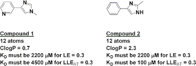
The index has some interesting implications. For example, the two fragments below have the same number of heavy atoms, and thus if they had the same activity they would have the same ligand efficiency; on this measure alone, neither would be preferred as a starting point for further work. However, because of their very different lipophilicities, fragment 2 would need to be 45 times more potent than fragment 1 in order to have the same LLEAT of at least 0.3.

A similar analysis can be done during optimization. For example, adding either a phenyl or a piperazinyl substituent should produce a 20-fold boost in potency in order to maintain ligand efficiency at 0.3, since both have 6 atoms. However, in order to maintain LLEAT at 0.3, the phenyl would need to produce a 460-fold boost in potency while the piperazinyl would need to improve potency only 3-fold. This is consistent with what other folks have reported qualitatively, but it’s nice to have a simple quantitative measure.

Although some people may groan at yet another index, and no metric is perfect, I like the fact that this one is intuitive and has the same range of “acceptable” values as ligand efficiency. What do you think – is it useful?

12 June 2011

Beware molecular obesity

Obesity in humans is a growing problem, and not just aesthetically: the condition may be responsible for millions of premature deaths. In a recent article in Med. Chem. Commun., Mike Hann of GlaxoSmithKline notes that “molecular obesity” is also leading to the untimely demise of far too many drug development programs.

Hann, who is especially known for his work on molecular complexity, defines molecular obesity as the “tendency to build potency into molecules by the inappropriate use of lipohilicity.” This is the result of an unhealthy “addiction” to potency. Hann suggests that since potency is easy to measure it is pursued preferentially to other factors, particularly early in a program. This is all too often achieved by adding mass, much of it lipophilic. The problem is that all this grease decreases solubility and increases the risks of off-target binding and toxic side effects.

Tools such as the Rule of 5 have been developed in part to avoid this problem, and a number of other indices have been introduced more recently. For example, lipophilic ligand efficiency (LLE) is defined as pIC50 – LogP; molecules with an LLE > 5 are likely to be more developable. Other guidelines that Hann mentions and that have been covered here include LELP, number of aromatic rings, and fraction of sp3 hybridized carbon atoms. But this is not to say that metrics will save the day:
The problem with the proliferation of so many “rules” is the trend to slavishly apply them without really understanding their required context for use and subsequent limitations.
Starting a program with the smallest possible lead should in theory lead to smaller drugs, and this is one of the key justifications for fragment-based approaches, though even here it is important that the molecules do not become obese during optimization. One of the themes at the 6th annual FBDD conference was to do a bit of optimization around the fragment itself before growing or linking. On a related note, Rod Hubbard warned in his opening presentation at the conference to “beware the super-sized fragment.” Not only are larger fragments likely to be less complementary to the target, the number of possibilities increases (and thus the coverage of chemical space drops) by roughly ten-fold with each atom added to a fragment.

Hann also argues that potency itself is over-rated: many teams seek single digit nanomolar binders even though approved drugs have average potencies of 20 nM to 200 nM.

The paper is a fun read (and is free after registration too). Also, Hann one-ups Donald Rumsfield’s (in)famous “known knowns, known unknowns, and unknown unknowns” by pointing out that much of this information falls into the category of “unknown knowns”:
Those things that are known but have become unknown, either because we have never learnt them, or forgotten about them, or more dangerously chosen to ignore
This review is an excellent corrective to the first two problems, and a clear warning about the third.

30 May 2011

Perverse trade-offs: the maximal enthalpy of ligands

Most readers of this blog are familiar with the concept of ligand efficiency:

LE = (free energy of ligand binding) / (number of heavy atoms)

The metric is easy to calculate, intuitive, and useful for evaluating fragments. Weirdly, as ligands get larger, the ligand efficiencies tend to decrease. This is a consequence of the fact that the maximal affinities of ligands also start to plateau as their sizes increase, a fact noted more than a decade ago by Kuntz and colleagues. The reason for this has never been satisfactorily explained, but the effect is so pronounced that some researchers have proposed alternatives to LE that take it into account, for example %LE and fit quality. In a paper published online in ACS Med. Chem. Lett., Charles Reynolds and M. Katherine Holloway have delved into the origins of declining LE with increasing size in more detail.

The researchers analyzed 102 ligand-protein complexes representing 14 target classes for which thermodynamic data had been collected using isothermal titration calorimetry (ITC). They calculated ligand efficiency as well as “enthalpy efficiency” and “entropy efficiency”, where enthalpy or entropy takes the place of free energy in the numerator. In the case of enthalpy efficiency, the trend was the same as for ligand efficiency: as molecules got larger, the enthalpy efficiencies tended to decrease. For entropy efficiencies, however, there was essentially no trend. In other words, the leveling out of binding energy is due to enthalpic effects more than to entropic effects. The authors suggest that:

The size effect on enthalpy is a result of increasing ligand complexity and the need to satisfy multiple geometric constraints simultaneously.

The findings thus provide further support for trying to identify fragments whose binding is largely enthalpically driven, but, as usual in the real world, things are not quite so easy. One of the frustrating things about medicinal chemistry is the phenomenon of enthalpy-entropy compensation: if you try to improve the enthalpy of binding, say by adding a hydrogen-bond acceptor, you may end up paying an entropic penalty such that overall binding increases only modestly, if at all. Indeed, the data in the paper show a very strong correlation between enthalpy and entropy: enthalpic binders tend to have negative (unfavorable) entropy, and the most entropic binders actually have positive (highly unfavorable) enthalpies of binding.

Worse, despite the striking correlation between enthalpy and entropy, there is almost no correlation between free energy of binding and enthalpy or entropy. The researchers suggest that these perverse trends (or lack thereof) explain why computational approaches to drug discovery are not more successful: most modeling relies on one or a few low-energy conformations, in effect ignoring entropy. However, there is a correlation between overall binding energy and enthalpy for certain targets, such as HIV protease and aldose reductase; perhaps such targets are more amenable to modeling.

In summary, the message seems to be that you should try to find enthalpic binders if you can, though the binding energetics may change unpredictably during optimization. For modelers, the research suggests that ignoring entropy is unwise. For medicinal chemists, I’m not sure how much of this is actionable, but sometimes it’s nice to know the origins behind the perversity of the universe.

16 May 2011

Native MS: turning up the voltage

At the FBDD meeting in San Diego last month there was some discussion of the connection between protein-ligand binding in the gas phase (ie, in native mass-spectrometry experiments) vs in solution; the discussion continued in the comment section here. Valerie Vivat, head of the mass spectrometry, molecular & cell biology department at NovAliX, has now provided a thorough response.

When analyzing non-covalent complexes by mass spectrometry under non-denaturing conditions, one must keep in mind indeed how interactions stabilizing the complex in solution will be affected by the ion transfer in the gas phase. Basically, interaction based on hydrophobic effect is lost while the electrostatic-based interactions (Van der Waals, H-bonds, ionic interaction) are preserved (and are even strengthened due to the absence of solvent shielding). Regarding water molecules now, during the ESI process, biomolecules are transferred in the gas phase as partially hydrated ions and complete ion desolvation is subsequently achieved through low energy collisions with the residual gas molecules in the mass spectrometer interface. This complete desolvation is required for accurate mass measurements. Consequently, in most cases, molecular mass measurement of the complexes is accurate enough to rule out the possibility that water molecules remain after ions are transferred from the atmospheric pressure to the deep vacuum of the TOF analyzer, even in the case of crystallographic water molecules.

Regarding gas phase stability : Reports in the literature mention that collision-induced dissociation experiments and the determination of Vc50s can be used to monitor gain or loss in polar interaction. Indeed, we have now accumulated a variety of in house examples showing that increase in the gas phase stability of a complex is correlated with a gain in polar interactions as shown by X-ray or ITC. Monitoring the complex gas phase stability is thus an attractive feature to quickly evaluate, for example, gain or loss in polar interaction of analog series targeting the same protein binding site.

Importantly, the gas phase stability is not an indication of the complex binding affinity. Evaluation of complex affinity (after complexes are formed in solution) is done under instrumental conditions showing no dissociation of the non-covalent complexes. For complexes stabilized by electrostatic-based interactions (and thus rather stable in the gas phase), it is indeed much easier to find optimal instrument settings compared to complexes stabilized by hydrophobic effect. However, this does not preclude complexes mainly stabilized in solution by hydrophobic effect to be detected at all. For example, nuclear hormone receptors in complex with fatty acids or phospholipids are readily detected with appropriate settings. In this case, the polar head of the ligands is likely to act as a stable anchor in the gas phase, resulting in non-covalent complexes which remain intact during the ca. 1 millisecond flight of the ion from the ionization source to the detector.
To sum up, native MS can be run under two different modes: 
1.     Low energy conditions where no complex dissociation occurs are used to assess the binding affinity of protein / ligand complexes,
2.     High energy conditions inducing complex dissociation provide insight into the extent of polar interaction involved between the protein and the ligand. This type of experiment makes sense to compare a series of molecules binding to the same protein binding site.

Valerie makes some interesting points here, in particular the statement that "the gas phase stability is not an indication of the complex binding affinity." Still, I do like the fact that native MS could be used as a quick way to sort enthalpic from entropic binders. What do you think?

15 April 2011

Sixth Annual Fragment-Based Drug Discovery

The only US-based conference completely devoted to fragment-based drug discovery ended in San Diego this week. As with last year, I won’t attempt to summarize all of the talks – there was far more information presented than I have time to write (or that you probably have patience to read!) For those of you who were there, please feel free to mention some of the things I missed.

One of the points that Don Huddler (GlaxoSmithKline) and I (Carmot) made in the pre-conference short-course is that finding fragments is a solved problem. As Rod Hubbard (Vernalis, University of York) noted in his opening presentation, “it’s pretty simple to find fragments that bind; a graduate student can do it in a couple months.” Even membrane proteins are starting to yield to fragment-based screening, as Gregg Siegal (ZoBio, Leiden University) discussed in his closing session (see also here).

That’s not to say that new methods for finding fragments aren’t useful, particularly if they open new target space, are faster or more reliable, or provide new information. An example of the latter was the presentation by Denis Zeyer (NovAliX) on native mass-spectrometry (see also here). Because hydrophobic interactions are weaker in the gas phase than in water, it should be possible to select for molecules that bind predominantly through polar interactions. In fact, by gradually increasing the voltage in their MS instrument, Zeyer and colleagues generated “VC50” curves, the voltage at which half the compound dissociates from the protein. At least in one case, a higher VC50 correlated with the presence of an additional hydrogen bond to the protein compared with related molecules.

Polar contacts are generally associated with enthalpic rather than entropic interactions, and whether such fragments are preferable was the subject of some discussion, particularly at a breakfast round-table discussion. In contrast to a meeting just last year, several participants were actively collecting thermodynamic data, though there was some uncertainty as to what to do with it. This is a controversial subject; one person suggested that enthalpic binders are likely to be more hydrophilic than entropic binders, so just keeping an eye on lipophilicity is likely to be just as useful and far easier than actually measuring thermodynamic parameters. Charles Reynolds (Ansaris) provided an analysis that illustrates some of the difficulties in using thermodynamic data – I’ll follow up on this in a later post.

The shape of fragments has been previously discussed, and Ivan Efremov (Pfizer) gave a nice case study of a strikingly three-dimensional fragment: an X-ray screen of 340 molecules against BACE resulted in a single hit, a spirocyclic pyrrolidine. The electron density of this was so clear that it didn’t even need to be deconvoluted from the other three compounds in the pool, and medicinal chemistry ultimately led to low micromolar inhibitors.

There was general consensus that ligand efficiency (and various lipophilicity adjusted versions) is a helpful metric. One practitioner said that his company had sometimes pursued more chemically tractable but less ligand efficient fragments and generally came to regret those decisions. But a fragment with lower ligand efficiency could still be interesting: with fragments, even small changes could have huge effects on binding (see for example AT13387, which was discussed by Chris Murray of Astex). Thus, a bit of initial fragment optimization could be a good investment before pursuing more intensive chemistry, particularly if commercial or in-house analogs are available. Interestingly, I couldn’t find anyone who uses either fit quality or %LE.

In the early days of fragment-based lead discovery a common selling point was that it sped up drug discovery, but a theme in this meeting was that it is not necessarily faster but can provide leads against more difficult targets or better leads against “normal” targets. Of course, one has to be wary of taking a good fragment, slapping a bunch of grease on it, and turning it into a lipophilic monster.

Indeed, an analysis of fragment-derived leads published a couple years ago was not flattering. Taking up the thrown gauntlet on behalf of fragments, Chris Murray presented a retrospective analysis of all 42 fragment to lead programs at Astex (including 21 kinases and 9 proteases). The average parameters of these leads were considerably more attractive in terms of both molecular weight and ClogP that the published values of the HTS hits. At least according to this analysis, fragment approaches have the potential to deliver superior molecules, as long as one is disciplined and creative in how these approaches are applied.

10 March 2011

Growing into closed pockets

Fragment-growing is a popular way to increase the activity of fragments, all the more so when there is an obvious place towards which to grow. In a paper published online in the J. Am. Chem. Soc., Iwan de Esch and colleagues at VU University Amsterdam describe the structural and thermodynamic consequences of one such effort, and conclude that binding in a certain normally closed pocket is enthalpically rather than entropically driven.

The researchers were interested in acetylcholine-binding protein (AChBP), a soluble and crystallizable homolog of an important class of ligand-gated ion channels. This is the same protein (and the same group) highlighted last year in the context of ligand efficiency hot spots. In the current work, a fairly potent fragment, compound 1, was co-crystallized with AChBP and the structure solved. This fragment binds in roughly the same position as the more potent natural product lobeline (compound 2), but lobeline contains a hydroxyphenethyl group that the fragment lacks (see figure). This moiety binds in a hydrophobic pocket that does not appear in the fragment complex due to the movement of a tyrosine residue. Recognizing the potential for the pocket to form, the researchers introduced this moiety into their own molecule, producing compound 3.

Gratifyingly, compound 3 binds about 50-fold more tightly than the initial fragment. This molecule was also co-crystallized with AChBP, and, as designed, the phenyl group binds in the “lobeline pocket". Moreover, compound 3 does not show a corresponding increase in affinity towards a version of AChBP from a different organism that does not have this pocket.

To correlate binding mode with thermodynamics, the researchers also characterized the binding of their compounds using surface plasmon resonance (SPR) and isothermal titration calorimetry (ITC). ITC measures enthalpy directly, and performing SPR analyses at different temperatures can also be used to dissect enthalpic and entropic terms. The two methods generally concurred, though the numbers did jump around a bit, and in a few cases SPR predicted negative (ie, unfavorable) entropy where ITC suggested positive (favorable) entropy for the same compound.

Both SPR and ITC indicated that the increase in potency for compound 3 over compound 1 is driven by enthalpy, not entropy. This is somewhat unexpected, as the contacts made by compound 3 are largely hydrophobic, and the simplistic view is that such contacts are usually entropy-dominated. (The added hydroxyl in compound 3 doesn’t appear to be doing anything useful, and in fact removing it increases potency roughly three-fold). The researchers suggest that, for poorly solvated hidden pockets such as this, enthalpy may dominate. Perhaps also the protein rearrangement necessary to open the pocket is entropically costly.

There is much more data in the paper than can be summarized here, and the notion that ligands that induce conformational changes in proteins could be enthalpic rather than entropic binders is an intriguing hypothesis. However, as a vigorous debate last year demonstrates, it is still unclear whether knowing the answer – as scientifically interesting as it may be – will have practical implications for drug discovery.