30 August 2021

Covalent fragments meet high-throughput synthesis and crystallography

Two hot areas of FBLD include covalent fragments (see here for example) and high-throughput crystallography (see here and here). Two new papers add to the excitement but also reveal some of the challenges.
 
The first, published open access in J. Med. Chem. by Charles Eyermann (University of Cape Town), Christopher Schofield (Ineos Oxford Institute of Antimicrobial Research), Christopher Dowson (University of Warwick) and collaborators at Diamond Light Source focuses on an antibacterial target, the penicillin binding protein PaPBP3 from Pseudomonas aeruginosa.
 
Some members of the group had previously screened a library of 40 compounds against this protein and identified a single hit that bound covalently. Inspired by this result, they assembled a library of 262 commercial covalent fragments, 152 of which contained boron, an element known to react with serine and threonine hydrolases. These were screened crystallographically at 250 mM, resulting in 34 boron-containing hits “with various levels of electron density observed at the active site.” Many of these seem to have given ambiguous density, and only a handful of fragment structures are reported.
 
Next, the researchers attempted fragment growing; the team ultimately obtained 10 structures, drawing from the original fragments and the elaborated molecules. Interestingly, these showed three different binding modes: a monocovalent complex with the active-site serine, a dicovalent complex with the active-site serine and a neighboring serine, and a tricovalent complex with these two serine residues and a nearby lysine side chain.
 
Despite having up to three covalent bonds to the enzyme, even the elaborated molecules showed at best double-digit micromolar inhibition of PaPBP3, and none showed antimicrobial activity. The bond between boron and serine or lysine is reversible, so while disappointing, this weak activity is not entirely surprising. The result is reminiscent of efforts against the SARS-CoV-2 main protease, Mpro or 3CLpro, where many of the crystallographic hits turned out to be very weak binders.
 
The second paper, published open access in Angew. Chem. Int. Ed. by Alexander Dömling and collaborators at University of Groningen and the Paul Scherrer Institute, also looked at 3CLpro. Here though, rather than starting with commercial fragment libraries the researchers used high-throughput synthesis to make their own.
 
The Dömling lab has had a long-standing interest in multi-component reactions in which several reagents are combined to generate products. The researchers explored the Passerini reaction (which uses an aldehyde or ketone, a carboxylic acid, and an isocyanide) and the Ugi reaction (which uses all of these as well as an amine). For the carboxylic acid, they chose an electrophile such as acrylic acid. They initially worked out the conditions at 0.5 mmol scale in 96-well plates and then purified the products using chromatography or precipitation. The majority of wells yielded product in good yields and purity.
 
Next, the researchers turned to high-throughput methods, performing the reactions in 384-well plates using acoustic liquid handling. This is similar to the approach I wrote about here that was used to discover covalent KRAS inhibitors, ultimately leading to the approved drug sotorasib.
 
The researchers then soaked 181 of their compounds against the SARS-CoV-2 protein 3CLpro, with each compound at 10 mM. Unlike the crude reaction screening described here, the researchers used pure compounds. This effort resulted in five hits, though one of them turned out to bind noncovalently. Three of the molecules had low micromolar activity.
 
In the end, while both of the papers do report the discovery of covalent modifiers, the functional activities in the first paper are modest, and the active warheads in the second paper (chloroacetamides and acrylates) are likely too reactive to be advanceable. A nice feature of crystallography is that it can provide structural information for extremely weak hits. But as we’ve asked previously, how weak is too weak? Getting more crystal structures more rapidly is one thing, but figuring out which ones are useful requires even more skill, creativity, and luck.

23 August 2021

Fragments vs DYRK1A and DYRK1B: Part 2

Last week we highlighted work out of Vernalis and Servier in which fragment-based methods were used to identify potent and selective inhibitors of DYRK1A and 1B, potential targets for cancer and neurodegenerative diseases. The NMR screens yielded 166 hits, only one of which was advanced in that paper. A second J. Med. Chem. paper by Andras Kotschy and collaborators describes the optimization of another fragment.
 
Compound 1 is a whoppingly potent fragment with impressive ligand efficiency. If you’ve ever worked on kinases you probably think you know how it binds, as the diaminopyrimidine moiety is a common hinge-binding motif. In fact, crystallography revealed that the molecule binds in a completely different orientation and that the methoxy group makes a single hydrogen bond to the hinge amide NH. Cyclizing the molecule led to compound 10, with a satisfying boost in affinity.

 
Unfortunately, compound 10 was also a potent inhibitor of the kinase CKD9. To gain selectivity, the researchers took advantage of the fact that one of the backbone carbonyl oxygens in the hinge adopts an unusual orientation in DYRK1A, making room for the methyl group in compound 33. Next, the researchers replaced the benzofuran core for reasons of “synthetic tractability, metabolic stability, and freedom to operate.” This exercise ultimately led to compound 40.
 
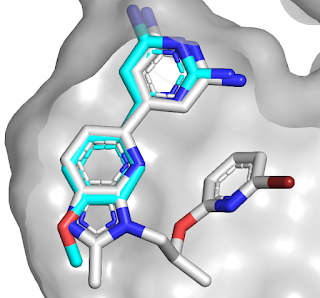
This compound was profiled against 442 kinases and found to be quite selective, with only 8 kinases significantly inhibited at 1 µM. One of these was the related kinase DYRK2, but further growing led to selective compound 58. An overlay of the initial fragment (blue) with compound 58 (gray) reveals how the binding mode has been maintained, in contrast to the series described last week.

Compound 40 had only modest antiproliferative activity against human cancer cell lines that were grown in 2D culture but was more active when the cells were grown in 3D culture. The molecule had good oral bioavailability in mice, and xenograft studies revealed that it inhibited tumor growth, though it was also toxic at higher doses. The researchers do not mention brain penetration, though given the number of hydrogen bond donors I would be surprised if it crosses the blood-brain barrier.
 
This paper is a nice example of how getting high affinity is often only the beginning of a long journey. In combination with the story from last week it is also a useful reminder of how many starting points a single fragment screen can provide: just two fragments led to two completely independent series. Whether molecules from these series advance to the clinic, they provide useful tools to further understand the biology of DYRK1A.

16 August 2021

Fragments vs DYRK1A and DYRK1B: Part 1

The dual-specificity tyrosine-phosphorylation-regulated kinases 1A and 1B (DYRK1A and DYRK1B) belong to a family of five serine/threonine kinases implicated in several cancers as well as Down’s syndrome and other neurodegenerative diseases. For the latter indications in particular, brain penetration would be essential for any inhibitor, just as in the LRRK2 story last week. In a new (open access) J. Med. Chem. paper, Rod Hubbard and collaborators at Vernalis and Servier describe the discovery of a chemical probe.
 
The researchers started by testing their in-house library of 1063 fragments in pools of six, each at 500 µM, in three ligand-detected NMR screens. This resulted in a whopping 166 hits. Crystal structures of the eight most ligand-efficient fragments bound to DYRK1A were obtained, including compound 5. Fragment growing led to compound 16, which bound the kinase 200-fold more tightly. 
 

The crystal structure of compound 16 bound to DYRK1A was compared to structures of other known ligands and suggested the possibility for an alternative binding mode. This led to the synthesis of compound 24, with low nanomolar affinity against both DYRK1A and DYRK1B (only the former is shown in the figure). This compound turned out to be surprisingly unstable in slightly acidic aqueous solution (below pH 5), but replacing the oxygen with a nitrogen fixed this, and further tweaking ultimately led to compound 34.
 
Compound 34 was profiled at 1 µM against a panel of 442 kinases and found to be fairly selective, with only 15 kinases inhibited by at least 50%. It is orally bioavailable in mice, brain penetrant, and inhibited the proliferation of glioblastoma cells, although the potency was significantly attenuated by serum. In a xenograft study the compound caused tumor growth delays and was well-tolerated.
 
This is a nice example of fragment-based lead discovery heavily dependent on structural information. Comparing the binding mode of compound 34 (gray) with that of compound 5 (light blue) reveals the significant shift in binding mode of the initial fragment.

The paper is also a useful reminder of how long it can take for industry research to be published. Work began in 2009, and Rod presented some of it at the CHI FBDD conference in 2019. But this is not the end of the DYRK1A story: stay tuned for next week!

09 August 2021

Fragments vs LRRK2 with the help of a surrogate and a magic methyl

Leucine-rich repeat kinase 2 (LRRK2) has been implicated in Parkinson’s disease and has thus long been targeted by drug hunters. Dozens of inhibitors have been approved for other kinases, making the protein class appear “easy”, but LRRK2 is particularly challenging. First, it is a large multidomain protein that has resisted crystallography. Second, an inhibitor for a chronic disease such as Parkinson’s will need to be highly selective. Finally, the fact that LRRK2 is in the brain means that inhibitors will need to cross the treacherous blood-brain barrier (BBB), whose function is to exclude anything unusual. A recent J. Med. Chem. paper by Douglas Williamson and collaborators at Vernalis and Lundbeck addresses the first two of these issues.
 
The researchers started by screening 1313 fragments (at 200 µM each) against the disease-relevant G2019S mutant of LRRK2. The screen was run using the DiscoveRx KINOMEscan, which relies on displacement of a kinase from an immobilized ligand. Some 80 hits were then triaged in a kinase activity assay.
 
Rather than banging their heads against the wall that has blocked X-ray structures of LRRK2, the researchers turned to a crystallographic surrogate. The readily crystallizable kinase CHK1 has some similarity to LRRK2, and some inhibitors bind to both kinases. Introducing ten mutations into CHK1 around the ATP-binding site led to a LRRK2 surrogate – an approach we’ve previously mentioned.
 
Among the fragment hits were adenine (compound 7) and two closely related molecules. Crystallization of compound 7 with wild-type CHK1 and the LRRK2 surrogate revealed two different binding modes. Similarity-based searches of in-house and literature compounds led to molecules with nanomolar potency, and subsequent optimization led to compound 17 (all molecules were tested against both wild-type LRRK2 as well as the G2019S and had similar affinities).
 

Interestingly, crystal structures of these molecules revealed them to bind more similarly to the complex of compound 7 bound to wild-type CHK1 rather than the surrogate. Adding a methyl group to compound 17 led to compound 18 with a satisfying 100-fold boost in potency: a true magic methyl, as the other enantiomer had slightly worse affinity than compound 17. Crystallography with the surrogate revealed hydrophobic interactions between the methyl and an alanine side chain. It is worth noting that compound 18 is still fragment-sized and yet has high picomolar affinity for LRRK2.
 
Extensive medicinal chemistry followed, ultimately leading to compound 45. Profiling against 468 kinases in the KINOMEscan assay demonstrated it was quite selective, binding only three other targets with dissociation constants less than 1 µM. The compound was active in cells containing either wild-type or G2019S LRRK2. Unfortunately, while the compound showed good oral bioavailability in dogs, it was not orally bioavailable in rats. Moreover, brain to plasma ratios were low in mice, and the molecule was a substrate for the human protein BCRP, a transporter that pumps small molecules across the BBB.
 
Although these liabilities halted further work on the series, this is nonetheless a nice fragment to lead story that highlights the utility of crystallographic surrogates. But the different binding modes for the initial fragment are a reminder that multiple binding modes are not uncommon, and it is best to employ crystallography not just early but often, when you can.

02 August 2021

Linking fragments on DNA, revisited

Three years ago we discussed using DNA-encoded libraries to find and link fragments. In a new open-access Bioorg. Med. Chem. article, Nicolas Winssinger and colleagues at University of Geneva report a different version of this approach.
 
Rather than using DNA, the researchers constructed their libraries with peptide nucleic acids (PNAs), which can be assembled using traditional solid-phase peptide synthesis and will also hybridize to DNA. Each PNA is coupled to a different fragment, and the fragment-PNA molecules are then bound to microarrays of DNA such that two fragment-PNA molecules bind to a single DNA strand. In this case the researchers used 250,000 combinations of fragment pairs.
 
Next, a protein of interest (here the anti-apoptotic cancer target BCL-xL) was screened at 50 nM. Binding to specific pairs of fragments was assessed by fluorescent detection of the protein at various spots on the microarray.
 
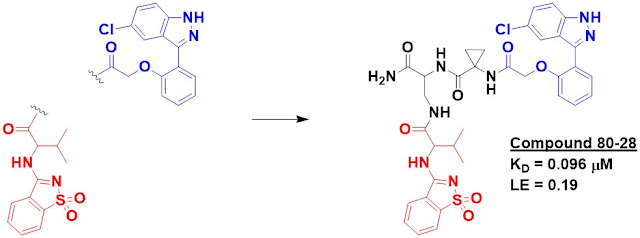
Trying to figure out which of the fragments are best – and how to link them – is “not trivial,” so the researchers took a combinatorial approach. Based on the first screen, they generated a new library of 10,000 molecules in which 10 sulfonamide-containing fragments were linked to 100 heterocycle-containing fragments using 10 different linkers. These compounds were screened using the same microarray technology, and the best binders were then resynthesized with a biotin tag rather than the PNA.
 
The biotinylated molecules were able to pull down recombinant BCL-xL in solution. Two of them, including compound 80-28, were even able to pull down recombinant protein that was spiked into cell lysate. Importantly, neither fragments 80 nor 28 did this by themselves. The affinity of 80-28 was measured by SPR to be 96 nM. 
 
 
Finally, the researchers tested 80-28 in K562 cells and found that it was cytotoxic with EC50 = 1.7 µM. They compare this favorably to venetoclax, the second fragment-based drug to be approved. However, this is a disingenuous comparison: venetoclax was specifically designed to bind less tightly to BCL-xL than to the related protein BCL-2. A more appropriate comparison would be a molecule such as navitoclax or the specific BCL-xLbinder A-1155463.
 
Like most BCL-family binders, compound 80-28 is also a rather unusual looking molecule, with a high molecular weight of 735. Unlike navitoclax and venetoclax, it also has 7 hydrogen bond donors and many more rotatable bonds. The long floppy linker in particular is something the earlier DNA-based fragment-linking work sought to fix. As we noted then, such linkers may be an inherent liability with the approach. From a technology perspective this is interesting work. But from a drug discovery perspective it still has some way to go to prove itself practical.