01 August 2022

What rings are found in drugs?

Recently we highlighted the “Ring Replacement Recommender,” which provides suggestions for how to improve affinity by replacing one ring with another. The recommendations are based on an analysis of hundreds of thousands of molecules. But what about the rings found in actual drugs? This is the focus of a J. Med. Chem. paper by Richard Taylor and collaborators at UCB and Bohicket Pharma Consulting.
 
The researchers examined FDA-approved and investigational drugs with disclosed structures as of January 2020. These were fragmented into component “ring systems” for analysis. (Ring systems include not just monocycles but fused rings, such as purine. For example, sotorasib consists of four ring systems: benzene, pyridine, piperazine, and pyrido[2,3-d]pyrimidin-2-one.) More than 90% of drugs contain at least one ring.
 
Approved drugs have just 378 unique ring systems in total – a small increase from when the researchers examined approved drugs in 2014. The phenyl ring is found 727 times, with pyridyl (86 examples) a distant second, followed by piperidine (76 examples) piperazine (65 examples) and cyclohexane (47 examples). After that the numbers drop off sharply, with pyrazine in 50th place with just six examples and fluorene in 100th place with three examples.
 
Investigational drugs at first appear to be more diverse, with 450 unique ring systems, 280 of which are not found in approved drugs. Of these 280, pyridazine is the most common, with nine examples, followed by oxetane, with seven, but things quickly become less common from there, with 271 of the ring systems found just once. In contrast, ring systems found in drugs are found in multiple compounds, and in fact two thirds of investigational drugs only contain previously used ring systems.
 
Many of the new ring systems are closely related to those found in approved drugs, with nearly half differing by at most two atoms. Perhaps because of this the overall properties of the ring systems are similar between approved and investigational drugs, with no significant differences in heteroatom ratio, percentage of sp3 centers, or number of rings per system.
 
What new opportunities exist? The researchers identified nearly half a million synthetically accessible ring systems and winnowed these down to 3902 ring systems that have similar heteroatom ratios to those found in drugs and differ by at most two atoms. This attempt to explore new chemical space is similar to earlier work from the same group (here) as well as that from others (here, here and here).
 
The researchers also examined growth vectors and combinations of rings, the latter by using graph theory. These analyses suggest that investigational drugs do have greater variety. In other words, even if the component rings are shared with approved drugs, they might be combined in new ways.
 
Whether certain ring systems are more likely to fail in the clinic was intentionally not addressed, due to the difficulty of assessing why the failures occurred. For example, drugs can fail for commercial reasons; a company may choose to drop a drug against a particular target rather than be tenth to market. And even when the failure is due to the science, it might not be an indictment of the drug itself. Verubecestat did lower β-amyloid levels in people as designed, but had no effect on Alzheimer’s disease.
 
This paper is a fun read, and it will likely provide ideas for scaffold hopping and library design. It is also a reminder of how much chemical space remains to be explored.

25 July 2022

Fragments vs TEAD: noncovalent this time

Last week we described a fragment-derived covalent probe that targets the four closely related TEAD transcription factors, which are part of the Hippo signaling pathway implicated in some cancers. A new paper in J. Med. Chem. by Timo Heinrich and collaborators at Merck KGa, iBET, and Cancer Research Horizons brings us another fragment-derived probe, this one noncovalent.
 
The researchers started by screening 1930 fragments, each at 2 mM, against TEAD1 and TEAD3 using SPR. Perhaps not surprisingly given the high concentration used, this led to a whopping 560 hits. These were then tested in dose-response format against TEAD1 with or without the coactivator YAP; 254 compounds showed differential affinity, among them compound 1. This molecule was crystallized bound to TEAD3, which revealed that it binds to the hydrophobic pocket normally occupied by a covalently-bound palmitoyl group required for activity. Despite being a fragment, compound 1 was active in a cell reporter assay, and the researchers state that further optimization was done using cellular assays rather than biophysical or biochemical experiments.
 

Analysis of the crystal structure suggested that enlarging the cyclopentyl moiety could fit more snugly into a hydrophobic pocket, while adding a small propyl moiety could extend into a separate pocket, leading to compound 6, with a 10-fold boost in activity. Replacing the propyl with an additional ring led to sub-micromolar compound 9. Finally, replacing the saturated ring with a substituted phenyl moiety led to MSC-4106, with low nanomolar activity in the cell reporter assay.
 
Thermal stabilization (specifically, nanoDSF) assays showed that MSC-4106 stabilized TEAD1 and TEAD3 but not TEAD2 or TEAD4. Palmitoylation assays confirmed this selectivity profile. The paper also includes a nice table comparing experimental selectivities of seven other non-covalent TEAD inhibitors, which vary from having activity only against TEAD1 to activity against all four homologs.
 
MSC-4106 was clean when tested at 10 µM against a panel of 58 receptors and 1 µM against nearly 400 kinases. It did not inhibit hERG or any of the common CYP450s. Finally, PK studies in mice, rats, and dogs showed that the compound is orally bioavailable with a long half-life. Given these favorable properties it was taken into xenograft studies, where it showed tumor growth inhibition at 5 mg/kg and tumor regression at 100 mg/kg. Analysis of tumor tissue showed downregulation of a TEAD-regulated gene, Cyr61.
 
Can we draw any lessons from comparing covalent MYF-03-176 (discussed last week) with non-covalent MSC-4106? Probably not, given that the former hits all TEAD homologs while the latter is selective for TEAD1 and TEAD3. Both molecules look to be excellent chemical probes for further dissecting Hippo signaling. I look forward to seeing how TEAD inhibitors ultimately fare in the clinic.

18 July 2022

From covalent fragment to lead against TEAD

As noted just last month, covalent fragment-based drug discovery is becoming ever more popular. However, many papers report relatively weak hits with little or no optimization. A new preprint posted to bioRxiv (HT Covalent Modifiers) by Tinghu Zhang, Nathanael Gray, and collaborators at Stanford and elsewhere describes a fragment-to-lead story for the TEAD family of transcription factors.
 
The four highly homologous members of the TEAD family play a role in the Hippo signaling pathway. When spurred by the coactivator YAP they cause gene expression that has been implicated in certain cancers, particularly mesothelioma. To bind YAP, TEAD needs to be palmitoylated on a specific cysteine residue. A covalent inhibitor that binds to this cysteine could prevent palmitoylation and thus block Hippo signaling.
 
Multiple academic and industrial groups have been pursuing this target, and one previously reported inhibitor is flufenamic acid. This molecule was used in the new paper to design a small library of analogs each functionalized with an acrylamide moiety. These were screened against TEAD2 and analyzed by mass spectrometry; MYF-01-37 modified the protein (though unfortunately time and exact concentrations are not specified). Proteolysis and tandem mass spectrometry confirmed that the molecule binds to C380, the site of palmitoylation.
 
Analysis of previously published crystal structures revealed a side pocket off the main hydrophobic channel that normally binds the palmitoyl group. The researchers created a focused library of analogs to try to access this pocket, which led to molecules such as MYF-03-69. This compound was active in a biochemical assay and showed rapid labeling of the protein as assessed by mass spectrometry. A crystal structure of the compound bound to TEAD1 confirmed the molecule forms a covalent bond to the target cysteine and does in fact bind in both pockets. 
 

MYF-03-69 inhibited palmitoylation of all four TEAD paralogs in biochemical assays. More importantly, it showed activity in several cell assays, including blocking palmitoylation and disrupting the interaction between TEAD and YAP. The molecule downregulated YAP-TEAD transcription in reporter gene assays as well as RNA sequencing assays. Finally, MYF-03-69 showed mid-nanomolar antiproliferative activity in mesothelioma cells but not in non-cancerous cell lines.
 
Despite this promising activity, MYF-03-69 lacked acceptable oral bioavailability. Further medicinal chemistry led to MYF-03-176, which has improved bioavailability and showed even better activity in reporter gene assays and better antiproliferative activity in mesothelioma cell lines. The molecule also led to tumor regression in a mouse xenograft model when dosed orally.
 
This is a nice story with lots of information, though were I a reviewer I would ask for the kinact/KI values for the molecules. This ratio describes the rate of covalent modification and is time and concentration independent, which makes comparisons with other molecules more straightforward (see this 2017 open-access paper for a good discussion). Since this is a preprint hopefully the final published paper will include these values. 
 
Regardless, MYF-03-176 looks like an excellent chemical probe for studying the effect of irreversible inhibition of Hippo signaling.

11 July 2022

Fragments in the clinic: HTL9936

Of the 50+ fragment-derived drugs that have entered the clinic, only two (both from Sosei Heptares) target transmembrane proteins, reflecting the difficulty of structure-based design for this hard-to-crystallize class of proteins. The story behind one of them was published late last year in Cell by Malcom Weir, Andrew Tobin, and a large group of collaborators.
 
The researchers were interested in the M1 muscarinic acetylcholine receptor, which is involved in memory and learning. By activating the receptor the hope is to be able to treat symptoms associated with Alzheimer’s disease. The M1 receptor has been a long-standing target for this disease, but previous drugs have caused side effects ranging from salivation and sweating to gastrointestinal distress and seizures. The M1 receptor is one of five closely related subtypes, and some of the side effects have been attributed to hitting the M2 and M3 receptors. However, the M1 receptor itself may also not be entirely innocent, so the goal was to develop a partial agonist, the idea being that this may be more effective in the brain, where the M1 receptor is highly expressed, while sparing other tissues where the M1 receptor is rarer.
 
The campaign began with a virtual screen of 1.6 million molecules (with molecular weights up to 400 Da) against a homology model of the human M1 receptor bound to a known agonist. This led to the purchase of 322 compounds, of which 16 were active in a cell-based functional assay, including compound 4. Fragment growing led to compound 6 and ultimately to HTL9936, which is selective for M1 over M2, M3, and M4 receptors. It also showed no significant agonism against a panel of 62 GPCRs even at 10 µM concentration.
 

Sosei Heptares pioneered the use of mutagenesis to stabilize specific conformational states of GPCRs, and this process was used to produce co-crystals with HTL9936 to understand its binding mode. Like other reported agonists, which were also characterized crystallographically, HTL9936 binds in the orthosteric site of the M1 receptor, but the increased size of the homopiperidine ring relative to other ligands provides selectivity over other receptors such as M2.
 
HTL9936 was tested in mice, rats, dogs, and cynomolgus monkeys, and in general showed good safety and brain penetration. The molecule even showed cognitive benefits in a mouse model of neurodegeneration and in aged beagles. It did cause an increase in heart rate and blood pressure in dogs, and there was a single convulsive episode, but only at a very high dose.
 
The paper also summarizes the results of human clinical trials which demonstrated that HTL9936 is well tolerated up to 100 mg doses, though at higher doses sweating, salivation, and changes in heart rate and blood pressure were observed. A small trial in healthy elderly people did not show any improvement in memory tasks, though functional magnetic resonance imaging studies did show that the molecule activated regions of the brain associated with cognition.
 
And that’s where the story ends. The Sosei Heptares website does not list HTL9936, though a different M1 receptor agonist (HTL0018318) is described. This paper also illustrates the long gap that can occur between research and publication: ClinicalTrials.gov lists three Phase 1 studies for HTL0009936, one of which began in 2013, and all of which ended by early 2017. Like most approaches to Alzheimer’s disease that have been tested, perhaps targeting the M1 receptor is a dead end. But reaching that conclusion requires highly selective chemical probes. Kudos to the team at Sosei Hetpares for their efforts.

03 July 2022

What belongs in the Protein Data Bank?

The rise of high-throughput crystallography is among the most exciting recent developments for fragment finding. Historically deemed too slow for primary screening, crystallography was reserved for select hits from an assay cascade. Now crystallographic screens up-front sometimes yield hundreds of hits. Many have been deposited in the Protein Data Bank (PDB). In a recent (open access) Protein Sci. commentary, Mariusz Jaskolski (Mickiewicz University), Bernhard Rupp (Medical University Innsbruck), and collaborators in the US question this practice.
 
In particular, the researchers ask whether molecules processed using Pan-Dataset Density Analysis (PanDDA) belong in the PDB. The method, which we described here, is typically used when hundreds of compounds have been soaked into crystals of the same protein. Most molecules will not bind, and these empty structures can be averaged to provide a background map to better identify weakly-bound ligands that may have only partial occupancy.
 
The researchers seem suspicious of this technique, referring to “supposed ligands” that may “confuse most biomedical researchers” and “degrade the PDB integrity,” the effect of which “could be disastrous.” To support their argument, they provide two examples from the PDB where the atomic models diverge from the electron density calculated using conventional methods and one with wonky statistics.
 
To avoid “contamination of the PDB by suboptimal structures,” the researchers suggest depositing structures from large-scale crystallographic screens in a separate database. Alternatively, they suggest clearer annotation. (To be fair, all three of the examples cited are already prominently marked “PanDDA analysis group deposition.”)
 
Needless to say, this is controversial. In a bioRxiv preprint, Manfred Weiss (Helmholtz-Zentrum Berlin) and collaborators in the US, Germany, Sweden, and the Netherlands, some of whom co-developed PanDDA, take a different view.
 
The researchers agree that group depositions need to be marked clearly, but they argue that they squarely belong in the PDB rather than in a separate repository. Moreover, “commentaries that underestimate the knowledge of PDB users, that ignore the opportunities present in heterogenous crystallographic data, and that miss out on chances for education on structure quality do more harm than good.”
 
The three examples described by Jaskolski and colleagues are re-examined, and while it is true that two of them do show poor occupancy using conventional methods, the ligands are clearly visible when PanDDA is used. (In the third case, there was an error in the resolution cutoff during automated processing, but the data could be successfully reprocessed manually.)
 
PanDDA was developed specifically to identify small, low occupancy ligands, so the researchers argue that these entries “cannot and should not be treated in the same way” as other ligands. Banning them from the PDB would potentially impede future research.
 
Weiss and colleagues refer to the Structural Genomics campaign of the late 1990s and early 2000s to solve myriad structures of diverse proteins, most of which were not being otherwise studied. At the time some commentators derided this effort as “stamp collecting.” Yet the number and diversity of structures thus deposited into the PDB likely contributed to the success of automated protein folding algorithms such as AlphaFold2.
 
Similarly, including structures from PanDDA processing could lead to unforeseen advances. For example, Weiss and colleagues suggest we may be able to “extract all aspects of conformational as well as of compositional heterogeneity out of all these data sets.” A better understanding of the role of protein dynamics in ligand binding is likely to require thousands of similar datasets of the kind being uploaded.
 
Personally, I believe that scientists should be wary of all published information. As the old saying goes, trust, but verify. As evidenced by my five-part series “Getting misled by crystal structures,” even conventional structures in the PDB should not necessarily be taken at face value. With that precaution, I’ll hold with the conclusion of Weiss and colleagues: “As long as the data is there, let’s embrace it and make it available!”

27 June 2022

CovPDB: a free, searchable database of covalent protein-ligand structures

Last week we highlighted KinaFrag, a database of kinase-fragment complexes. Continuing the theme, this week brings us CovPDB, a database of high-resolution covalent protein-ligand structures. The database was described by Stefan Günther and colleagues at Albert-Ludwigs-Universität Freiburg in an open-access Nucleic Acids. Res. paper earlier this year.
 
The researchers downloaded all structures from the protein data bank (PDB) as of 31 August 2020 and extracted those with covalently bound ligands refined to at least 2.5 Å resolution. These were then manually curated to remove cofactors (such as retinal) and crosslinkers. Next, the chemical structures of the pre-reacted ligands were extracted from the primary citations. Everything was then combined into an easy-to-use database, and all the contents can also be downloaded.
 
CovPDB contains 2,294 unique protein-ligand complexes, with 733 different proteins and 1501 different ligands. A total of 93 different types of warheads are represented, from exotic (arsine oxide) to conventional (vinyl carbonyl, including acrylamides). These are further grouped into 21 covalent mechanisms. 
 
As expected, covalent bonds to cysteine and serine are most common, with 959 and 830 examples, respectively. Lysine, with 205 representatives, is a distant third, but I was surprised that various unreactive amino acid residues such as glycine, valine, and proline also showed up. Closer inspection revealed that these are N-terminal residues; the ligand reacts with the free amine. Though these sorts of bonds occur with several drugs, including carfilzomib and voxelotor, it might be nice to have separate annotations to keep these from being confused with residues that react exclusively at the side chain.
 
Browsing by ligand, protein, complex, warhead, covalent mechanism, or targeted residue is straightforward, as is searching by multiple methods, including ligand similarity and substructure. Each entry has its own page with a wealth of information, including an interactive 3D-viewer. Here’s the entry for one of the Tethering hits that ultimately led to sotorasib.


CovPDB should be especially useful to computational folks looking to build models based on high-quality data, but it's also fun to browse for new ideas and inspiration.
 
Importantly, the researchers state that they will update this database annually. As covalent drug discovery (including with fragments) becomes increasingly prominent, I expect the size of CovPDB to grow rapidly.

20 June 2022

KinaFrag: a free, searchable database of kinase fragments

Four of the six approved fragment-derived drugs are kinase inhibitors, and three of these bind in the active site. Despite these successes, there are plenty of opportunities for new kinase-directed drugs, particularly those targeting cancer resistance mutations. In a recent Brief Bioinform. article, Guang-Fu Yang and colleagues at Central China Normal University describe a new tool to facilitate these discoveries.
 
The researchers started by trawling multiple databases such as kinase.com, DrugBank, ChEMBL, and the Protein Data Bank for kinase inhibitors. The results were combined and collated to yield a set of 7783 kinase-inhibitor fragment complexes, with more than 3000 unique fragments. Most of these bind in the “front cleft” of the active site, where the adenine of ATP normally binds, but several hundred also sit in the so-called back pocket or the intervening area.
 
What’s nice is that all this information is available on a free website called KinaFrag. You can download the structures yourself, but the site can also be browsed or searched. Fragments are annotated with links to various databases; here’s an example.
 
 
There are some bugs. While I was able to search by physicochemical parameters such as molecular weight and number of hydrogen bond donors, I could not get the substructure search to work. I’d be curious as to whether readers could do so.
 
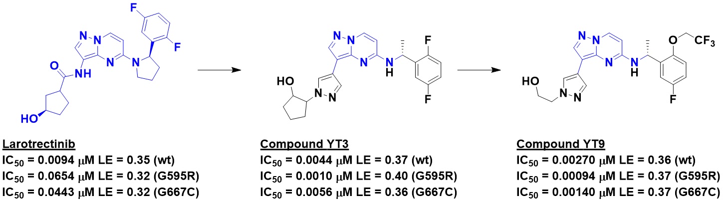
To demonstrate the utility of KinaFrag, the researchers describe a case study in which they started with the anticancer drug larotrectinib, which inhibits TRK family kinases. However, the molecule is less effective against several mutations observed in the clinic. Examining the bound structure revealed that the mutations introduce steric clashes. Retaining the hinge-binding fragment while performing virtual screening of fragments from KinaFrag led to molecules such as YT3, potent against both wild type TRKA and two resistance mutants, and further optimization resulted in YT9. 
 

Not only was YT9 active against the wild type and mutant forms of TRKA, it showed good oral bioavailability and pharmacokinetics in rats. Encouragingly, the molecule slowed tumor growth in both wild type and mutant TRKA mouse xenograft models.
 
One could debate whether this is an example of FBLD; the discovery of YT9 could also be considered a classic case of scaffold hopping. But semantics aside, this is a nice example of thinking in terms of fragmenting molecules. More broadly, KinaFrag looks like a useful tool for work on kinases – especially if the substructure search works.

13 June 2022

Fragments vs HIV-1 Protease: Pocket-to-Lead

The drugging of HIV-1 protease is a classic structure-based design success story, as discussed in a guest post by Glyn Williams from the early days of the SARS-CoV-2 pandemic. The peptide origins of approved inhibitors such as saquinavir are obvious, and the residual structural features can present problems for oral bioavailability. Although there have been fragment screens against the enzyme, the hits do not seem to have been pursued, perhaps in part to the number of approved drugs. But viruses never stop mutating, and developing new chemical matter is prudent. In a recent J. Med. Chem. paper, Yuki Tachibana and colleagues at Shionogi describe a fragment-based approach.
 
The researchers started by performing a virtual screen, but none of the hits were active when tested in a biochemical assay. The active site of HIV-1 protease contains four hydrophobic subsites, and none of the virtual hits filled all four of them. Thus, the researchers chose to focus on fragments that could make some of the interactions while providing growth vectors to additional subsites. They call this a “pocket-to-lead” strategy.
 
Fragment 5 docked nicely into the active site; the hydroxyl group makes interactions with the catalytic aspartic acid residues, while the phenyl ring tucks into the S2 pocket. Growing into the S2’ and S1’ pockets led to molecules such as compound 9, which showed weak but detectable activity. (Astute readers will notice that the stereochemistry around the hydroxyl moiety has changed; both diastereomers are active.) A crystal structure of compound 9 bound to HIV-1 protease confirmed the predicted binding mode

 
Examination of the crystal structure revealed that the parafluorobenzyl substituent was not completely filling the S1’ pocket, and was also in a strained conformation. Replacing this with an alkyl substituent led to low micromolar compound 12. Finally, growing into the S1 subsite led to compound 14, a low nanomolar inhibitor with sub-micromolar antiviral activity.
 
This is a nice example of structure-guided, computationally-enabled fragment-based lead discovery that bears some similarity to the V-SYNTHES method we highlighted earlier this year. As the researchers note, the cyclic lactam found in fragment 5 had been used previously in HIV-1 protease inhibitors. It might have been possible to get to something similar to compound 14 from that earlier molecule. But regardless, compound 14 is emphatically non-peptidic. Whether it will lead to superior drugs remains to be seen, but the paper does say that further optimization is underway.

06 June 2022

What to make first? A new “Ring Replacement Recommender” provides suggestions

So you’ve run a fragment screen, gotten some hits, and validated them. What then? Looking for in-house or commercial analogs is always a good idea, but if you’re serious about a project you’ll eventually need to do chemistry, for example replacing one ring with another (say, a pyridyl for a phenyl). The possibilities are almost endless, especially if you don’t know how your fragment binds. In a new Eur. J. Med. Chem. paper, Peter Ertl and colleagues at Novartis describe a “Ring Replacement Recommender” to rapidly improve biological activity.
 
To determine which replacements are likely to improve affinity, the researchers turned to ChEMBL, a database of more than 2 million molecules and associated biological activity extracted from tens of thousands of publications. From these, more than 68,000 chemical series were chosen for analysis. Each series had on average 16 members, and at least three. The biological activity of each member of a series was compared with other members of the same series. (Importantly, the researchers intentionally excluded anti-targets such as hERG and CYPs so the tool wouldn’t inadvertently improve binding to these.) Focusing only on ring replacements that were reported in at least five publications led to a set of 26,762 changes. Changes could be as modest as adding a methyl substituent or more elaborate such as changing a single aromatic ring to a fused aromatic-aliphatic ring system.
 
One would think that most changes would have little effect, as had previously been seen in the case of methyl additions. Indeed about 65% of the replacements caused shifts in potency of 2-fold or less, which is probably within experimental error. However, 2860 replacements of 245 rings improved affinity at least 2-fold (averaging 3.5-fold), with 223 cases yielding greater than ten-fold improvements.
 
Analyzing the data further, the researchers found 80 ring systems that frequently led to improvements in affinity, and they suggest these could be used as “universal” or privileged building blocks. Strikingly, 74 of these are aromatic, confirming work from Cohen we highlighted in 2020 that proteins may favor “flat” rather than shapely molecules.
 
The researchers also extracted 9515 drugs and clinical compounds from ChEMBL and examined the component fragments. Of the 80 ring systems in the universal set, 19 are found in 50 or more drugs, with another 37 found in at least 5 drugs. This set may be a particularly attractive go-to list.
 
Importantly, not only are all the replacements available in the Supporting Information, the researchers have created a handy and free online tool. Just click on a ring of interest and the Ring Replacement Recommender provides suggestions, along with the average fold improvement observed and the number of publications used for the calculation.
 
To see how well it works, I looked at a couple recent examples which entailed ring changes. The indole to indazole replacement used in the TLR7/8 work described last month was not suggested by the Recommender, though in that case the researchers had the benefit of a crystal structure. On the other hand, a cyclobutyl to phenyl substitution for SARS-CoV-2-3CLp was correctly predicted to be beneficial.
 
Of course, as we’ve said repeatedly, affinity is only part of the battle in drug discovery, and the researchers emphasize that their recommendations may not improve physicochemical or pharmacokinetic properties. But for the earliest stage of a program, and especially in the absence of other data, it’s worth giving the Recommender a try.

30 May 2022

Covalent fragments vs Rgl2

Just over a year ago the FDA granted accelerated approval to sotorasib, the first marketed inhibitor of KRAS and the first approved fragment-derived covalent drug. In a recent ChemMedChem paper, Samy Meroueh and colleagues at Indiana University School of Medicine describe their efforts against a protein in a related pathway.
 
KRAS is a GTPase which cycles between an “on” state, where GTP is bound, and an “off” state, where GTP is hydrolyzed to GDP. KRAS is just one member of a superfamily of GTPases. Two other members also associated with cancer include RalA and RalB. Sotorasib acts by binding to a mutant form of KRAS in which a glycine is replaced by a cysteine, but this mutation does not occur in RalA or RalB. An alternative approach to targeting GTPases is to prevent them from becoming activated by guanine exchange factors (GEFs), which help exchange GDP to GTP. We’ve previously written about how fragments have led to noncovalent inhibitors of the GEF SOS1, which activates RAS proteins.
 
To sum up, there’s more than one way to block GTPase activity: directly, or by preventing activation by an associated GEF. The new paper focuses on Rgl2, a GEF that serves RalA and RalB.
 
Rgl2 sports four surface-exposed cysteine residues, so the researchers screened the protein against a library of 1260 electrophilic fragments at 75 µM for 24 hours at 4 °C and then assessed whether it could still activate RalB. 50 fragments inhibited guanine nucleotide exchange by at least 30%, and a dozen were studied in detail. All were time-dependent inhibitors and had EC50 values from 2.6 to 120 µM at 24 hours.
 
Next, the researchers mutated each of the four surface-exposed cysteine residues to serine. The twelve fragments still inhibited all the mutants except C284S. SOS1 does not contain a cysteine at the position corresponding to C284, and indeed none of the twelve fragments significantly inhibited SOS1 activation of KRAS. All this suggests the fragments act via modification of C284.
 
The easiest and most direct measurement of covalent binding is with intact protein mass spectrometry, and the researchers confirmed that 10 of the 12 fragments did in fact form adducts. Interestingly, Rgl2 was modified two or three times by each fragment, which is perhaps not surprising given that they had relatively reactive warheads (chloroacetamides or propiolamides). Mass-spec studies with the mutants revealed that most of the modifications were at C284 and C508.
 
Whether or not these fragments are advanceable, the discovery that modification of C284 inhibits Rgl2 is useful. Interestingly, C284 is near but not at the Ral binding interface, and the researchers suggest that their fragments block protein activity allosterically. I believe such allosteric sites are common throughout the proteome, and readily addressable using covalent approaches. Watch this space!

23 May 2022

A fragment-sized chemical probe for Notum

Practical Fragments has written previously (here and here) about the enzyme Notum, which shuts down Wnt signaling by removing a palmitoyl group. Aberrant Wnt signaling is implicated in maladies from cancer to osteoporosis, but Paul Fish has been particularly focused on neurological conditions such as Alzheimer’s disease. In a paper just published in J. Med. Chem., Fish and collaborators at University College London, University of Oxford, and The Francis Crick Institute describe their discovery of a chemical probe for this target.
 
As we discussed last year, the researchers conducted a crystallographic screen of the 768-member Diamond-SGC Poised Library, which resulted in 59 hits. Biochemical confirmation studies revealed that fragment 6b, a close analog of a fragment described earlier, is remarkably potent. The substituted phenyl ring nicely fills the lipophilic active site, and the triazole forms a hydrogen bond with a backbone amide of the protein. Structure-based design subsequently led to compound 7y, with low nanomolar potency.
 

The previous fragment-based efforts against Notum also yielded potent molecules, but they had poor brain-penetration. In contrast, compound 7y has a high brain-to-plasma ratio, though the compound also has high clearance, which was attributed to phase 2 metabolism at the hydroxyl. The researchers explored a variety of replacements and substitutions, all of which led to loss in potency, but interestingly removing the hydroxymethyl substituent altogether was tolerated.
 
The resulting molecule, ARUK3001185, is a potent inhibitor of Notum both in biochemical and cell assays. It has good oral bioavailability and pharmacokinetics in mouse and rat. Importantly, it also has excellent brain penetration in both species. The molecule showed virtually no inhibition of 39 other serine hydrolases or 485 kinases and was fairly clean in a safety panel of some four-dozen human targets, including hERG. In other words, ARUK3001185 appears to be an excellent chemical probe.
 
This is a nice example of how a fragment-sized molecule can nonetheless achieve high affinity and selectivity. As we’ve seen repeatedly, potency is not enough; one often needs to spend considerable effort to optimize other properties such as brain penetration. It will be fun to see what this new probe can teach us about Wnt signaling in the brain.

16 May 2022

SAMPL7: Epic computational fail or just no solution?

Every few years computational chemists are invited to compete in the Statistical Assessment of Proteins and Ligands (SAMPL) challenges. Researchers are asked to solve a problem for which the solution is known but not yet published; this blinded format allows a more rigorous test of methods than the typical retrospective studies. SAMPL7 focused on fragments binding to proteins, and the results have been published (open access) in J. Comp. Aided Mol. Des. by Philip Biggin and collaborators at University of Oxford and elsewhere.
 
The subject of this challenge was PHIP, a multidomain protein implicated in insulin signaling and tumor metastasis, though the biology is a bit complicated. PHIP contains two bromodomains, small modules that act as epigenetic readers by binding to acetylated lysine residues (Kac), and the researchers chose to focus on the second bromodomain (PHIP2). Bromodomains have proven to be highly ligandable, though this one is unusual in having a threonine in place of a conserved asparagine.
 
The experimental results that contestants were challenged to predict came from fragment screening using high-throughput crystallography at Diamond Light Source’s XChem. PHIP2 crystals diffracted to high resolution (1.2 Å) and were soaked with 20 mM fragment for 2 hours at 5 °C. In total 799 fragments were screened: 768 from the DSI-poised library (see here) and 31 FragLites (see here). The team took great pains to gather high-quality data, screening the FragLites twice and re-soaking 202 fragments that produced poor R factors or resolution worse than 2 Å. This resulted in 52 hits, a hit rate of 6.5%, consistent with the 2-15% typically seen at XChem. Most (47) of these were in the Kac-binding site, and these were the focus of the SAMPL7 challenge.
 
The first task was for modelers to simply predict which of the 799 fragments bound and which did not. Full experimental details were provided, including pH and the crystallization conditions. Entrants were given 1 month. There were eight submissions plus a control, which randomly selected compounds as binders or non-binders. Most of the contestants used some sort of docking strategy; details are provided in the paper.
 
Shockingly, none of the submissions scored better than random. Three of the entrants failed to correctly identify a single binder, and four identified between 1 and 5 of the 47.
 
The second task was to predict the binding modes of the crystallographically identified ligands. Contestants were provided with the 47 hits and asked to submit up to five poses for each. Perhaps stung from their performance on the first task, or perhaps put off by the two-week requested turnaround time, only five groups submitted entries.
 
Performance was assessed by calculating the root mean square deviation (RMSD) between the experimental and docked structure(s), with RMSD ≤ 2 Å considered successful. Despite this fairly lenient cutoff, “the performance of the methods was disappointing.” The best scored 24%, while two methods scored 2% and 0%. I’ll leave it to chemists to opine whether even a 24% success rate for docking would give confidence to embark on analog synthesis.
 
The third task was to select follow-up molecules from a large database for experimental validation, but alas “the COVID-19 pandemic resulted in a diversion of funds before this follow-up study could be done.” Nonetheless, four intrepid groups submitted entries, and these are discussed in the paper.
 
Taken at face value, this is downright damning for computational chemists. It is also at odds with many nice success stories, for example those described at last month’s DDC conference. So what’s going on?
 
For one thing, not everyone paid attention to the information provided. The crystals were at pH 5.6, but some of the entrants nonetheless assumed pH 7.4.
 
This raises a second and more important point. As the researchers acknowledge, “there is the possibility that our fragments do not necessarily bind in solution, whereas scoring functions are almost always calibrated and validated against solution and structural data.” In other words, perhaps the fragments were not identified computationally because they only bind extremely weakly to a crystalline protein soaking in dilute acid.
 
This highlights perhaps the biggest drawback of fragment screening by crystallography: no matter how beautiful the structure may appear, you get no measure of affinity. Indeed, a paper we highlighted last year was able to confirm binding by NMR for only a minority of crystallographically identified fragments against the SARS-CoV-2 main protease. This does not mean that the crystal structures are “wrong,” but the ligands may be so weak as to be unadvanceable.
 
A picture can be worth a thousand words, but it can also be misleading. Advancing fragments is best done with the help of multiple orthogonal methods.

09 May 2022

Fragments vs TLR7/8, starting from HTS

The toll-like receptors TLR7 and TLR8 are closely related proteins that respond to single-stranded RNA, often associated with viral infection, to activate the immune system. While this is useful to ward off disease, when the proteins become overactivated they can lead to autoimmune disorders such as lupus (see here for a recent discussion by Derek Lowe). In a recent ACS Med. Chem. Lett. paper Claudia Betschart and colleagues at Novartis describe advancing a fragment to a potent inhibitor of both proteins.
 
The researchers built a biochemical (specifically, a TR-FRET competition) assay in which they screened 50,000 molecules, each at 20 µM. The campaign yielded some 1500 hits, and this 2020 paper describes the optimization of one of these.
 
The new paper describes the optimization of a completely different molecule, compound 2. This rule-of-three compliant fragment was not only potent in the biochemical assay, it also showed low micromolar cell activity. A crystal structure of the compound bound to TLR8 revealed that it binds at the interface of a homodimer, making hydrogen bonds to both monomers and stabilizing an inactive conformation of the receptor. 
 

A carbon atom in compound 2 was replaced with a nitrogen in compound 3 in the hopes of picking up an additional hydrogen bond, and this led to a ten-fold increase in potency. TLR8 is located in acidic endosomes, and adding a basic piperidine moiety to try to optimize the subcellular localization did in fact improve cellular potency for compound 5. However, basic amines are often associated with hERG binding, which can cause cardiac problems, and this turned out to be the case for this series. This liability was addressed by adding a fluorine to lower the pKa of the amine. Further addition of small moieties to complement the protein led to additional increases in potency, ultimately yielding compound 15.
 
In addition to low nanomolar and even picomolar cellular activity against TLR7 and TLR8, respectively, compound 15 is selective against other TLRs as well as a panel of 100 off-targets. The compound has good DMPK properties in mice and reduced TLR7-dependent interferon-α release in a mouse model.
 
This is a nice medicinal chemistry story focusing on all aspects of optimization, not just potency. Like last month’s Notum and SARM1 posts, it is also another example of a fragment rising to the top of a high-throughput screen. Fragments don't have to be weak.

02 May 2022

Fragment events in 2022 and 2023

The first third of 2022 has already been graced with two major fragment conferences. Two more have recently been added, and 2023 is starting to take shape.

May 9-11:  While not exclusively fragment-focused, the Eighth NovAliX Conference on Biophysics in Drug Discovery will have several relevant talks, and for the first time will use a hybrid model, both online and in Munich. You can read my impressions of the 2018 Boston event here, the 2017 Strasbourg event here, and Teddy's impressions of the 2013 event herehere, and here.
 
May 24-25:  BioSolveIT is holding a DrugSpace Symposium, with a heavy emphasis on fragments. It's both virtual and free, with an impressive lineup of speakers.

September 28-30: FBDD Down Under 2022 will take place in beautiful Melbourne. If you've been longing to travel, Australia has recently opened its borders. This is the fourth major FBDD event in the country, and given the success of the first and third, it should be excellent.
 
October 17-20: CHI’s Twentieth Annual Discovery on Target will be held both virtually and in Boston, as it was last year. As the name implies this event is more target-focused than chemistry-focused, but there are always plenty of FBDD-related talks. You can read my impressions of the 2020 virtual event here, the 2019 event here, and the 2018 event here.
 
 
2023
April 10-13: CHI’s Eighteenth Annual Fragment-Based Drug Discovery, the longest-running fragment event, has already been scheduled for 2023 in San Diego. This is part of the larger Drug Discovery Chemistry meeting. You can read impressions of the 2022 event here, the 2021 virtual meeting here, the 2020 virtual meeting here, the 2019 meeting here, the 2018 meeting here, the 2017 meeting here, the 2016 meeting here; the 2015 meeting herehere, and here; the 2014 meeting here and here; the 2013 meeting here and here; the 2012 meeting here; the 2011 meeting here; and 2010 here
 
September: FBLD 2020 was sadly canceled due to COVID-19, but FBLD 2023 is scheduled to be held in Boston (exact dates TBD). This will mark the eighth in an illustrious series of conferences organized by scientists for scientists. You can read impressions of FBLD 2018FBLD 2016FBLD 2014,  FBLD 2012FBLD 2010, and FBLD 2009.
 
Know of anything else? Please leave a comment or drop me a note!

25 April 2022

Seventeenth Annual Fragment-Based Drug Discovery Meeting

Last week the CHI Drug Discovery Chemistry (DDC) meeting returned triumphantly to San Diego. This was the best conference I’ve attended in years, which reflects not just the quality of the meeting itself but the fact that three-dimensional events are vastly superior to their 2D counterparts.
 
About 75% of the more than 700 attendees were physically present, though having a virtual option turned out to be wise; at least three of the speakers had COVID but were still able to present remotely. Although the FBDD track lasted just a day and a half, fragments were well-represented across the four days and ten tracks. I won’t attempt to summarize the more than 40 talks I attended but will just cover some broad themes.
 
Computational Methods
Seva Katritch (USC) described the V-SYNTHES approach we highlighted in January. This modular method enables computational fragment growing, in effect facilitating a search of 11 billion molecules from just 600,000 scaffolds. The method as described makes heavy use of Enamine’s make-on-demand molecules, and I think everyone in the audience was excited to hear that the company has started making and shipping compounds from Kyiv again.
 
In the comments to the blog post on V-SYNTHESES someone mentioned BioSolveIT, and Paul Beroza (Genentech) described using their software for a similar approach. One of the targets they investigated, ROCK1, was also investigated with V-SYNTHES, and both techniques yielded unique nanomolar inhibitors.
 
Jan Wollenhaupt (Helmholtz Zentrum Berlin) also mentioned BioSolveIT in the context of fragment growing by catalog. Fragments identified crystallographically from their F2X libraries (see here) were grown to low micromolar endothiapepsin binders. Interestingly an unbiased docking screen did not find these molecules, illustrating the utility of stepwise computational approaches.
 
DOTS is another approach to computational growing and docking enabled by rapid synthesis we’ve previously written about, and Xavier Morelli (CNRS) gave an update, including the fact that they plan to launch a webserver soon.
 
Physical Methods
Tim Kaminski (InSingulo) described an intriguing method for screening liposome-bound proteins such as GPCRs. Dyes incorporated into the liposome are visualized using single molecule microscopy, and the liposomes can be observed in real time binding to immobilized targets in 384-well plates. Tim mentioned that the instrument should be available for purchase next year.
 
A new take on an old method was described by Félix Torres (ETH), who discussed using photochemically induced dynamic nuclear polarization (photo-CIDNP) to increase the sensitivity of NMR, thereby reducing experimental times by a factor of 100. The method requires specialized fragments and a customized NMR, but they can currently screen 1500 fragments per day, and the approach could be particularly valuable for screening hard-to-express proteins.
 
Sticking with the theme of photochemistry, Rod Hubbard (Vernalis by way of Hitgen) discussed a DNA-encoded library of more than 130,000 fragment-linker combinations each containing a photoaffinity tag. Screening this against PAK4 yielded 425 hits, and of the 30 chosen for validation more than 90% confirmed by NMR or crystallography. As we noted in 2020, combining DEL and FBLD provides new opportunities for exploring chemical space.
 
It’s been a few years since we discussed weak affinity chromatography, and Kirill Popov (WAC) provided an update. They’ve applied the approach to more than 50 targets and have obtained hit rates up to 20%. An example against SMARCA4 yielded hits that were subsequently found to bind at two sites, one of which had not previously been described.
 
Covalent fragments continue to increase in popularity. FragNet alum Lena Muenzker (BI) described an intact-protein mass spectrometry screen of the E3 ligase SIAH1 against 1260 acrylamides, resulting in 214 hits. Crystallography has been successful, and they are planning to use these to generate covalent PROTACs.
 
We’ve previously written about screening covalent fragments in cells, and Benjamin Horning (Vividion) and Madeline Kavanagh (Scripps) described a nice chemoproteomics case study in which an alkynamide-containing fragment was identified that binds to cysteine 817 in the kinase JAK1. Optimization led to a low nanomolar binder that inhibits JAK1 signaling.
 
Cysteine is not the only amino acid amenable to covalent modification. Plenary keynote speaker Laura Kiessling (MIT) described squarate derivatives as tunable “Goldilocks” warheads for lysine, with the right balance of reactivity and stability.
 
Success Stories
Cases studies were abundant, including some new disclosures that I’ll hold off describing until they publish. Of course, drugs are the ultimate success stories, and several of these were presented. Svitlana Kulyk recounted the discovery of MRTX1719, Mirati’s MTA-cooperative PRMT5 inhibitor, including some interesting tangents not discussed in the publication.
 
Steve Fesik (Vanderbilt) gave two presentations on near-clinical compounds, one targeting MCL1 and the other WDR5. In both cases weak fragments were advanced to picomolar binders within one to two years, but it has taken much longer to optimize other properties of the molecules.
 
Indeed, this turned out to be something of a theme. Valerio Berdini (Astex) discussed the discovery of erdafitinib, the third approved FBLD-derived drug. The program started in 2006, and it took just nine months to go from the fragment hit to late lead optimization. But the compound didn’t enter the clinic until 2012, and it took until 2019 to be approved.
 
Similarly, Wolfgang Jahnke (Novartis) described the story of the sixth approved fragment-based drug. The fragment screen against ABL was conducted in 2006, but the project went through two near-death experiences. Asciminib finally entered the clinic in 2014, and it was approved last year.
 
But timelines are not destined to be long. We’ve previously written about vemurafenib, the first FBLD-derived drug, which took just six years from project initiation to approval. Ryan Wurz (Amgen) gave a retrospective on sotorasib, the fifth approved FBLD-derived drug. Amgen started the program in August 2012, sotorasib was first synthesized in early 2017, first dosed in humans in 2018, and approved in May of last year. Fast doesn’t mean easy: it took 110 co-crystal structures, and I counted more than 100 names on the acknowledgement slide. But success against KRAS is a welcome reminder that sometimes we really can accomplish the impossible when we work together.
 
This is a good point on which to close. Assuming SARS-CoV-2 doesn’t intervene, DDC is scheduled to return to San Diego April 10-13 next year. I hope to see you there!

18 April 2022

Fragments win in a virtual screen against Notum

Wnt proteins are implicated in a variety of diseases, from Alzheimer’s to colorectal cancer. The enzyme Notum shuts down signaling by removing a palmitoyl group from Wnt. Last year Practical Fragments highlighted several series of Notum inhibitors identified from biochemical and crystallographic fragment screens. The researchers behind those efforts, including Paul Fish and Fredrik Svensson (University College London), have now published a successful virtual screen against the enzyme in J. Med. Chem.
 
Starting with 1.5 million compounds available from ChemDiv, the researchers chose 534,804 based on a variety of computational filters including molecular weight (200-500 Da), number of hydrogen bond donors (<=2) and ClogD (-4 to 5). A virtual screen of these (using Glide) produced 1330 high-scoring hits, of which 1088 were chosen for purchase. Of these, 952 were available, a much higher percentage than the ZINC15-reliant paper we wrote about earlier this year.
 
All 952 compounds were tested in a biochemical assay, and the 44 that gave >50% inhibition at 1 µM were then tested in dose-response format. This yielded 31 compounds with IC50 values < 500 nM. These could be subdivided into four structurally related clusters and eight singletons. Further triaging removed compounds likely to cause assay interference as well as those similar to known Notum inhibitors. This left two clusters and two singletons.
 

Compound 1f was the most potent member of a series of 9 related (and possibly covalent) inhibitors. Although these strongly inhibited the enzyme in the biochemical assay, they were essentially inactive in a cell-based assay. They were also highly insoluble and showed low cell permeability, and were thus dropped.
 
Compound 2a was one of two related molecules that were also quite potent when initially tested. Unfortunately, when the molecules were resynthesized they turned out to be significantly weaker and were also not very soluble, so this series was also halted.
 
The singleton compound 3 turned out to be a covalent inhibitor; the catalytic serine formed an ester with the molecule. The mechanism is more fully described in this open-access J. Med. Chem. paper.
 
That leaves the second singleton. Compound 4d was not just active in the biochemical assay, it also showed sub-micromolar cell activity. SAR, guided by crystallography, ultimately led to low nanomolar inhibitors. The pKa of compound 4d was measured to be 7.9, which is less acidic than many previously reported Notum inhibitors and thus more likely to be cell permeable. This turned out to be the case experimentally, and the compound was also stable in mouse liver microsomes. Pharmacokinetics in mice were promising for several compounds, but unfortunately brain penetration – which the researchers were hoping for – was negligible. (This could be an advantage for peripheral diseases.)
 
This is a nice example of lead discovery in academia. Like last week’s post, it also illustrates that fragments themselves can be quite potent. Indeed, although the researchers were looking for molecules up to 500 Da in their virtual screen, all of the best hits were fragment-sized. Another illustration that small is beautiful.

11 April 2022

Nucleophilic fragments vs SARM1: in situ inhibitor assembly

Recently Practical Fragments wrote about nucleophilic fragments that could react with proteins or cofactors. Previously we’ve also written about in situ chemistry, in which a protein catalyzes the formation of an inhibitor. An interesting marriage of these concepts has just been published (open access) in Mol. Cell by Robert Hughes (Disarm Therapeutics), Thomas Ve (Griffith University) and a group of international collaborators.
 
The researchers were interested in the protein SARM1, which is implicated in the axon degeneration associated with several neurodegenerative disorders. Last year the researchers published a Cell Rep. paper (also open access) in which a biochemical screen of roughly 200,000 molecules led to the discovery of isoquinoline as a 10 µM inhibitor of SARM1. Optimization led to 5-iodoisoqinoline, dubbed DSRM-3716, a 75 nM fragment-sized inhibitor. The paper goes on to demonstrate that the molecule not only prevents axonal degeneration but can even promote recovery of injured axons. The new paper explores the mechanism of action.
 
SARM1 is an NADase: it cleaves the critical cofactor nicotinamide adenine dinucleotide (NAD+). While using NMR to study the mechanism of inhibition, the researchers found that DSRM-3716 reacts with NAD+ to form the new compound shown. In this sense, DSRM-3716 acts as a prodrug, somewhat analogous to sulfanilamide antibiotics which act as PABA mimics to block folate biosynthesis.
 

What’s behind the inhibition of SARM1? A series of crystallographic and cryo-EM studies of SARM1 reveal that the protein can self-associate into multimers which are either inactive or active depending on the relative orientations of the individual proteins. NAD+ normally binds at the interface between two SARM1 proteins. The compound made from NAD+ and DSRM-3716 binds here as well, blocking further activity. The crystal structures also revealed a clear halogen bond (see here) with the iodine in DSRM-3716, explaining the increased activity over isoquinoline itself.
 
Unlike the nucleophilic fragments we wrote about last month, isoquinoline probably won’t raise too many eyebrows among medicinal chemists, as the moiety is found in a handful of approved drugs. The researchers also demonstrated that DSRM-3716 itself is selective for SARM1 in a panel of other enzymes that use NAD+.
 
This is a lovely case of high-throughput screening in which the hit turns out to be a fragment. Indeed, the highly charged compound that actually inhibits SARM1 would not be cell-permeable, but that's just fine since it is formed inside cells. It is worth noting that nearly 1000 approved drugs could be classified as fragments in terms of molecular weight. In the case of CNS drugs, small is beautiful, and it will be fun to watch how far DSRM-3716 derivatives will be able to advance.

01 April 2022

Fragments in space!

Practical Fragments has discussed fragments on Mars and Venus, but those planets are just two small specks in a vast universe. Always thinking big, the luminaries at DREADCO (who previously brought us fragment screening in cells using cryo-EM) have set their sights on deep space. Their theoretical proposal has just been published in the Journal of Extraterrestrial and Space Technologies.
 
One of the big unknowns in molecular recognition is precisely how small molecule ligands approach proteins. To find out, the researchers propose creating a library of fragments, each of which is attached to a very tiny mirror. Proteins of interest would also have tiny mirrors affixed to them. Laser interferometry would be used to study the interactions of proteins and ligands in extremely dilute solutions.
 
One potential problem with this approach is gravity, which is hard to escape on Earth, so the researchers propose running their experiment at a Lagrange point. They had hoped to catch a ride on the James Webb Space Telescope, but the mirror fabrication has taken longer than expected.
 
Even for a secretive multinational megacorporation like DREADCO this will be an expensive endeavor, so they’ll probably have to wait until they’ve eradicated human disease before launching this project. In the meantime, they’re taking suggestions for protein targets – feel free to leave yours in the comments!