14 September 2012

Fragments vs CYPs – on purpose

The cytochrome P450 enzymes, or CYPs, are a huge class of oxidizing enzymes found across all kingdoms of life. In humans these enzymes metabolize many drugs, and to avoid drug-drug interactions, drug hunters generally shun or re-engineer molecules that inhibit CYPs. But microorganisms such as Mycobacterium tuberculosis (Mtb), the causative agent of tuberculosis, also contain CYPs, and targeting these could lead to a sorely needed new treatment for this disease. A team led by Chris Abell at the University of Cambridge has published just such a strategy in Angew. Chem. Int. Ed.

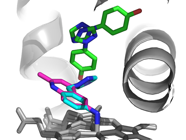
The researchers were interested in CYP121, which is unique to Mtb and important for its viability. They used a thermal-shift assay to screen 665 commercial fragments at 5 mM, of which 66 increased the melting point by at least 0.8 ˚C. 56 of these were further characterized by STD and WaterLOGSY NMR, and 26 showed interactions with the protein and could also be competed with a known substrate. Eight of the most soluble of these were then soaked into crystals of CYP121, leading to four high-resolution structures, three of which are shown here. Isothermal titration calorimetry was used to determine dissociation constants.


 Interestingly, two of these fragments (1 and 2 in blue and red, respectively) coordinate to the heme iron, while the other two do not. Fragment 4 (green in figure above) showed two binding modes in the crystal structure, leading the researchers to make molecules that merge both binding modes. Although the resulting molecules bind in a similar fashion as compound 4 as judged crystallographically, they show at best marginal improvements in affinity and sizable losses in ligand efficiency. Quantum mechanical calculations suggested that this lack of improvement was due to conformational strain within the molecules.

Happily, merging compounds 1 and 2 was much more successful, leading to compound 14, which maintained ligand efficiency and improved affinity. A crystal structure revealed that, as designed, compound 14 binds in a very similar manner as the initial fragments. The molecule was also selective for CYP121 over a different CYP from Mtb as well as several human CYPs.


This is a nice paper not only because it reports a successful example of fragment merging on a new class of targets, but because it also describes several approaches that didn’t work. Fragment merging and fragment linking probably fail more often than they succeed, and this report really digs into the SAR and addresses why merging can be so challenging.

Of course, what would be really cool would be to link compound 14 with compound 4 (ie, link all of the fragments in the top figure), and the paper ends with the statement that this is currently ongoing. It will be fun to see the results.

10 September 2012

Fluorine Fetish

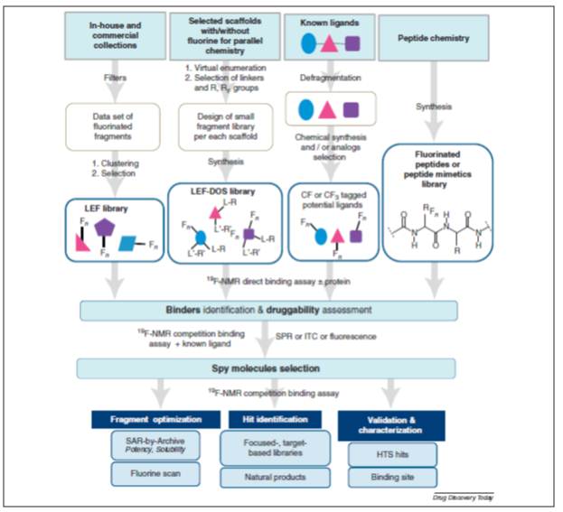
As readers of this blog may know, I love 19F NMR. 19F NMR is well known in screening applications, like FAXS (fluorine chemical shift anisotropy and exchange for screening) and FABS (fluorine atoms for biochemical screening). Back in January, I reviewed a very nice paper from Amgen on the subject. Now, Anna Vulpetti and Claudio Dalvit, the father of 19F in industry, have a review out in Drug Discovery Today on Post Screen applications. This review tries to make the case for ubiquitous use of 19F.
They propose a complete workflow for using 19F NMR (Figure Above). They point out the variety of ways that fluorine can impact the H2L stage. Flourine has many advantages when replacing proton: increased metabolic stability, increased acidity, and decreased basicity. Lipophilicity is increased if the fluorine is near a basic nitrogen or on a aromatic ring. Permeability can be increased. Fluorine can also be a powerful binding moiety by means of H-bond interactions, lipophilic contacts and multipolar and/or antiparallel polar interactions, indirect contact with the protein mediated by water molecules, or by preorganizing the ligand conformation as needed for the protein recognition.

However, this leads to a point Dan made almost three years ago:
I wondered why fluorine-labeled fragments are not used more widely; Fluorine has a strong NMR signal and is very sensitive to the local environment, so when a fluorine-containing fragment binds to a protein this can be easily detected. In fact, the dynamic range for this type of assay is so great that fragment binding can be detected at concentrations several orders of magnitude lower than their dissociation binding constants.

This seems like a very powerful approach, but I haven’t seen many other people using it. Are folks concerned about the need for fluorine in every fragment (although many are commercially available) or is there something else I’m missing?
To my eyes, I would think the following (far from an exhaustive list) could each or together be reasons why 19F hasn't received wider uptake:
  1. Not everyone has 19F probe they can dedicate to screening, follow up, etc.
  2. 19F libraries require a lot of work to put together
  3. 19F is magic methyl by another name
  4. See 1.

Let us know your thoughts in the comments.


29 August 2012

Poll Results: Do you Need Structure

The poll question, Do you Need Structure To Prosecute Fragments is closed. We had 27 votes. 6 (22%) people voted for "Absolutely. No X-ray, no fragments." 6 (22%) voted for "Nope". And the majority 55% (15/27) voted for "Yes, but I am flexible as to what structure is." So, ~80% of the people who voted (self-selection?) think you do not need a X-ray structure to move forward.

So, what kind of methods can we put in the "structure" bin that is not X-ray? I think we need to define structure to get to that first question. In my eyes (and I hope this generates some dialog in the comments), structural information is data that informs on how the ligand and target interact. In my eyes, non-structural structural information is SAR without the guessing. Typical SAR: let's walk this methyl (hopefully its a magic methyl) around this ring, then ethyl, propyl, butyl, futile. Change the ring from phenyl to pyridyl...and so on.

Epitope mapping is one well established method to establish interactions between ligand and target. I am partial to this, but I am an NMR jock. To me, this is as instructive as a crystal structure. It tell you which part of the molecule is in closest contact with the target, which aren't. It leads to imminently testable medchem hypotheses.

Hydrogen-deuterium exchange (HDX) is another method which could inform on the target side of the ligand-target interaction. HDX is most robustly performed by Mass spectrometry, but can also be done by NMR (if the protein is amenable, blah^3). Is this a robust (and SENSITIVE!!!) enough method for routine mapping of ligand-protein interactions?

I ask this out of ignorance, not out of general gadfly-ness, but what methods do those people who are "flexible to structure" use to generate non-structure structural information?

24 August 2012

ACS Fall Meeting 2012


I recently returned from Philadelphia, where the American Chemical Society held its 244th national fall meeting. As always this was a massive affair, but fragments were well-represented, particularly in a nice session organized by Percy Carter, Debbie Loughney, and Romyr Dominique.

I opened the session by giving an introduction to fragment-screening, as well as an overview of some of the work we’re doing at Carmot. Andrew Good had perhaps the best title (“Fragment fat wobbles too”), and discussed some of the work done at Genzyme on Pim-1 kinase. Eric Manas next described some of the computational tools being used at GlaxoSmithKline, in particular strategies to deal with water. He also discussed the utility of looking for fragment analogs early in a project. In the last talk before the intermission, Chris Abell from the University of Cambridge described a number of projects from his group, starting with antimicrobial targets (such as this one); we’ll cover another in a separate post. Chris is unabashedly going after difficult targets, not just protein-protein interactions, but oligonucleotides – specifically riboswitches. There is only limited precedent for targeting RNA with fragments, so it will be fun to see how this progresses.

Francisco Talamas next described a nice example from Roche using FBLD to discover hepatitis C NS5B Palm I allosteric inhibitors. An HTS campaign of around 900,000 molecules yielded just 3 hits, none of which were advanced. A fragment screen of about 2700 fragments gave a better hit rate (5.9%), but of the 29 co-crystal structures attempted only a single structure was obtained. However, by combining the information from this crystal structure with information from other crystal structures, both proprietary and public, the researchers put together a set of rules to design a de novo fragment library tailored to this protein. This effort ultimately yielded compounds that were optimized to a clinical candidate.

Next, Nick Wurtz from Bristol-Myers Squibb described his company’s approach to discover neutral Factor VIIa inhibitors. The researchers used a combination of computational, functional, and biophysical approaches to find uncharged fragments that would bind in the P1 pocket, leading to a couple dozen crystal structures. Despite the low affinities of these fragments (typically mM), many of them could successfully be merged onto an existing series, replacing a positively charged moiety to yield potent molecules with better permeability. This is the first time I’ve seen a fragment story out of BMS, so I'm glad to see that they’re active in this area. This is also a prime example of what has been described as fragment-assisted drug discovery.

Finally, Prabha Ibrahim of Plexxikon gave a lovely overview of the discovery and development of vemurafenib, including a more detailed description of the SAR than has been presented in their earlier papers.

In addition to this dedicated session, there was a scattering of other talks and posters, including a notable poster from Timothy Rooney at the University of Oxford using fragment-based approaches to discover bromodomain inhibitors, a target class we’ve previously discussed.

A session entitled “A medicinal chemist’s toolbox” ranged over several topics of interest. Ernesto Freire of Johns Hopkins gave a great overview of thermodynamics in drug discovery, a topic we’ve previously covered. Most readers are probably familiar with the concept of enthalpy-entropy compensation, in which (for example) an added hydrogen bond fails to achieve the desired boost in potency due to unfavorable entropy. Recognizing this, he suggested that one should target groups in proteins that are already well-structured, so you don’t have to pay the cost of structuring a disordered part of the protein. He also suggested that if you introduce one hydrogen bond, you might be better off introducing a second one too, as the incremental entropic cost is likely to be low.

György Keserű of Gedeon Richter discussed the importance of avoiding lipophilicity by using tools such as LELP, which we’ve covered here and here. Continuing this theme, Kevin Freeman-Cook of Pfizer described two examples of using LLE in lead discovery programs, in particular calculating LLE values before making compounds. Although this may seem obvious, what was quite striking was the dramatic effect subtle changes in structure could make to ClogP values.

Of course, these are just a few of thousands of presentations. Please feel free to point out any that caught your eye, or expand on some of those mentioned above. And just a reminder, it’s only 4 weeks to FBLD 2012 in San Francisco – the biggest fragment event of the year!
15 August 2012

Two types of hot spots


Practical Fragments has previously written about the concept of hot spots – regions on a protein where fragments are particularly prone to bind. It’s always nice to have one of these when starting a program, since it’s a good indication that the protein is ligandable.

But there’s another type of hot spot too. When two proteins interact, they often do so through very large interfaces comprising dozens of amino acid residues. This is daunting from a molecular recognition perspective, but it turns out that most of these residues contribute very little energetically to the binding affinity, as assessed by alanine scanning mutagenesis. The few residues that do matter often cluster into hot spots, which are generally much smaller than the entire interface.

In a new paper in J. Chem. Inf. Model., Sandor Vajda, Adrian Whitty, Dima Kozakov, and colleagues at Boston University ask how these two types of hot spots are related.

The researchers used the program FTMap to look for fragment-binding hot spots on the protein ribonuclease A (RNase A). They found four in the vicinity of the binding site for the protein ribonuclease inhibitor (RNI), three of which had also been shown experimentally to bind small organic (solvent) molecules.

Having shown that FTMap can find fragment hot spots, they next turned to a set of 15 protein-protein complexes for which alanine scanning data were available; amino acid side chains were considered hot spots if mutation to alanine decreased binding by more than 2 kcal/mol. Applying FTMap to the receptor of each protein-protein pair showed that 92% of alanine-scanning hot spot residues map onto FTMap hot spots. Moreover, there were very few false negatives: 92% of “unimportant” residues did not map onto FTMap hot spots. In other words, the two types of hot spots seem to overlap considerably.

Although these results may make sense intuitively, the researchers discuss several reasons why this was not a foregone conclusion. First, a residue identified as a hot spot by alanine scanning might not be important for binding per se, but may instead be important for imposing long-range structure on the protein. Second, alanine scanning only identifies important side chains; backbone atoms are not considered, so a fragment may bind to a hot spot that is invisible by alanine scanning. Finally, hot spots identified by alanine scanning reflect the interactions between two proteins, whereas hot spots identified by fragments (or their virtual equivalents) look at only a single protein. This is important because if a residue protrudes from one protein into a cavity on the other, the protruding residue may be a hot spot for interactions but, because of its geometry, not be a good binding site for small molecules. As the authors put it:

A convex surface site on a protein typically will not bind small molecules strongly no matter how much binding energy the region generates in an interaction with a complementary concave site on its protein binding partner. Thus, observation of a hot spot by alanine scanning mutagenesis does not necessarily imply the existence of a small molecule fragment consensus site at that region.

And of course, as we’ve noted before, proteins can be wriggly little things, with new pockets opening up where you least expect them. So despite all these caveats, it’s reassuring to see that, for the most part, there is some constancy in the hotness of spots.

13 August 2012

Deadlines approaching for FBLD 2012


It is just under six weeks to the start of the conference – which will be the fragment event of the year. Please see here for the fantastic lineup of speakers and exhibitors assembling September 23-26 in San Francisco.

This post is to remind you of two deadlines:

September 1 is the closing date for poster abstracts. This is to allow time for the booklet to be assembled and plan for poster space.

Registrations will continue to be accepted until the meeting; however, the much reduced rate on hotel rooms is not guaranteed beyond September 1.

Looking forward to a great conference and hoping to meet many of you again or for the first time. If you want a sense of what to expect please see here and here.

01 August 2012

From nanomolar to picomolar, via fragments


It’s not every day that you see a picomolar inhibitor. This is all the more true for membrane proteins. And fragment-based lead discovery is rarely attempted with membrane proteins. For all these reasons, a new paper by Guang-Fu Yang at Central China Normal University, Jia-Wei Wu at Tsinghua University, and co-workers in J. Am. Chem. Soc. caught my eye.

The researchers were interested in the cytochrome bc1 complex, which is essential for cellular respiration and a validated antifungal target. Starting with the co-crystal structures of molecules such as azoxystrobin bound to the enzyme complex, they computationally replaced the pyrimidine-containing moiety (red in figure below) with a library of 1735 fragments and calculated the binding energies, in a process called pharmacophore-linked fragment virtual screening (PFVS). Several of the top ten hits were synthesized and tested, and all of these had nanomolar potency. Compound 4 was further optimized, again with the aid of computational chemistry, leading ultimately to picomolar inhibitors such as compound 4f.


Those of you of a suspicious nature may be concerned that the methoxyacrylate moiety looks like a PAINfully reactive electrophile. Happily, the researchers were able to obtain a crystal structure of a molecule in this series bound to the cytochrome bc1 complex, showing that the molecule binds non-covalently and in close agreement to the predicted structure.

In some ways PFVS is reminiscent of Silverman’s fragment hopping, another computational screening and linking approach. Such techniques work best when the protein-ligand complex is relatively rigid, making modeling more straightforward than it would be for a more flexible system

A medicinal chemist could argue that traditional techniques may well have arrived at similarly potent molecules without fragments or fancy modeling. Still, the fact remains that fragments and modeling were used to discover impressively tight-binding compounds, illustrating again the versatility and increasing application of fragment-based techniques.

24 July 2012

There and Back Again (and Back Again)

BACE is a very popular target (there are potentially 20 Billion reasons for it). As we noted in April, Pfizer has entered the contest (publicly now). Pfizer utilized a subset (340 fragments) of their recently described Fragment library (GFI) used X-ray as the discovery platform soaked in 4 at a time. At an intial concentration of 20 mM, 58 of the 85 mixes yielded useable diffraction data. They then repeated the screen at 2mM and 200uM to attempt to gather data on compounds which disrupted the crystal lattice at higher concentrations. All of this led to the discovery of one fragment (the spiropyrrolidine).
They then threw the biophysical and biochemical book at this compound to establish it as a bona fide inhibitor: Octet (1.4mM Kd, 0.31 kcal/mol/atom), STD-NMR, 1H-15N HSQC NMR, functional NMR assay to determine weak Kds, and a BACE inhibition assay (1.1mM). It was found that this fragment had excellent permeability and low potential PGP efflux.
They then did their SAR to optimize the core and develop "growth" vectors. They ended up improving potency by three orders of magnitude with seriously affecting the ligand efficiency nor the in vitro properties predictive of good brain penetration.
To me, this is the most interesting point in the paper. Does ontogeny recapitulate phylogeny for drugs? If you start with good properties, do you keep them? I know there is a good amount of debate on whether or not a fragment will keep its binding mode as it is optimized/expanded. What is the general thinking on properties? Do people screen (at least in silico) their libraries for things like permeability?

18 July 2012

Finding cryptic pockets computationally


The mobility of proteins is a constant source of wonder. I enjoy looking at experimentally-determined structures of small molecules bound to proteins, and it’s even more fun when the protein undergoes dramatic conformational changes to accommodate the ligand. But what may be fun for a chemist is a considerable challenge for molecular modeling: it’s hard enough to dock small molecules to a rigid model of a protein, and all the more so when an apparently flat protein surface yawns open to reveal a new pocket. In a recent paper in J. Comp. Chem., Olgun Guvench and collaborators at the University of New England College of Pharmacy and the University of Maryland look specifically for such “cryptic” binding sites.

The researchers used the cytokine IL-2, which is known to have cryptic pockets. In fact, small molecule inhibitors have been found that target the IL-2 receptor binding site in part by binding to cryptic pockets in the cytokine. The apo form of IL-2 (ie, without any small molecule bound) was used as a starting structure in a computational technique called Site Identification by Ligand Competitive Saturation (SILCS). In this approach, multiple molecular dynamic simulations are carried out with the protein “soaked” in a virtual solution of water and ligand (in this case very simple molecules such as benzene, propane, or acetonitrile). The idea is to let pockets form and see if the ligands bind in the pockets.

Molecular dynamics simulations, in which individual atoms within a protein are allowed to move, run the risk that the protein will deviate too far from a stable structure and denature completely. This can be avoided by introducing various restraints to keep atoms from moving too much, but if the restraints are too severe the protein is too rigid and you won’t see pockets form.

Also, as many people are painfully aware, small molecules can form aggregates in aqueous solution, and the same thing can happen in virtual water. In SILCS, the virtual fragments are programmed to repulse each other, keeping the fragments more or less distributed in solution.

The researchers found that they could in fact identify the cryptic pockets in IL-2, either by using relatively loose restraints or by running multiple unrestrained simulations and simply discarding those in which the protein denatured dramatically. Only the hydrophobic fragments found the cryptic binding sites, perhaps reflecting the relatively hydrophobic nature of the pockets. Additional pockets were also found, though whether these are real or not is unclear.

It’s still a long way from simulations with propane to running molecular dynamics screening simulations on hundreds or thousands of unique fragments, but given the increasing speed of processing power, perhaps the gap will be bridged sooner than expected.

10 July 2012

Another (impractical) NMR Screening Method

NMR has a checkered history in drug discovery. In the 90s, it promised to deliver structures just like X-ray. Strike 1! After that, especially after the advent of SAR by NMR, it promised to deliver boatloads of hits from screening. Strike 2! After that, pharmaceutical NMR worked hard to make sure that it was impactful and value-added. It found niches in which it thrives, e.g. a variety of -omics. In drug discovery, NMR still needs to realize it is living with two strikes. How can NMR survive and even thrive? Quite simply. NMR needs to provide rapid, robust, and easily understandable data to medchemists that leads to decisions. Data that results in no action has no value.

In this paper, Salvia et al. present a ligand-based NMR screening method using "long-lived states (LLS)" of the ligand to boost the sensitivity of ligand-based screening. This new method provides 25x better signal-to-noise than established (T1rho) methods and uses less protein. One of the benefits of this method is it allows NMR to study interactions as tight as 100nM and up to 1 mM.

The graphical abstract (above) shows that while this method is very similar in concept to other ligand-based methods (TOP: equilibrium between NMR differentiated states) it requires much more work than these other methods (Bottom: titrations of ligands). The data (Below) does generate quite satisfying curves, and as noted by the authors, are in agreement with previously published values.
I think this work, while an interesting application of Long Lived States, has really no practical value to the screening world. The strength of the binding can be too strong, making the bound lifetime too long, and thus there is a practical floor for Kd. Of course, because it is based upon kinetics, it can be very different for every system.

If you want to determine Kds for a complex < 10uM there are better, far more robust methods (SPR, for one). The amount of time and effort required to generate Kds from this method seems to run contrary to the tenets I described above (rapid, robust, and (most importantly) easily understandable). To me, the title of the paper simply does not deliver. This method is NOT a screening application. A screening application is one experiment (NMR or otherwise) from which you can determine whether a compound is binding or not, ideally from a mixture of compounds.

I would be curious to see in the comments if anyone (especially our NMR savvy readers, you know who you are) think that this method has practical applications.


05 July 2012

Fragments vs membrane proteins with SPR


Membrane proteins such as GPCRs account for something like half of all drug targets, but they present a serious challenge for fragment-based approaches. This is partly because the biophysical methods usually used for fragment screening often don’t work for membrane proteins, and partly because, in the absence of structural information, it’s hard to know what to do with fragment hits. But there is progress. We’ve highlighted a couple papers that use functional screens or TINS to find fragments against membrane proteins, and in a recent issue of Biochem. Pharmacol. U. Helena Danielson and colleagues at Beactica and two academic institutes show how surface-plasmon resonance (SPR) can also be applied.

The researchers were interested in GABAA receptors, a class of ion channels involved in multiple physiological functions. The receptors normally form hetero-pentamers, but for simplicity the researchers used a homo-oligomeric receptor, consisting of five β3 subunits. Each β3 subunit carried a tag containing eight histidine residues that could be recognized by antibodies immobilized to the surface of the SPR chip. The GABAA receptors were detergent-solubilized; control channels contained antibodies and detergent with no receptors. The resulting GABAA-modified chips were quite stable; the researchers report being able to run roughly 200 samples over the course of 20 hours with a single chip. (The specific detergents and conditions are critical, so if you’re interested in pursuing this yourself the experimental section is invaluable.)

A set of 51 histaminergic and 15 GABAergic ligands were tested for binding, resulting in nearly two dozen hits with dissociation constants (KDs) between 13 and 300 micromolar. Some of these are exceptionally small: for example, histamine, with a molecular weight of 111 g/mol and just 8 heavy atoms showed a KD of 98 micromolar, which is consistent with published results using different methods. A number of other ligands were also identified, some for the first time, though other previously reported ligands did not repeat in this system.

It will be fun to see the screening results of a larger, unbiased library. Of course, finding fragment hits against a membrane protein is only the first step to developing drugs – one still needs to figure out how to improve potency, most likely in the absence of structure. But, to paraphrase Churchill, at least this paper and related research represent the end of the beginning.

27 June 2012

FBLD 2012 Early Registration


40 superb speakers

The most beautiful city on the planet

And you


The biggest fragment conference of the year is happening in San Francisco this September.

We are still accepting posters, so if you have a fragment story to tell, this is the venue.

Early registration ends July 1, so sign up today!

To read about previous events in this series see here. And for a complete list of upcoming events, please see here.

26 June 2012

Do You Need Structure To Prosecute Fragments?

As pointed out here, the majority of people think you need to have structure to successfully move fragments forward.  I, as has been noted previously, disagree and vehemently so.  However, if you fall in the category where you need that crutch you may be familiar with INPHARMA.  X-ray crystallography is the workhorse of structures, but in many cases (far more as we get into more and more complex targets), X-ray fails.  Old-fashioned solution NMR (with 15N, 13C, etc.) is just not fast enough to be a viable tool.  A few years back, INPHARMA was introduced to try to bridge the gap between the two methods.
  
INPHARMA (INter-ligand mapping for PHARmacophore MApping) is based upon robust ligand-based screening data.  Two weakly binding, and competitive, fragments are put into solution with the target.  If the compounds are weak enough (>10uM) it is possible to detect a NOE between protons on the two ligands.  This NOE is mediated by the active site, so the compounds must be binding in the active together (see below).  It is then possible to run very intense computational methods to determine the orientation of the ligands in the binding site (if the structure of the target is known).  


In this paper, Isabelle Krimm looks at INPHARMA's ability to discriminate different binding sites on a single target.  For this she uses, glycogen phosphorylase (a type 2 diabetes target) that has four distinct binding sites: active site, inhibitor site, allosteric site, and new allosteric site. 
Cpd 1 and 2 bind the inhibitor site, where Cpd 3 binds the new allosteric site.  Cpds 4 and 5 are proposed analogs of 2, Cpds 6 and 7 are analogs of 3, and Cpds 8 and 9 are "frequent-hitters".  NOESY experiments were recorded for the six fragments in the presence of 2 and 3.  All compounds exhibited intramolecular NOEs upon binding to the target.  Additionally, Cpds 2 and 4/5 showed intermolecular NOEs, as did Cpd 3 with 6, 7, 8, and 9.  Cpds 4 and 5 did not shows NOEs with 3, nor did 6, 7, 8, and 9 show NOEs to Cpd 2.  For fragments 4-7, competition data support that these are inter-ligand NOEs and that Cpds 8 and 9 are non-specific binders.  No intermolecular NOEs were seen to Cpd 1.  Cpd 1 has a IC50 of 1 uM, while Cpd 2 is 100uM.  This supports theoretical calculations that the two competitive binders must have binding constants no more than 8x different.  

In this paper, Lee et al. demonstrate the use of hyperpolarization to increase the sensitivity of INPHARMA. Dynamic Nuclear Polarization (DNP) uses electrons to transfer magnetization to nuclei leading to orders of magnitude increases in signal.  Lee et al. use DNP to increase the signal in INPHARMA significantly by hyperpolarizing one of the two competitive compounds, in a method they call HYPER-BIPO-NOE (Hyperpolarized binding pocket NOE)[which may be one of the worst not-even-acronyms ever]. 
1D HYPER-BIPO-NOE spectra, a) in full scale and
b) expanded to show transferred signals. Stacked spectra are, from top
to bottom: hyperpolarized ligand 1 with ligand 2 and protein (HYPERBIPO-
NOE); hyperpolarized ligand 1 with ligand 2, but without protein
(Control 1); hyperpolarized [D6]DMSO/D2O, with only ligand 2 and
protein (Control 2); Thermal spectrum of the HYPER-BIPO-NOE
sample (Thermal). The resonance from DMSO, which was suppressed
using presaturation, is designated by *.
This tremendous increase in signal can be extraordinarily useful for INPHARMA applications because they are able to obtain the inter-ligand NOE in a single scan.  The authors then go on to demonstrate that HYPER-BIPO-NOE-INPHARMA data is similar to STD-INPHARMA and can be used to generate binding poses. 

These two papers show that INPHARMA can be a useful tool to orient fragments similarly in a binding pocket.  INPHARMA requires that you have a weak binder that binds in a known site on the target.  For the current generation of targets this may exclude INPHARMA from being used.  It is also important to note that the choice of mixing time (which can range from 70-800ms) is a critical parameter.  The incorrect choice of the mixing time can lead to poor signal intensity and false negative results.  DNP, while turnkey if you have enough money, is not common in industry at all.  I would be surprised if HYPER-BIPO-NOE-INPHARMA actually gets any traction there at all.  There is also a poll with this post, please read and answer.

21 June 2012

Fragments versus CDK4 and CDK6


The cyclin-dependent kinases (CDKs) were some of the earliest protein kinases targeted for drug discovery. They are important for cell-cycle progression, and thus cancer. However, selectivity among the multiple CDK family members has been challenging. In the June issue of ACS Med. Chem. Lett., researchers from Astex and Novartis describe the optimization of a fragment to a selective inhibitor of CDK4 and CDK6.

Astex has been working on CDKs for some time; one of Practical Fragments’ first posts described AT7519, an inhibitor of CDK1, 2, 4, 5, and 9 that is in multiple phase 2 clinical trials. In the new paper, the researchers start with a fairly potent CDK6 hit (fragment A). Crystallography suggested that replacing the pyrrole with a pyridine would provide better vectors from which to grow the fragment, leading to Compound B, which was still active. Growing in two directions then led to Compound 1, and extensive structure-based design led ultimately to Compound 6, which is selective for CDK4 and CDK6 over CDK1 and CDK2. In a panel of 35 additional off-target kinases, the compound displayed IC50 values of 5 micromolar or worse. Compound 6 also showed target modulation in mice and tumor xenograft activity, albeit at fairly high doses.


The authors note that selectivity was a key goal, and that in the course of optimization they were willing to sacrifice potency against their desired targets in order to avoid hitting CDK1 and CDK2. The success of this strategy illustrates again the importance of maximizing ligand efficiency at the outset, as drops in LE can then be used to “pay” for other desirable properties. (Note also that the drop in LLEAT is not quite as severe as the drop in LE.)

Some enthusiasts have argued that fragments provide a more efficient path to the clinic, and this can certainly be the case, as illustrated by the rapid progress of vemurafenib from fragment to drug. However, the current paper illustrates that advancing fragments can still require considerable resources: with 36 authors on two continents, it is clear that this project was not a walk in the park. It is, however, another illustration of starting with a fragment to develop a useful molecule.

12 June 2012

Capillary Electrophoresis


One of the fun aspects of fragment-based lead discovery is the number of ingenious biophysical methods for finding low-affinity fragments. In a recent issue of J. Biomol. Screen., Carol Austin and colleagues at Selcia describe their approach, capillary electrophoresis, which they term CEfrag.

Capillary electrophoresis itself has been around for quite a while. It involves applying a high voltage across a thin capillary filled with liquid; a solution to be analyzed is injected, and the voltage causes migration of analytes (for example, proteins or small molecules). Analyte movement through the capillary depends on charge and “hydrodynamic radius,” which is a function of molecular size and shape. In the case of CEfrag, the idea is to start with a reporter molecule that can be readily detected, for example via UV absorbance. Under a standard set of conditions, this “probe ligand” will have a characteristic mobility. If an excess of protein that binds to the probe ligand is present in the running buffer, the migration time will shift. If an inhibitor is also present in the running buffer, this will prevent the probe ligand from binding to the protein, also causing the migration time to change. By running different concentrations of inhibitor and measuring the changes in mobility, the inhibition constant can be determined.

The researchers demonstrated their approach using that old work-horse of FBLD, the cancer target Hsp90. The known Hsp90 inhibitor radicicol was used as the probe ligand. A total of 609 fragments were screened individually at an initial concentration of 0.5 mM, yielding 42 fragments that reproducibly inhibited radicicol mobility by 20% or more. This ~7% hit rate is similar to that found by others for this target.

Only 12 of the 42 hits identified by CEfrag were also detected in a confirmatory fluorescence polarization (FP) assay, of which only 5 gave measurable IC50 values. However, FP is not ideal for evaluating fragments. In fact, one of the CE hits that didn’t reproduce by FP was ethamivan, the starting fragment for the program that ultimately led to Astex’s AT13387, now in a phase 2 clinical trial for GIST.

To get a better sense of the quality of the CE hits, the researchers put 6 fragments into crystallography trials: 3 hits from both CE and FP, two from CE alone, and one that hit neither. The negative control didn’t produce a structure, whereas two of the FP-confirmed hits produced co-crystal structures (the one that did not had solubility issues). One of the two CE-only hits (ethamivan) also did.

With a throughput of 100 compounds per day per instrument, this is not a high-throughput method, but it is comparable to many other biophysical approaches. Also, the low protein consumption and ability to use unmodified protein are selling points. Have you tried CEfrag? If so, what do you think?

06 June 2012

Fragments versus Ras – Part 2


Practical Fragments recently highlighted a paper from Genentech in which researchers there discovered fragments that block the activity of the prominent oncology target Ras. Illustrating just how much interest there is in this protein, Stephen Fesik and colleagues at Vanderbilt University have just reported results of their own work in Angew. Chem. Int. Ed.

Fesik is famous for SAR by NMR, the first truly practical approach to fragment-based lead discovery. In the current work, the researchers also used NMR (HSQC with 15N-labeled protein) to screen 11,000 fragments, yielding about 140 binders to the GDP-bound form of K-Ras. A number of these were then further characterized crystallographically: of 20 cocrystal structures obtained, all of them were found to bind in the same hydrophobic pocket identified by the Genentech researchers. Fesik and colleagues also noticed a nearby, electronegative cleft, and grew one of their fragments (compound 1) to take advantage of this. This led to compound 12, the most potent compound reported. In addition to binding to the GDP-bound form of K-Ras as assessed by NMR, this compound also inhibited Sos-mediated nucleotide exchange in a functional assay.


Overlaying one of these compounds (blue – similar to compound 12) with the Genentech compound DCAI (red) reveals that while both compounds bind in the same hydrophobic pocket, they make very different contacts.


Of course, it still remains to be determined whether this is a ligandable site on the protein (ie, whether these – or any – molecules can be advanced to high potency). Given the importance of Ras, it’s certain that lots of people are doing their best to find out.