18 December 2023

Review of 2023 reviews

The annual Practical Fragments look-back on the preceding year may not be the most highly anticipated year-end tradition, but I hope you find something of interest in this twelfth edition.
 
I was fortunate to attend several conferences and wrote about CHI’s Discovery on Target in Boston and Drug Discovery Chemistry in San Diego. As for reviews, Louise Walsh and collaborators at Astex, Vrije Universiteit Amsterdam, Novartis, and Frontier Medicines (me!) published our annual analysis of fragment-to-lead success stories in J. Med. Chem., this one covering the year 2021. Some twenty other reviews of interest to this readership were also published. I’ll cover them thematically below.
 
Methods
Crystallography is the most popular fragment-finding technique, and in Expert Opin. Drug Disc. Wladek Minor and collaborators at University of Virginia and Jagiellonian University examine “the current role and evolution of X-ray crystallography in drug discovery and development.” At the start of 2023 the Protein Data Bank (PDB) contained more than 200,000 structures, which sounds impressive until you learn that the AlphaFold database contains more than 200 million predicted protein structures. But this is not experimentalist vs machine: the researchers note how machine learning approaches can be used to more rapidly refine and improve experimental data with resources such as CheckMyBlob and PDB-REDO.
 
For those wishing to dig deeper, two papers in Methods Enzymol. go into experimental detail. In the first, Natalie Tatum and colleagues at Newcastle University describe “crystallographic fragment screening in academic drug discovery.” May Sharpe and collaborators at the Swiss Light Source and University of Hohenheim describe their fast fragment-screening pipeline in a comprehensive (49 page) guide. The focus is on reproducibility, and there is plenty of practical advice. For example, “the authors have even been successful in flying with crystal plates,” though getting these through airport security may be easier in some countries than others.
 
Protein-detected NMR was the first truly practical fragment-based approach, and another paper in Methods Enzymol. by Brian Volkman, Brian Smith, and colleagues at Medical College of Wisconsin describes “fragment-screening by protein-detected NMR.” This distills eight years of effort building their internal protein-detected NMR fragment screening platform that has been applied to 16 proteins thus far. The chapter is particularly detailed on protein and library preparation and screening.
 
Compared with crystallography and NMR, virtual screening can be dramatically faster; we’ve highlighted multibillion-compound screens. In WIREs Comput. Mol. Sci., Artem Cherkasov, Francesco Gentile, and colleagues at University of British Columbia and University of Ottawa discuss (open access) how computational methods are “keeping pace with the explosive growth of chemical libraries.” They cover brute force methods, fragment-based virtual screening, and machine-learning based methods, all while avoiding hype, and conclude that it will take time for these methods to “have a real impact on practical drug discovery.”
 
Finally, Marianne Fillet and collaborators at University of Liege and University of Namur provide a general review in Trends Anal. Chem. covering multiple methods to detect non-covalent fragments. These include established techniques such as biochemical assays, ligand-observed NMR, crystallography, thermal shifts, and SPR, as well as less common ones such as WAC, microscale thermophoresis, ACE, and DEL. The paper includes several nice tables and even a decision tree to help choose among the various approaches.
 
Covalent fragments
Many techniques to detect noncovalent interactions also apply to reversible covalent inhibitors, the subject of a review in Med. Chem. Res. by Faridoon and collaborators at Genhouse Bio and Olema Oncology. The researchers focus on various warheads including cyanoacrylamides, nitriles, ketones and aldehydes, boronic acids, and others, and provide multiple examples for each.
 
In contrast, an open-access review in Pharmaceuticals by Monique Multeder and collaborators at Leiden University Medical Center discusses methods to detect both reversible as well as irreversible covalent protein-drug adducts. Crystallography is the most informative, but the researchers also delve into various mass-spectrometry techniques including top-down (with intact proteins) and bottom-up (after digestion of modified proteins). Also covered are activity-based protein profiling (ABPP) methods, NMR, and fluorescence-based approaches. The nearly 300 references make a useful compendium.
 
One of the most exciting recent developments is “proteome-wide fragment-based ligand and target discovery,” the subject of an open-access review in Isr. J. Chem. by Ines Forrest and Christopher Parker, both at Scripps. This concise, highly readable account covers a lot of ground, from ABPP to fully functionalized fragments (FFFs) to phenotypic screening.
 
If you’re doing covalent FBLD you’ll need a library of covalent fragments, and if you’re building one, I’d recommend a review in Prog. Med. Chem. by David Mann and colleagues at Imperial College London. The paper nicely summarizes design principles such as choice of warhead and the fact that reactivity can vary considerably even among compounds with the same warhead. Synthetic methods and screening approaches are also well covered, along with methods to distinguish specific binding from nonspecific reactivity.
 
Most covalent fragments target cysteine residues, but there at least nine other potentially reactive amino acids, and these are the subject of an open-access review by György Keserű and colleagues at Budapest University of Technology and Economics in Trends Pharm. Sci. Lysine, serine, threonine, tyrosine, and histidine are the most common targets, though some of the warheads are so reactive that specificity will be challenging, let alone reasonable pharmacokinetic properties. This is especially true for aspartic and glutamic acids, methionine, and tryptophan.
 
Finally, another article in Trends Pharm. Sci. by Carlo Ballatore and colleagues at University of California San Diego describes using covalent strategies to develop stabilizers and inhibitors of protein-protein interactions (PPIs). Site-directed fragment tethering with disulfide and imine chemistry is a focus, particularly in the context of 14-3-3 proteins. Proximity-enabled covalent strategies, in which warheads are grafted onto non-covalent molecules, are also covered. There is also a short section on covalent PROTACs – more on that topic below.
 
Targets
Keeping with the theme of protein-protein interactions, Ge-Fei Hao, Guang-Fu Yang, and collaborators at Central China Normal University and Guizhou University discuss fragment-based approaches against “undruggable” PPIs in Trends Biochem. Sci. After describing why protein-protein interactions can be difficult, the paper presents several successful case studies, including venetoclax, sotorasib, and targeting 14-3-3 proteins.
 
Targeted protein degradation continues to be a major focus for drug discovery, and this is commonly achieved by hijacking E3 ligases to cause them to ubiquitinate a target of interest. Iacovos Michaelides and Gavin Collie (AstraZeneca) describe how FBLD has been used to find ligands against E3s in an open-access J. Med. Chem. paper. There are more than 600 E3s, and because their biology relies on protein-protein interactions they are often tough targets. Fragment hits can be weak and difficult to advance, though the researchers do describe several success stories including against KEAP1 and XIAP/cIAP. Covalent fragments have the potential to permanently reprogram E3 ligases, and these are covered well too.
 
Another difficult type of target is RNA, the topic of two reviews. In an open-access Curr. Opin. Struct. Biol. paper Kevin Weeks and colleagues at University of North Carolina Chapel Hill provide a concise and beautifully illustrated overview of the field. They note that “RNA-targeted FBLD is in its infancy,” but given that the first report dates to 2002 it is a long childhood, and the paper does a good job of describing the challenges.
 
A more extensive treatment of “fragment-based approaches to identify RNA binders” is provided by Matthew Disney and colleagues at UF Scripps in J. Med. Chem. The paper describes many case studies, some of which we’ve covered, and also contains a handy table comparing the pros and cons of a dozen different methods for finding RNA-binding fragments.
 
Tuberculosis kills more than 1.5 million people each year, and fragment-based approaches have been applied against multiple targets within the pathogen, as reviewed by Baptiste Villemagne and colleagues at University Lille in Eur. J. Med. Chem. We’ve covered many of these studies on Practical Fragments, but as the paper notes none have advanced to the clinic. This is attributed in part to cell permeability, and the researchers suggest turning to phenotypic screens (see below).
 
Other
Fragment linking can be difficult but highly effective, especially for difficult targets. An overview of published linkers is provided by Isabelle Krimm and collaborators at Université Claude Bernard Lyon and Université Montpellier in Expert Opin. Drug Disc. The paper includes a table summarizing 40 fragment linking stories, noting that most linkers are short and flexible. Another table summarizes 19 examples of target-guided synthesis, including dynamic combinatorial chemistry. As the paper notes, all of these are small model studies based on known compounds. In silico approaches, the last topic covered, will probably prove more practical.
 
And on the subject of practical, Dean Brown (Jnana Therapeutics) provides an “analysis of successful hit-to-clinical candidate pairs” in J. Med. Chem. This is an update to his 2018 article and captures 156 clinical candidates reported in the journal between 2018 and 2021. Of these, 14 had fragments in their lineage. Most of these drugs appear in our list of fragment-derived clinical candidates (though berotralstat does not – I’ll need to look closer). The paper contains lots of interesting analyses. For example, of the 138 oral drugs, 39 had a molecular weight > 500 Da, 24 had Clog > 5, and 17 had more than 10 hydrogen bond acceptors (HBA). On the other hand, none had more than 5 HBD, emphasizing that you should be parsimonious with hydrogen bond donors.
 
Finally, veteran drug hunter Nicholas Meanwell provides “reflections on a 40-year career in drug design and discovery” (open access) in Med. Chem. Rev. Those of you who saw his talk earlier this year at the CHI DDC meeting will know what to expect, and those of you who didn’t will be in for a treat. A personal and entertaining romp through pharma starting in the early 1980s, the paper is full of surprises, such as the pursuit of minor impurities in a phenotypic screen that ultimately led to the hepatitis C drug daclatasvir. Nicholas notes that “you discover what you screen for, so screen design is of paramount importance.”
 
The paper also reveals a passion for medicinal chemistry: “In a search for inspiration for design concepts, I sat down one Saturday afternoon in early October of 1987 and perused every molecule in the United States Adopted Names (USAN) dictionary.” And, as he notes near the end, “Decision making in drug discovery and development is a delicate balancing act, inherently flawed based on absence of predictive accuracy, and knowing when to conclude a discovery program with grace is also an important trait.” That said, he provides examples of successful programs that were almost killed multiple times – and others that were killed at Bristol Myers Squibb but subsequently succeeded elsewhere. While this is frustrating on one level, Nicholas takes satisfaction in the fact that “the science that we conducted and the molecules and pharmacophores that we defined have been of benefit to mankind.”
 
There are still a couple weeks left in the year, but that’s it for Practical Fragments for 2023. Thanks for reading, and special thanks for commenting. And if you live in one of the 70+ countries with elections in 2024, please vote.
11 December 2023

Fragments vs IP6K1 without using structural information

The three members of the inositol hexakisphophate kinase family are potential targets for a wide range of diseases, from Alzheimer’s to cancer to metabolic disease. However, current inhibitors are not specific for individual isoforms. Also, the most potent compounds contain a carboxylic acid moiety, which is usually at odds with brain penetration. In a new ACS Med. Chem. Lett. paper, James Barrow and collaborators at Johns Hopkins School of Medicine, the Lieber Institute for Brain Development, and AstraZeneca describe neutral, selective inhibitors.
 
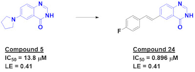
The researchers started with a high-throughput biochemical screen of 17,000 fragments, each at 100 µM, against IP6K1. The library itself is available here. After dose-response follow-up studies, 90 hits confirmed, with IC50 values as good as 2 µM. Most of the hits contained carboxylic acids, but compound 5 did not, and also had good ligand efficiency.
 
No crystal structures of IP6K1 have been reported, so the researchers used an AlphaFold model for docking compound 5. This suggested a fragment growing approach. A variety of replacements for the pyrrolidine were attempted, and while some of these had improved activity many also proved to be chemically unstable. Removing the nitrogen and growing led to compound 24, which was both chemically stable and had sub-micromolar activity.
 
The quinazolinone core itself was associated with poor solubility, and the researchers made multiple attempts to modify it, such as introducing additional nitrogen atoms or methylating to remove a hydrogen-bond donor. Unfortunately, all these modifications led to significant losses in potency.
 
Compound 24 is highly selective for IP6K1 over IP6K2 and somewhat selective over IP6K3. Unfortunately, it showed no cellular activity, possibly due to modest biochemical potency and solubility. Nonetheless, this brief paper illustrates that starting with a larger than normal fragment library can lead to new chemotypes. Screening the larger library gave the researchers more chances to find fragments that did not contain carboxylic acids. Indeed, the difficulty of modifying the quinazolinone moiety demonstrates the utility of screening more molecules. Had the closely related molecules been in the library, they might not have turned up as hits, but their presence would suggest that relevant chemical space had been interrogated.
 
The paper is also a nice example of optimizing hits in the absence of structural information. Although much needs to be done to turn compound 24 into a chemical probe, the fact that it is still so small (almost rule-of-three compliant) provides hope that this can be done.

04 December 2023

Screening tough proteins by SPR

Surface-plasmon resonance (SPR) is among the most popular methods for finding fragments. However, as we have noted, SPR can be very prone to operator error and misinterpretation. In a recent (open access) SLAS Discovery paper, U. Helena Danielson (Uppsala University) and a who’s-who team of biophysicists from across Europe provide experimental strategies for screening difficult proteins.
 
The researchers chose five different proteins, some of which were screened in two or three different forms for a total of nine protein constructs. Six of these were screened against their FL1056 library, a custom-built 1056-member library which include molecules from the FragNet program. The library includes a number of “three dimensional” molecules as assessed by principal moment of inertia (PMI). The other library, FL90, is a small set of commercially available fragments we highlighted here.
 
Before screening compounds against proteins, the researchers conducted a “clean screen.” This involved injecting fragments (at 500 µM each) over the sensor surface using the same buffer that would be used in the actual screen to pre-identify fragments that stick to the surface. This typically disqualified about 1% of fragments, though for one set of conditions the number was closer to 3%.
 
That work done, the researchers turned to the actual screens. After proteins were immobilized on the sensor chips, the fragments were typically screened at a single concentration of 250 µM each. The threshold for the initial hit cutoff was set low, often around 10% of the library, to minimize false negatives. Subsequent follow-up studies at varying concentrations were used for confirmation. This led to a significant winnowing, with the final number of confirmed hits between 0.5 and 7% of the library.
 
The proteins themselves were intentionally chosen to present various difficulties. Acetylcholine binding protein (AChBP, which we wrote about here) forms a large (125 kDa) pentameric complex with multiple binding sites. Lysine demethylase 1 (LSD1) is a multidomain, cofactor-dependent protein that requires a partner protein, CoREST, for activity. LSD1 was screened in the presence or absence of CoREST. Farnesyl pyrophosphate synthase (FPPS, which we wrote about here) is a target for cancer and osteoporosis, and the microbial forms are targets for trypanosomiasis drugs. Human as well as Trypanosoma cruzi and Trypanosoma brucei proteins were screened. Protein tyrosine phosphatase 1B (PTP1B, which we recently wrote about here) is a difficult enzyme with a couple allosteric sites. The C-terminal region is intrinsically disordered, and the protein was screened with or without this region. Finally, human tau is both intrinsically disordered and prone to aggregation. As we noted earlier this year it is of interest due to its potential role in Alzheimer’s disease.
 
Happily, hits were identified against all the proteins, some with ligand efficiency values above 0.5 kcal/mol per heavy atom. The chemical structures for selected hits are shown, and the researchers do appropriately caution that validating them using orthogonal (non-SPR) methods is essential before further studies.
 
I do wish the researchers had noted whether shapely hits were enriched or depleted among the confirmed hits. To my eye most seemed fairly flat, and some seemed dubiously PAINS-like, including an eyebrow-raising dinitro-catechol. Nonetheless, the paper is a nice summary of multiple SPR campaigns. If you’re about to embark on one yourself, it is worth carefully perusing.

27 November 2023

Beware of fused tetrahydroquinolines

Practical Fragments has written frequently about pan-assay interference compounds, or PAINS. These molecules contain substructures that frequently show up in hits that tend not to be advanceable, often wasting considerable effort. One criticism of the PAINS concept is that the original definitions were based on a limited number of screens in one assay format. In a new (open-access) J. Med. Chem. paper, Alison Axtman and collaborators at University of North Carolina Chapel Hill, Emory University, and Oxford University characterize one class of PAINS in more detail.
 
The researchers focused on fused tetrahydroquinolines, or THQs. Of the 51 molecules containing this substructure in the original 2010 PAINS paper, 34 hit in at least one of the assays, and one hit all six. At the time Jonathan Baell and Georgina Holloway noted that “it is not clear for some PAINS, such as the fused tetrahydroquinolines, what the relevant mechanisms of interference may be.” 


The new paper notes that fused THQs are common in screening libraries, with more than 15,000 commercially available. They also frequently show up as hits: the researchers summarize more than two dozen examples against a wide variety of targets including phosphatases, kinases, protein-protein-interactions, and more. In most cases the hits are modestly active, with low to mid micromolar IC50 values, though a few are high nanomolar. 
 
Promiscuity per se is not necessarily bad. Just last week we noted that the 7-azaindole fragment was the starting point for three approved drugs. However, despite showing up as hits in so many screens, only one peer-reviewed paper reports a crystal structure of a fused THQ bound to a protein, and the researchers note that “no optimized chemical probes or approved drugs contain the chemotype.”
 
Importantly, fused THQs hit in a variety of different assay formats, including spectrophotometric, chemiluminescent, SPR, and radiochemical. Thus, these are not merely problematic in the AlphaScreen format studied in the original PAINS paper.
 
So what’s going on? The researchers found that, while molecules containing the fused THQ core were initially colorless, they darkened when dissolved in DMSO or chloroform, turning purple within 72 hours. Interestingly, the reaction seems to be light-dependent: solutions stored in the dark remained colorless. Thin layer chromatography and NMR showed new species forming, and mass spectrometry revealed oxidation with loss of two or four hydrogen atoms. The isolated double bond in the cyclopentene ring seemed to be the culprit, as the saturated analog was stable. Indeed, all of the hits shown in the paper contain the double bond, so fused THQs that lack this feature may be fine – if they ever show up in your assay.
 
It is still not clear exactly how the decomposition products light up so many assays, but in general it’s a good idea to steer clear of molecules that fall apart in ambient light, unless you’re trying to make a photosensitizer.
 
The researchers conclude that “it is tragic to continue to watch groups invest time and resources in dead-end hit-to-lead campaigns, and the medicinal chemistry community will benefit everyone if the cycle stops.”
 
This concludes our public service announcement.

20 November 2023

Capivasertib: the seventh approved fragment-derived drug

On Thursday last week the FDA approved capivasertib for certain breast cancer patients. This marks the seventh fragment-derived drug to be approved. It is also the first approved drug targeting the kinase AKT.
 
Practical Fragments first wrote about capivasertib, then called AZD5363, way back in 2013, where we described the decade-long odyssey from fragment to drug. Interestingly that fragment, 7-azaindole, was also the starting point for two other approved drugs, pexidartinib and vemurafenib. As we noted at the time, “high-affinity molecules were obtained relatively quickly, but these still required a huge amount of effort to achieve selectivity, oral bioavailability, and other properties.”
 
What happened next is a poster child to counter one of the false beliefs Christopher Austin noted as being widespread outside industry: “Once an investigational therapy gets into humans for the first time, regulatory approval and marketing are all but assured.”
 
Capivasertib entered the clinic in 2010 in the first of more than 30 studies listed on ClinicalTrials.gov to date. One challenge was finding patients that would benefit sufficiently to offset a long list of side effects, including diarrhea and glucose fluctuations. In the end, the current approval is in combination with fulvestrant for “adult patients with hormone receptor (HR)-positive, human epidermal growth factor receptor 2 (HER2)-negative locally advanced or metastatic breast cancer with one or more PIK3CA/AKT1/PTEN-alterations, as detected by an FDA-approved test, following progression on at least one endocrine-based regimen in the metastatic setting or recurrence on or within 12 months of completing adjuvant therapy.”
 
Needless to say, these were not the first patients tested. Use of genetic testing to match patients with a drug likely to help them is not routine even today, let alone in 2010. Managing side effects also required figuring out how much of the drug to dose and how often. But additional combination trials are ongoing. Perhaps, as with venetoclax, capivasertib will eventually prove to be useful for a wider range of patients.
 
The first marketed fragment-derived drug, vemurafenib, sprinted from program initiation to approval in just six years. Capivasertib took twenty. As we previously noted, success in drug discovery is not necessarily fast or inevitable. Every year more than 40,000 people die of breast cancer in the US alone, but the death rate has slowly been declining. Hopefully the introduction of capivasertib will continue to reduce this.
 
Congratulations to all the researchers at AstraZeneca, Astex, and the Institute for Cancer Research for participating and persisting in this 20-year marathon to bring a new treatment to people with cancer.

13 November 2023

An update on the COVID Moonshot

On March 18, 2020, a group called the COVID Moonshot released crystal structures of 71 fragments bound to the SARS-CoV2 Mpro protein. The same day, they launched an online crowdsourcing initiative seeking ideas for how to advance these fragments, none of which had activity in an enzymatic assay. The results of this experiment in open science have just been published in Science, appropriately open-access.
 
Within the first week, the group received more than 2000 submissions. Ultimately more than 20,000 molecules were submitted, and all of these were evaluated in “alchemical free-energy calculations.” These are computationally intensive, requiring ~80 GPU hours per compound, so the consortium used the volunteer-based distributed computing network Folding@home. Compounds were evaluated not just for potency but also synthetic accessibility, and those that passed were synthesized at Enamine and tested in various functional assays.
 
In addition to accepting submissions for how to advance fragments, a core group of researchers proposed their own ideas. Interestingly, at least in the early stages of the project, this elite group did no better at coming up with more potent or synthetically accessible molecules, despite being intimately involved with the project. This finding validates the open-sourcing of ideas from the larger scientific community.
 
Ultimately more than 2400 compounds were synthesized, and more than 500 crystal structures were determined. All experimental results were posted online to help guide the synthesis of additional compounds. Speed was consistently prioritized, not just with high-throughput crystallography but also high-throughput chemistry and "direct-to-biology" screening of crude reaction mixtures.
 
The paper highlights one lead series, which originated from a community submission (TRY-UNI-714a760b-6, itself fragment-sized) inspired by merging overlapping fragments. This mid micromolar inhibitor was ultimately optimized to MAT-POS-e194df51-1, with mid-nanomolar activity in both biochemical and cell assays. (Despite a chloroacetamide in one of the original fragments and a nitrile in the final molecule, which is the warhead found in the approved covalent Mpro inhibitor nirmatrelvir, MAT-POS-e194df51-1 is non-covalent.) 
 

The molecule is potent against known SARS-CoV-2 variants, including recent ones such as Omicron. A crystal structure of the final molecule also overlays remarkably well onto the initial fragments.
 
The paper notes that there is still considerable work to do, particularly optimizing the pharmacokinetics to lower clearance and improve bioavailability. These efforts can take vast sums of time and money, and the lead series has been adopted by the Drugs for Neglected Diseases initiative for further development. Although a handful of drugs are already approved against SARS-CoV-2, there is room for improvement: Derek Lowe posted a vivid personal account of his experience on nirmatrelvir here.
 
When we wrote about the COVID Moonshot in March of 2020, we correctly predicted that vaccines would be approved before drugs from this effort emerged. Fortunately, our warning that “there will be a SARS-CoV-3” has not proven correct – yet. But open science endeavors such as the COVID Moonshot will help us prepare for this eventuality. We may not have made it to the moon yet, but perhaps we’ve learned how to leave Earth’s orbit.

06 November 2023

Finding weak fragments for membrane proteins with WAC

Last week we wrote about NMR, one of the most popular fragment-finding methods. This week we turn to a less common technique: weak affinity chromatography, or WAC. As we’ve written previously, WAC involves immobilizing a protein of interest in a chromatography column and flowing a ligand-containing solution through the column. If the ligand interacts with the protein, its elution time will be delayed in proportion to its affinity. In a new (open-access) Molecules paper, Claire Demesmay and collaborators at Universite Claude Bernard Lyon and Ecole Supérieure de Biotechnologie de Strasbourg extend the technique to membrane proteins.
 
Membrane proteins are themselves tricky to study, since removing them from their membranes often denatures them. One trick is to use nanodiscs, which are tiny lipid bilayer islands surrounded by proteins that keep them soluble in water. These scaffolding proteins can also be biotinylated so that the nanondiscs can be attached to streptavidin, which itself can be linked to a surface or matrix. Each nanodisc holds one or at most a few membrane proteins.
 
When we first wrote about WAC in 2011 the technique used standard HPLC columns, which required non-negligible amounts of protein. Here, the technique has been miniaturized to use glass capillaries with volumes of less than 1 microliter, requiring only a few tens of picomoles of protein. The researchers fill the capillaries with a bio-compatible polymer, functionalize it with streptavidin, and then capture biotinylated nanodiscs containing the membrane protein of interest.
 
A long-recognized challenge with WAC is nonspecific binding of the fragments to the column or matrix. Here, the researchers chose a filling (or monolith) that is more hydrophilic (for aficionados, they picked poly(DHPMA-co-MBA)) and found it superior to the previous polymer both with regards to capacity and non-specific binding.
 
Another challenge with WAC is detecting low-affinity binders: because interactions with the protein are weak, the shift in retention time is harder to detect. One solution is to pack more protein in the column, and the researchers develop a clever way of doing this with a “multilayer grafting” approach in which successive injections of streptavidin and nanodiscs more effectively fill the capillary. The combination of a more hydrophilic filling and multilayer grafting increased the column capacity for nanodiscs by three-fold.
 
The researchers tested their approach on the adenosine-A2A receptor (AA2AR), which has frequently been used as a model GPCR. Two previously reported weak ligands, both with affinities around 0.2 mM, could be detected, and competition with an orthosteric binder revealed that they were binding specifically.
 
This is a nice, how-to guide for performing WAC on membrane proteins, and the paper includes detailed equations for calculating affinities from differences in retention times. I look forward to seeing the technique used in de novo screens.

30 October 2023

NMR for SAR: All about the ligand

In last week’s post we described a free online tool for predicting bad behavior of compounds in various assays. But as we noted, you often get what you pay for, and computational methods can’t (yet) take the place of experimentation. In a new (open-access) J. Med. Chem. paper, Steven LaPlante and collaborators at NMX and INRS describe a roadmap for discovering, validating, and advancing weak fragments. They call it NMR by SAR
 
Unlike SAR by NMR, the grand-daddy of fragment-finding techniques which involves protein-detected NMR, NMR for SAR focuses heavily on the ligand. The researchers illustrate the process by finding ligands for the protein HRAS, for which drug discovery has lagged in comparison to its sibling KRAS.
 
The researchers started by screening the G12V mutant form of HRAS in its inactive (GDP-bound) state. They screened their internal library of 461 fluorinated fragments in pools of 11-15 compounds (each at ~0.24 mM) using 19F NMR. An initial screen at 15 µM protein produced a very low hit rate, so the protein concentration was increased to 50 µM. After deconvolution, two hits confirmed, one of which was NMX-10001.
 
The affinity of the compound was found to be so low that 1H NMR experiments could not detect binding. Thus, the researchers kept to fluorine NMR to screen for commercial analogs. They used 19F-detected versions of differential line width (DLW) and CPMG experiments to rank affinities, and the latter technique was also used to test for compound aggregation using methodology we highlighted in 2019. Indeed, the researchers have developed multiple tools for detecting aggregators, such as those we wrote about in 2022.
 
Ligand concentrations were measured by NMR, which sometimes differed from the assumed concentrations. As the researchers note, these differences, which are normally not measured experimentally, can lead to errors in ranking the affinities of compounds. The researchers also examined the 1D spectra of the proteins to assess whether compounds caused dramatic changes via pathological mechanisms, such as precipitation.
 
The researchers turned to protein-detected 2D NMR for orthogonal validation and to determine the binding sites of their ligands. These experiments revealed that the compounds bind in a shallow pocket that has previously been targeted by several groups (see here for example). Optimization of their initial hit ultimately led to NMX-10095, which binds to the protein with low double digit micromolar affinity. This compound also blocked SOS-mediated nucleotide exchange and was cytotoxic, albeit at high concentrations.

I do wish the researchers had measured the affinity of their molecules towards other RAS isoforms as this binding pocket is conserved, and inhibiting all RAS activity in cells is generally toxic. Moreover, the best compound is reminiscent of a series reported by Steve Fesik back in 2012.
 
But this specific example is less important than the clear description of an NMR-heavy assay cascade that weeds out artifacts in the quest for true binders. The strategy is reminiscent of the “validation cross” we mentioned back in 2016. Perhaps someday computational methods will advance to the point where “wet” experiments become an afterthought. But in the meantime, this paper provides a nice set of tools to find and rigorously validate even weak binders.

23 October 2023

A Liability Predictor for avoiding artifacts?

False positives and artifacts are a constant source of irritation – and worse – in compound screening. We’ve written frequently about small molecule aggregation as well as generically reactive molecules that repeatedly come up as screening hits. It is possible to weed these out experimentally, but this can entail considerable effort, and for particularly difficult targets, false positives may dominate. Indeed, there may be no true hits at all, as we noted in this account of a five-year and ultimately fruitless hunt for prion protein binders.
 
A computational screen to rapidly assess small molecule hits as possible artifacts would be nice, and in fact several have been developed. Among the most popular are computational filters for pan-assay interference compounds, or PAINs. However, as Pete Kenny and others have pointed out, these were developed using data from a limited number of screens in one particular assay format. Now Alexander Tropsha and collaborators at University of North Carolina Chapel Hill and the National Center for Advancing Translational Science (NCATS) at the NIH have provided a broader resource in a new J. Med. Chem. paper.
 
The researchers experimentally screened around 5000 compounds, taken from the NCATS Pharmacologically Active Chemical Toolbox, in four different assays: a fluorescence-based thiol reactivity assay, an assay for redox activity, a firefly luciferase (FLuc) assay, and a nanoluciferase (NLux) assay. The latter two assays are commonly used in cell-based screens to measure gene transcription. The thiol reactivity assay yielded around 1000 interfering compounds, while the other three assays each produced from 97 to 142. Interestingly, there was little overlap among the problematic compounds.
 
These data were used to develop quantitative structure-interference relationship (QSIR) models. The NCATS library of nearly 64,000 compounds was virtually screened, and around 200 compounds were tested experimentally for interference in the four assays, with around half predicted to interfere and the other half predicted not to interfere. The researchers had also previously built a computational model to predict aggregation, and this – along with the four models discussed here – have been combined into a free web-based “Liability Predictor.”
 
So how well does it work? The researchers calculated the sensitivity, specificity, and balanced accuracy for each of the models and state that “they can detect around 55%-80% of interfering compounds.”
 
This sounded encouraging, so naturally I took it for a spin. Unfortunately, my mileage varied. Or, to pile on the metaphors, lots of wolves successfully passed themselves off as sheep. Iniparib was recognized correctly as a possible thiol interference compound. On the other hand, the known redox cycler toxoflavin was predicted not to be a redox cycler – with 97.12% confidence. Similarly, curcumin, which can form adducts with thiols as well as aggregate and redox cycle, was pronounced innocent. Quercetin was recognized as possibly thiol-reactive, but its known propensity to aggregate was not. Weirdly, Walrycin B, which the researchers note interferes with all the assays, got a clean bill of health. Perhaps the online tool is still being optimized.
 
At this point, perhaps the Liability Predictor is best treated as a cautionary tool: molecules that come up with a warning should be singled out for particular interrogation, but passing does not mean the molecule is innocent. Laudably, the researchers have made all the underlying data and models publicly available for others to build on, and I hope this happens. But for now, it seems that no computational tool can substitute for experimental (in)validation of hits.

16 October 2023

Spacial Scores: new metrics for measuring molecular complexity

Molecular complexity is one of the theoretical underpinnings for fragment-based drug discovery. Mike Hann and colleagues proposed two decades ago that very simple molecules may not have enough features to bind tightly to any proteins, whereas highly functionalized molecules may have extraneous spinach that keeps them from binding to any proteins. Fragments, being small and thus less complex, are in a sweet spot: just complex enough.
 
But what does it mean for one molecule to be more complex than another? Most chemists would agree that pyridine is more complex than methane, but is it more complex than benzene? To decide, you need a numerical metric, and there are plenty to choose from. The problem, as we discussed in 2017, is that they don’t correlate with one another, so it is not clear which one(s) to choose. In a new (open access) J. Med. Chem. paper, Adrian Krzyzanowski, Herbert Waldmann and colleagues at the Max Planck Institute Dortmund have provided another. (Derek Lowe also recently covered this paper.)
 
The researchers propose the Spacial Score, or SPS. This is calculated based on four molecular parameters for each atom in a given molecule. The term h is dependent on atom hybridization: 1 for sp-, 2 for sp2-, 3 for sp3-hybrized atoms, and 4 for all others. Stereogenic centers are assigned an s value of 2, while all other atoms are assigned a value of 1. Atoms that are part of non-aromatic rings are also assigned an r value of 2; those that are part of an aromatic ring or linear chain are set to 1. Finally, the n score is set to the number of heavy-atom neighbors.
 
For each atom in a molecule, h is multiplied by s, r, and n2. The SPS is calculated by summing the individual scores for all the atoms in a molecule. Because there is no upper limit, and because it is nice to be able to compare molecules of the same size, the researchers also define the nSPS, or normalized SPS, which is simply the SPS divided by the number of non-hydrogen atoms in the molecule. Although SPS can be calculated manually, the process is tedious and the researchers have kindly provided code to automate the process. Having defined SPS, the researchers compare it to other molecular complexity metrics, including the simple fraction  of sp3 carbons in a molecule, Fsp3, which we wrote about in 2009. 
 
The researchers next calculated nSPS for four sets of molecules including drugs, a screening library from Enamine, natural products, and so-called “dark chemical matter,” library compounds that have not hit in numerous screens. The results are equivocal. For example, the nSPS for dark chemical matter is very similar to that for drugs. On the other hand, natural products tend to have higher nSPS scores than drugs, as expected. Interestingly, the average nSPS score for compounds in the GDB-17 database, consisting of theoretical molecules having up to 17 atoms, is also quite high.
 
The researchers assessed whether nSPS correlated with biological properties, and found that compounds with lower nSPS tended to have lower potencies against fewer proteins, as predicted by theory. That said, this analysis was based on binning compounds into a small number of categories, and as Pete Kenny has repeatedly warned, this can lead to spurious trends.
 
The same issue of J. Med. Chem. carries an analysis of the paper by Tudor Oprea and Cristian Bologa, both at University of New Mexico. This contextualizes the work and confirms that drugs do not seem to be getting more complex over time, as measured by nSPS. This may seem odd, though Oprea and Cristian note that by “normalizing” for size, nSPS misses the increasing molecular weight of drugs.
 
This observation also raises other questions, such as the fact that SPS explicitly excludes element identity. Coming back to benzene and pyridine, both have identical SPS and nSPS, which does not seem chemically intuitive. One could quibble more: why square the value of n in the calculation of SPS? Why allow s to be only 1 and 2, as opposed to 1 and 5?
 
In the end I did enjoy reading this paper, and I do think having some metric of molecular complexity might be valuable. I’m just not sure where SPS will fit in with all the existing and conflicting metrics, and how such metrics can lead to practical applications.

09 October 2023

Fragments finger the BPTF PHD Finger

Plant homeodomain (PHD) fingers, despite their name, are found in nearly 300 human proteins. They are small (50-80 amino acid) domains that typically recognize post-translational modifications such as trimethylated lysine residues in histones. The PHD finger in BPTF is implicated in certain types of acute myeloid leukemia. However, because of the large number of PHD fingers as well as their small binding sites, few attempts have been made to develop corresponding chemical probes. (Indeed, the only mention of them on Practical Fragments was in 2014.) In a just-published ACS Med. Chem. Lett. paper, William Pomerantz and collaborators at University of Minnesota and St. Jude Children’s Research Hospital report the first steps.
 
The researchers started by screening a library of 1056 fragments (from Life Chemicals) against the BPTF PHD finger using ligand-observed (1H CPMG) NMR. Fragments were at 100 µM in pools of up to five. This gave a preliminary hit rate of 5.7%, but only ten compounds (<1%) reproduced when compounds were repurchased and retested individually.
 
These ten fragments were next tested by SPR (at 400 µM), which confirmed six of them. Also, all ten CPMG hits were tested in an AlphaScreen assay in which they competed with a known peptide binder. This confirmed nine, including the six that confirmed by SPR.
 
Interestingly, the most potent fragment in the AlphaScreen assay was the starting point for the KRAS inhibitor we highlighted last year. However, this fragment did not show binding to the BPTF PHD finger by SPR, and the researchers had identified the 2-aminothophene substructure as a hit against an unrelated protein. Whether this fragment is privileged or pathological may be context dependent.
 
This and the top three fragments that confirmed in all assays were used as starting points for SAR by catalog, and a handful of analogs were purchased. The researchers also resynthesized two of the compounds. Oddly, resynthesized F2 turned out to be three-fold more active in the AlphaScreen assay than the commercial material. One analog, compound F2.7, showed mid-micromolar activity.


Docking and two-dimensional protein-observed (1H,15N HSQC) NMR experiments suggest that most of the fragments bind in the “aromatic cage” which normally recognizes methylated lysine residues, but F2 may bind in an adjacent region. Both subpockets were also identified as being ligandable using the program FTMap.

This paper is a nice example of using orthogonal methods to find and carefully validate fragments against an underexplored class of targets. The researchers conclude by stating that “these hits are suitable for further SAR optimization and development into future methyl lysine reader chemical probes.” I look forward to seeing more publications.

02 October 2023

Discovery on Target 2023

Last week CHI’s Discovery on Target was held in Boston. This was the Twentieth Anniversary edition, though oddly last year also claimed to be the twentieth. Regardless, attendance surpassed pre-pandemic levels, with some 1200 attendees, 90% of them in person. Eight or nine concurrent tracks over the course of three days competed with one another, while a couple pre-conference symposia and a handful of short courses were held before the main event. Outside obligations kept me from seeing many talks, including plenary keynotes by Jay Bradner (Novartis), Anne Carpenter, and Shantanu Singh (both at the Broad Institute), but most of these were recorded and will be made available for a year, and I look forward to watching them. Here I’ll just touch on a few of the fragment-relevant talks I was able to attend.
 
“Protein Degraders and Molecular Glues” was a popular track during all three days of the main conference, and in a featured presentation Steve Fesik (Vanderbilt) described how he is using NMR-based FBLD to identify tissue-specific E3 ligases and β-catenin degraders. In the case of β-catenin, a difficult oncology target, a fragment screen identified a 500 µM hit that was optimized to 10-20 nM. This has no functional activity on its own, but combining it with a ligand for an E3 ligase to generate a bivalent PROTAC causes degradation of the protein. Steve is currently optimizing the pharmaceutical properties of these molecules.
 
One exciting application for PROTACs is tissue-specific targeted protein degradation, which could avoid systemic toxicity for proteins such as Bcl-xL. Steve said that for the past five years he has been pursuing ligands against E3 ligases preferentially expressed in certain tissues, and he presented brief vignettes for three of them. These came from an initial list of 20 E3 targets, but many of them turned out to be too difficult to express.
 
Steve typically screens a library of nearly 14,000 fragments, large according to our recent poll, but this has proven fruitful as only about 10% of proteins he has screened have turned out to be “teflon.” He noted the odd little fragment hit that proved so impactful to the KRAS program we highlighted last year as being something that might have been excluded from a smaller library.
 
We wrote last week about ligands for the E3 ligase DCAF1, and Rima Al-Awar (Ontario Institute for Cancer Research) described another series. She also described ligands against the oncology target WDR5, a target Steve Fesik has pursued as well.
 
Continuing the theme of targeted protein degradation, Jing Liu described Cullgen’s discovery of fragment-sized ligands for a broadly-expressed E3 ligase which could be an alternative to CRBN-targeting ligands when resistance (inevitably) arises. Although he did not specify the E3 ligase, Cullgen has filed a patent application for ligands targeting DCAF1.
 
Rounding out targeted protein degradation, Kevin Webster, my colleague at Frontier Medicines, described the discovery of covalent ligands for the E3 ligase DCAF2 (or DTL) using chemoproteomics and a variety of other techniques including cryo-electron microscopy. Consistent with Steve’s comments, considerable effort went into successfully obtaining a soluble, well-behaved protein.
 
The late Nobel laureate Sydney Brenner said that “progress in science depends on new techniques, new discoveries, and new ideas, probably in that order.” Harvard’s Steve Gygi, one of Frontier’s Scientific Advisory Board members, described multiple new techniques in a featured presentation focused on cysteine-based profiling. These included multiplexed methods to more rapidly find covalent ligands for targets across the proteome. A just-released mass spectrometry instrument made by Thermo Fisher called the Astral further accelerates the process with order-of-magnitude improvements in both speed and sensitivity compared to existing machines.
 
The cell-based covalent screening described by Steve Gygi is very powerful, but so is investigating a single protein, as demonstrated by the discovery of sotorasib. AstraZeneca did early work on covalent screening (which Teddy noted in 2015), and they have continued to build their platform, as described by Simon Lucas. The company has around 12,000 covalent fragments, some beyond the rule of three, with molecular weights between 200 and 400 Da and logP between 0 and 4. More than 90% are acrylamides, a clinically validated warhead, and the researchers are careful to avoid particularly reactive molecules that would be non-specific.
 
In contrast to the electrophilic fragments that comprise most covalent libraries, Megan Matthews (University of Pennsylvania) is exploring nucleophilic fragments for “reverse polarity activity-based protein profiling,” as we highlighted last year. This has led to the discovery of unusual post-translational modifications. For example, the sequence of the protein SCRN3 suggests that it should be a cysteine hydrolase, but the purified protein has no cysteine hydrolase activity, and in cells the N-terminal cysteine is processed to form a glyoxylyl moiety.
 
Finally, Alex Shaginian provided an overview of DNA-encoded library screening (DEL) at HitGen. The company currently has 1.2 trillion compounds spread across more than 1500 libraries, and an obvious question is whether this is overkill. Alex noted that one protein has been screened three times over the course of several years. In the original screen, a modest (30 µM) hit was found from 4.2 billion compounds screened. A later screen of 130 billion compounds produced nothing new, but a more recent screen of 1 trillion compounds led to four mid-nanomolar series. As Steve Fesik noted, screening larger libraries, whether experimentally or computationally, really can be helpful, especially for the hardest targets.
 
Despite only attending half the conference this post is getting long, but for those of you who were there, which talks would you recommend watching?

25 September 2023

Fragments vs DCAF1: a new tool for targeted protein degradation

Targeted protein degradation (TPD) goes beyond merely inhibiting a protein; it takes a protein out of commission entirely. This is frequently done using a bivalent ligand: one part binds to the protein of interest, while the other part binds to an E3 ligase, which ubiquitinates the protein of interest, targeting it for destruction in the proteasome. Human cells have hundreds of E3 ligase proteins, some of which may work better in certain situations, such as specific cell compartments or tissues. In a recent ACS Med. Chem. Lett. paper, Anna Vulpetti and colleagues at Novartis describe progress against DCAF1.
 
DCAF1 is one component of the Cullin4-RING E3 ubiquitin ligase complex. The C-terminus of the protein contains a WD40 repeat (WDR) domain, which in this case consists of seven “blades” arranged around a central cavity, or “donut hole”. WDR domains are relatively common, and indeed we wrote about a previous Novartis effort that identified chemical probes against another WDR domain in the protein EED. In the new work, the researchers took 21 EED binders and screened them using both protein-detected and ligand-detected NMR against DCAF1, identifying two hits. Crystallography revealed that compound 1 binds in the central cavity, which previous computational screening had suggested would be ligandable
 
 
Next, the researchers screened 30 related compounds from within Novartis. Two of them, including compound 4, had improved affinity (as assessed both by NMR and SPR) and could be characterized crystallographically. In addition to binding in the central cavity, these compounds also bound to a site in the blade region, which the researchers wanted to avoid. Adding a piperazine to compound 4 both improved affinity and disrupted binding to the blade region; further optimization and growing to better fill the central cavity led to compound 13, the most potent molecule in the paper.
 
A crystal structure of a closely related molecule reveals that the acetyl group is near the entrance to the donut hole, providing an easy synthetic attachment point to construct bivalent degraders. A separately published preprint revealed this to be successful, with degraders of BRD9, multiple tyrosine kinases, and BTK.
 
There are several takeaways from this nice fragment to lead story. First, despite the fact that compound 1 is clearly fragment-sized (albeit a bit too lipophilic to be fully rule-of-three compliant), the word fragment never appears in the article. FBLD has become so routine that researchers may not even mention it, which does mean that our list of fragment-derived drugs is destined to be incomplete.
 
Second, although DCAF1 and EED share less than 25% sequence similarity, screening EED hits turned out to be successful, which could argue for screening specific subsets of fragments (for example kinase-focused or, in this case, WDR-focused). On the other hand, compound 1 binds in a different manner to DCAF1 than it does to EED. Indeed, compound 1 actually binds in two different orientations to DCAF1, consistent with its low affinity. The researchers mention a paper published earlier this year that reports a successful DEL screen against the target. Perhaps DCAF1 is just very ligandable, and a naïve fragment screen would have worked just as well as the pre-selected set.
 
Finally, the fact that this program yielded bivalent degraders suggests that many E3 ligases might be coopted for drug discovery. The field of targeted protein degradation is just getting started.

18 September 2023

Fragments vs hIL-1β: Growing into a cryptic pocket to inhibit a protein-protein interaction

Protein-protein interactions have a well-deserved reputation for being difficult to drug with small molecules. This is particularly true for cytokine-receptor pairs, which are involved in a host of extracellular signaling functions. Human interleukin-1β (hIL-1β) plays a key role in inflammation by binding to its receptor IL-1R1. Biologics such as anakinra and canakinumab have been approved as drugs, but apart from some very low affinity fragments no small molecule inhibitors are known. In a new (open access) Nat. Commun. paper, Frédéric Bornancin, and collaborators at Novartis and University of Leicester report the first.
 
The researchers started by screening the 3452-compound LEF4000 library, which we described here, using 19F-NMR. After confirmation using protein-observed 2D NMR just a single super-sized fragment hit remained, consistent with the difficulty of the target. The individual enantiomers of this racemic compound were studied, and only (S)-1 was found to be active. Further characterization revealed that, despite weak affinity, this compound had both slow association and dissociation rates. More on that below.
 
Fragment growing in multiple directions led to mid-micromolar compounds such as 11 and 12. Combining elements from these molecules ultimately led to compound (S)-2, with low micromolar affinity as assessed by SPR
 
 
Compound (S)-2 specifically blocked the binding of hIL-1β with its receptor IL-1R1, but did not inhibit the binding of the related cytokine hIL-1α to IL-1R1. Even better, the compound blocked IL-1R-mediated signaling in cells at low micromolar concentrations in two different assays. The similar activity in biochemical and cell assays is likely due to the fact that the compound only needs to act at the cell surface, so permeability is not an issue, in contrast to our post last week.
 
A crystal structure of (S)-2 bound to hIL-1β revealed important interactions between the protein and both the phenol and lactam nitrogen, two contacts that were maintained during fragment optimization. The structure explains why only the (S)-enantiomer is active, as maintaining these contacts would cause clashes for the other enantiomer.
 
The structure also explains the mechanism of inhibition. (S)-2 binds to a cryptic pocket that forms in a region of hIL-1β important for interacting with IL-1R1, and formation of the pocket involves a loop movement that would be incompatible with the protein-protein interaction. The researchers argue convincingly that that the compound stabilizes the cryptic pocket, which naturally exists as a minor population within solution. This also explains the slow kinetics, which would be expected if the compound essentially has to wait until the cryptic pocket opens before it can bind.
 
There is still a long way to go to a drug. Not only is the affinity of (S)-2 modest, the two carboxylic acid moieties and the phenol are likely to impede oral bioavailability. Nonetheless, this is a lovely paper, and the researchers point out that cryptic pockets frequently involve “large movements of secondary structural elements” that could block biological function. Indeed, this is the case for approved drugs such as sotorasib. Don’t give up just because your protein of interest appears like a featureless billiard ball: there may well be opportunities hidden just beneath the surface.

11 September 2023

Fragments vs malarial DHFR

Malaria continues to be a worldwide scourge, with some quarter billion cases last year. A seventy-year-old drug called pyrimethamine targets the dihydrofolate reductase (DHFR) enzyme from Plasmodium falciparum, but resistance mutations have rendered this molecule mostly useless. An analog called P218 was developed to overcome this resistance and completed a handful of phase 1 clinical trials, but unfortunately the human pharmacokinetics were found lacking. In a new RSC Med. Chem. paper, Marie Hoarau and colleagues at the National Center for Genetic Engineering and Biotechnology in Thailand describe their efforts to improve this molecule.
 
The researchers recognized that the phenyl propanoate moiety of P218 was a metabolic liability and sought a replacement. They screened a library of 1163 fragments (from Key Organics) at 1 mM using a thermal shift assay. This resulted in 64 hits, 52 of which confirmed by SPR. Of these, 22 showed some level of inhibition at 0.5 mM against mutant PfDHFR.
 
Among the hits, five were “bi-aromatic carboxylates,” such as compound 136. These were prioritized because, while reminiscent of the phenyl propanoate in P218, they had fewer rotatable bonds. Some of them also showed slow off-rates by SPR, though in my opinion the sensorgrams look suspicious, perhaps due to excessive protein loading on the chip. (For example, the Kd for compound 136 calculated from the on and off rates comes in at 160 nM, unrealistically potent given that it shows only 20% enzymatic inhibition at 0.5 mM. Note – all values here and in the figure are for the mutant form of the enzyme.)
 

SAR by catalog was used to find additional analogs, such as compound AF10, which showed measurable inhibition of the enzyme. Next, the researchers tested hits in the presence of a pyrimidine fragment (L4) derived from P218, known to bind nearby. Compound AF10 showed greater inhibition than would be expected by simple additivity, perhaps suggesting some preorganization of the binding site, as in a different example discussed here.
 
Molecular modeling was used to link the carboxylate fragments with L4, and eight were made and tested. All inhibited both wild type and mutant PfDHFR, and compound 8 showed good selectivity over human DHFR too. A crystal structure confirmed that it bound as predicted. From a fragment-linking perspective, the sub-nanomolar affinity of compound 8 is impressively better than would be expected given the weak affinities of L4 and AF10.
 
Unfortunately, despite similar in vitro potency against the isolated enzymes, compound 8 and the other molecules tested showed “disappointing” activity against Plasmodium falciparum carrying either wild-type or mutant DHFR, roughly 100- to 1000-fold less potent than P218. The researchers suggest solubility may be a factor.
 
This paper is a useful reminder of the dramatic disconnects often seen between enzymatic and cell activity. Nonetheless, it is another good example of using fragment-based methods to replace one portion of an existing molecule.