26 December 2017

Review of 2017 reviews

The year is done, and the darkness
Falls from the wings of Night.

As we've done since 2012, Practical Fragments is using the last post of the year to highlight conferences as well as reviews not previously discussed.

Significant events included the venerable CHI FBDD meeting in San Diego, the NovAliX Biophysics conference in Strasbourg, and the first-ever fragment conference in Shanghai. We discussed a special issue of Essays in Biochemistry devoted to structure-based drug design, and Teddy came out of retirement to provide an entertaining summary of his experience putting together a book on biophysics in drug discovery - well worth reading if you're ever tempted to edit one yourself.

As in years past, several reviews were devoted to the broad topic of FBDD. Below, I’ll outline the general reviews, followed by those focusing on particular targets, techniques, and other topics.

György Keserű (Hungarian Academy of Sciences) and Mike Hann (GlaxoSmithKline) ask “what is the future for fragment-based drug discovery?” in Fut. Med. Chem. After a concise summary of the topic, they answer that it “includes target discovery and validation, the development of chemical biology probes, pharmacological tools and more importantly drug-like compounds.” In other words, the future looks bright.

FBDD is more comprehensively covered by Ben Davis and Stephen Roughley (Vernalis) in Ann. Reports Med. Chem. This is a complete, self-contained guide to the field, covering everything from history, theory, fragment library design, and fragment-to-lead approaches. It is ideal for a newcomer, but there are enough insights throughout that it makes a rewarding read for experts too.

Of the thirty-plus fragment-derived drugs that have made it to the clinic, none are directed against neglected diseases. Gustavo Henrique Goulart Trossini and colleagues at Universidade de São Paulo review some of the work that has been done in this area in Chem. Biol. Drug Des.

And rounding out general reviews, Christopher Johnson (Astex) and collaborators examined all 28 successful fragment-to-lead programs published in 2016, defined as at least a 100-fold improvement in affinity to a 2 µM or better compound. This is a sequel to our analysis of the 2015 literature, also published in J. Med. Chem., and many of the trends are similar. Interestingly, many leads maintained high ligand efficiencies, and there was no correlation between the “shapeliness” (deviation from planarity) of fragments and that of the resulting leads. Consistent with our recent poll on the importance of structural information, 25 of the 28 examples used crystallography at some point.

Targets
Three of the success stories from 2016 involved bromodomains, the subject of an entire month of Practical Fragments’ posts last year. In Arch. Pharm., Mostafa Radwan and Rabah Serya (Ain Shams University, Cairo) review this target class, with a particular emphasis on the four BET family proteins.

More than 30% of enzymes are metalloenzymes, yet these are targeted by fewer than 70 FDA-approved drugs. One of the first published examples of FBDD involved a metalloenzyme, but most efforts have been focused on a limited set of metal-binding pharmacophores, such as hydroxamic acids. Seth Cohen (University of California, San Diego) has been steadily building libraries of metallophilic fragments, and in Acc. Chem. Res. he describes how this approach can lead to new classes of inhibitors.

Protein-protein interaction inhibitors are another underrepresented class of drugs, though one approved FBDD-derived molecule falls into this category. In Methods, Daisuke Kihara and collaborators at Purdue University look at in silico methods to discover PPI inhibitors, including fragment-based approaches.

Unlike PPIs, kinases have been highly successful drug targets. We recently highlighted one review of cyclin-dependent kinases (CDKs), and in Eur. J. Med. Chem. Marco Tutone and Anna Maria Almerico (Università di Palermo) provide another. Although the main focus is on in silico methods, there is a section on FBDD.

Techniques
As noted above, X-ray crystallography has played a role in most successful fragment to lead programs. In the open-access journal IUCrJ, Sir Tom Blundell (University of Cambridge) provides an engaging and personal view of protein crystallography, a field in which he has played a starring role, starting with his early involvement in determining the crystal structure of insulin. He also notes that the interchange of ideas and techniques between academia and industry has long been a crucial driver of advances.

NMR was the first practical method used for FBDD, so it is not surprising that there are several reviews on the topic. In Arch. Biochem. Biophys., Michael Reily and colleagues at Bristol-Myers Squibb provide a detailed overview of NMR in drug design. This covers not just the ligand- and protein-detection methods often used in fragment screening, but also more intensive techniques to characterize protein-ligand interactions.

A briefer look at many of these topics is provided by Yan Li and Congbao Kang (A*STAR) in Molecules. This review also highlights more unusual approaches such as NMR experiments on living cells.

Artifacts are a fact of life in both FBDD and HTS, and it is always important to recognize these early. In J. Med. Chem. Anamarija Zega (University of Ljubljana) discusses how NMR can help. This includes methods to detect aggregators and covalent modifiers. Of course, NMR methods can introduce their own artifacts, and these are also covered.

Other topics
Speaking of artifacts, PAINS are responsible for quite a few. The term “PAINS” has also been somewhat controversial, and in a new paper in ACS Chem. Biol. Jonathan Baell (Monash University) and J. Willem Nissink (AstraZeneca) examine the “utility and limitations” of the term Jonathan coined seven years ago. As they acknowledge, the PAINS filters were derived from just 100,000 compounds run in a limited set of assays. This means that not every bad actor will be recognized by PAINS filters, and some compounds that are may only be PAINful in certain assay formats. Like Lipinski’s rule of 5, it is important to recognize the limits of applicability. As the authors note, “the key is to remain evidence-based.”

Another sometimes controversial topic is ligand efficiency and associated metrics, the subject of an analysis in Expert Opin. Drug Disc. by Giovanni Lentini and collaborators at the University of Bari Aldo Moro. This includes extensive tables of rules and metrics, both common and obscure. The authors note that, while metrics can be useful, it is important not to use them as a “magic box.” As they quote William Blake, “to generalize is to be an idiot.”

Shawn Johnstone and Jeffrey Albert (IntelliSyn Pharma) discuss pharmacological property optimization for allosteric ligands in a review in Bioorg. Med. Chem. Lett. As we recently noted, fragments are particularly suited for discovering allosteric sites, and this paper discusses how to characterize these.

Finally, Jörg Rademann and collaborators at Freie Universität Berlin discuss protein-templated fragment ligations in Angew. Chem. Int. Ed. Earlier this year we highlighted some of his work, and this review provides a thorough analysis of both reversible and irreversible approaches, with good discussions of detection methods, chemistries, and case studies.

That’s it for the year. Thanks for reading, and especially for commenting.

And may 2018 be filled with music, and light.

18 December 2017

New tools for NMR: 2017 edition

NMR was the first practical fragment-finding method, and continues to be popular. Just over the past year we’ve discussed several new techniques, (here, here, and here), and this post highlights three more.

In Angew. Chem. Int. Ed., Jesus Angulo and colleagues at the University of East Anglia describe differential epitope mapping by STD NMR (DEEP-STD NMR). STD NMR, the most popular of ligand-detected methods according to our poll, can provide some information as to which portions of a ligand are close to a protein, but doesn’t show where on a protein the ligand binds. In DEEP-STD NMR, two separate NMR experiments are conducted and the results compared to provide this information.

The researchers provide two implementation of the technique. In the first, the protein is “irradiated” at two different frequencies; for example, the aliphatic and aromatic regions. Protein residues that are directly irradiated will show a stronger STD to ligand protons than those that are indirectly irradiated, thus revealing whether one region of the ligand is closer to an aromatic or an aliphatic amino acid side chain. If the structure of the protein is known, this can then reveal the orientation of the ligand within the binding site. A similar experiment can be done using H2O vs D2O to determine whether a portion of a ligand is in close proximity to polar residues in the protein.

Water is the subject of the second paper, in J. Med. Chem., by Robert Konrat and colleagues at the University of Vienna and Boehringer Ingelheim. As we’ve previously noted, water often plays a critical role in protein-ligand interactions. The new method, called LOGSY titration, involves doing a series of WaterLOGSY experiments at different protein concentrations and plotting the signals for each proton in the ligand as a function of protein concentration; ligand protons close to the protein show steeper slopes. The researchers examine pairs of bromodomain ligands and demonstrate that LOGSY titration can confirm changes in binding mode previously seen by crystallography. The technique could also reveal what portions of the ligands make interactions with disordered water molecules, which are more difficult to detect in crystal structures.

Both of these techniques provide useful but incomplete information about ligand binding modes. A paper in J. Am. Chem. Soc. by Andreas Lingel and his Novartis colleagues describes how to generate more detailed models. The researchers used a deuterated protein in which all methyl groups (in methionine, isoleucine, leucine, valine, alanine, and threonine) were 13C-labeled. Multiple intermolecular NOEs between the protein and several previously characterized ligands were collected and the resulting distances fed into modeling software to produce good agreement with the known structures. More significantly, the researchers were able to use the method prospectively with two weak (0.9 and 2.8 mM) fragments. The binding models were sufficiently accurate to guide chemical optimization, resulting in molecules with 30-50 µM affinities. Subsequent crystal structures revealed that these bound as predicted. Impressively, this was done on a protein that forms 115 kD hexamers – larger than those typically tackled by NMR.

Teddy would normally close his NMR posts by stating – usually quite forcefully – whether he felt the technique was practical or not. I’m no NMR spectroscopist, so I’ll throw this question out to readers – do you plan to try any of these approaches?

11 December 2017

Flipping fragments in PDE2

A common assumption in fragment growing is that the binding mode of the fragment remains the same throughout optimization (for example here, here, and here). However, this is not always the case (as described here, here, and here). A recent paper in Bioorg. Med. Chem. Lett. by Ashley Forster and colleagues from Merck falls into this latter category.

The researchers were interested in phosphodiesterase 2 (PDE2), which hydrolyzes the cyclic nucleotides cAMP and cGMP. PDE2 is highly expressed in the frontal cortex and hippocampus and has been implicated in cognition and proposed as a target for Alzheimer’s Disease. But because PDE2 is just one member of a large class of enzymes, selectivity is important. Indeed, Merck researchers previously used fragment-based methods to discover selective inhibitors of another member of the family, PDE10A.

In this case the researchers used both high-concentration biochemical screens as well as an SPR screen of a library of 1940 fragments, all with molecular weights < 250 Da. This resulted in 54 competitive inhibitors of PDE2 with affinities better than 200 µM. (No details were provided on numbers of hits from each screen.) Compound 1 was progressed into lead optimization due to its high ligand efficiency and attractive physicochemical properties.


A crystal structure of compound 1 bound to PDE2 revealed the potential to grow into a hydrophobic pocket exploited by previously reported molecules, leading to compound 5. Modeling suggested that bulking up the benzylic linker could improve the binding mode, and indeed compound 8 had submicromolar affinity. Surprisingly however, a crystal structure of a related molecule (having a single methyl group off the linker instead of two) revealed that the initial fragment had flipped orientation.

Further modeling suggested replacing the two methyl groups with a cyclopropyl group, as in compound 12. This simple change gave a 100-fold boost in potency, which was attributed to the free form of the compound more closely matching the bound form. Finally, the remaining methyl group was removed to reduce lipophilicity and remove a potential metabolic liability, leading to compound 16. Crystallography revealed that this binds as expected (gray), with the fragment moiety in the “flipped” conformation.

Compound 16 is at least 100-fold selective for PDE2 against a panel of other PDEs. The attention to physicochemical properties paid off in the form of good oral bioavailability, low clearance, and a satisfactory half life in rats. Although the paper does not mention how long the program took, it does state that only 25 analogs were made to get from the initial fragment to compound 16, and also mentions further optimization. This is another nice example of how the union of crystallography, modeling, and medicinal chemistry can rapidly lead to useful molecules.

04 December 2017

Fragment activators of AMPK

Kinase inhibitors are common. Some 40% of the fragment-derived clinical compounds in our latest list target kinases. Kinase activators, on the other hand, are rare. It is easier to interfere with something than to enhance it, and of the 630+ posts on Practical Fragments, I believe only two discuss enzyme activators. A new paper (here) by Ping Lan, Iyassu Sebhat, and colleagues at Merck and Metabasis provides a third example.

The researchers were interested in adenosine monophosphate-activated protein kinase (AMPK), which plays a critical role in metabolism. As its name suggests, this kinase has naturally occurring activators, though these are nucleotides and thus not particularly useful as chemical probes. It also has fiendishly complex biology: the active enzyme is a heterotrimer of three distinct proteins, each of which comes in two or three flavors, leading to 12 different isoforms with their own unique tissue distributions – which vary among different animals.

After multiple HTS screens failed to produce anything of value, the researchers turned to fragments. Realizing that AMPK was a tough target, they assembled a library of 25,000 highly diverse fragments tending towards super-sized (all of them were greater than 200 Da, and they had up to 22 non-hydrogen atoms). A biochemical screen yielded just three hits, including compound 4.


Despite the generally larger size of fragments in the library, it is interesting that compound 4 follows the rule of three, with just 16 non-hydrogen atoms. Although only modestly active, the small size gave an impressive ligand efficiency, and the activation was nearly 80% that of the natural ligand AMP.

Rigidification of the linker between the benzimidazole and the acid moiety led to compounds such as 27, with low micromolar potency, while growing from the benzimidazole itself led to high nanomolar compounds such as compound 36. Combining these two modifications and further optimization for pharmacokinetic properties led to MK-3903, which was chosen as a development candidate.

MK-3903 activates 10 of the 12 AMPK isoforms and is fairly selective against a panel of off-targets. As predicted mechanistically, administering the compound to mice increases the phosphorylation of downstream substrates of AMPK. It also causes decreased fatty acid synthesis and increased insulin sensitivity. However, a related molecule causes cardiac hypertrophy in rats and monkeys.

In addition to the fact that MK-3903 is an enzyme activator, there are several other notable features about this story. First, despite the difficulty of the target, the team made rapid progress, moving from the initial screen to useful tool compounds in less than a year. Second, as near as I can tell, this optimization was done in the absence of direct structural information on how the compounds bind. (A publication by a separate team, who was closely monitoring the patent literature, describes the crystal structure and mechanistic analysis of a related molecule.) Third, all of this work stemmed from a single fragment: although more ligandable targets may produce lots of hits, in the end you only need one.

Finally, this paper illustrates the lag time that can occur between research and publication: several of the authors are from Metabasis, which was acquired by Ligand Pharmaceuticals way back in 2010. That was also when the patent publication describing these molecules was filed, suggesting the work could have been done a decade ago. That’s something to keep in mind when using the literature to guess who is working on fragments.

27 November 2017

Fragments in China

The 2017 International Symposium on Fragment Based Lead Discovery (pdf here) was held in Shanghai, China last week. I was fortunate to be able to attend what I believe was the first significant FBLD meeting in Asia. Antimicrobials were a major theme, particularly against drug-resistant pathogens. The two days were filled with nearly 20 talks, so I’ll just try to capture a few impressions.

Ian Gilbert discussed the fragment-based efforts underway at the University of Dundee, focusing especially on library design. Among initially purchased commercial compounds, only 56% passed quality control, with 26% insufficiently soluble (at least 2 mM in water) and most of the rest either unstable or impure, similar to what has been seen by others. Ian has also enlisted undergraduate students to make “capped” fragments ready for optimization, as well as novel heterocycles.

Biophysics was a major theme of the conference, and Ian made a strong case for biolayer interferometry (BLI), one of the lesser-used fragment finding techniques. A screen can be completed in just a few days with less than a milligram of protein. In particular, BLI may be useful for assessing ligandability: Ian tested 31 targets, 13 known to be ligandable and 5 known to be not ligandable, and found good agreement with previous research. Ligandable targets generally gave primary hit rates >4.5%.

Ismail Moarefi (Crelux, now part of WuXi AppTec) highlighted microscale thermophoresis (MST) and differential scanning fluorimetry (DSF). NMR had identified ten hits against Pim1, but only six had yielded crystal structures, despite considerable effort. Of the four that didn’t, three had no activity by MST, while the fourth was very weak. Ismail also discussed the Prometheus nanoDSF instrument, which is sufficiently sensitive that it can resolve two-stage melting curves for a two-domain protein.

Another lesser used fragment-finding technique, affinity mass spectrometry, was described by Wenqing Shui (ShanghaiTech University). This uses ultrafiltration to separate protein-bound ligands from unbound molecules and mass spectrometry to identify hits; up to 1000 molecules can be screened in a single assay! Wenqing provided several success stories, including fragment hits with very weak (millimolar) affinity. She also demonstrated that the technique works against a membrane preparation of a GPCR.

Among more common biophysical methods, NMR was represented by Ke Ruan (University of Science and Technology of China). The challenge was characterizing a low-solubility ligand which caused extensive line-broadening of the protein due to intermediate exchange rates. This was solved by examining the distance between a fluorinated ligand and a paramagnetic label on the protein and using this to model the binding mode.

But by far the star of the show was crystallography. We’ve previously mentioned the high-throughput capabilities developed at the Diamond Light Source, and part of the impetus for this conference was to bring these technologies to China. Frank von Delft (Diamond and University of Oxford) noted that since the XChem platform launched in late 2015 more than 50,000 crystals have been screened against more than 40 targets, resulting in more than 1000 fragment structures. The group is committed to removing barriers and bottlenecks and today can process 1000 crystals per week through compound soaking, harvesting, data collection, and processing (using specially developed programs such as PanDDA). More than 30 external groups have used the facility, and every target has yielded at least one hit.

Of course, to collect data on 1000 crystals requires you to reproducibly grow lots of well-diffracting crystals that can handle the rigors of soaking, and Diamond has released a handy list of tips and tricks. Getting the right crystals was also the theme of two talks, one by Sheng Ye (Chinese Academy of Sciences) and the other by Carien Dekker (Novartis). Sheng emphasized the importance of optimizing the protein construct, which could include trimming flexible termini or disordered loops, mutating flexible surface residues, or considering different species. He also noted that adding heavy metal ions can actually improve the quality of the crystals as well as making the structures easier to solve. Carien also emphasized the importance of getting the construct right and discussed how seeding (crushing a hard-won crystal and using this to seed new drops) can be very useful. As we’ve noted, screening fragments at extremely high concentrations seems to be the current state of the art, with Novartis moving to 50 mM in the final soak and Diamond going beyond 200 mM! (In contrast to other types of screens at high concentrations, crystallography should not yield false positives, though hits might bind so weakly as to be undetectable by any other method.)

Such a wealth of structures can be daunting, and Anthony Bradley (Diamond) described the construction and use of a “poised library” for follow-up studies. The 768 fragments are (mostly) soluble to 500 mM in DMSO and are designed such that simple chemistry could generate 1.4 million analogs based on reagents currently in stock at Enamine. Potential analogs can be searched using the Fragment Network approach described here, and I was happy to see that Diamond has released their own open-source version (updated link as of 3 Jan 2018).

Jianhua He (Chinese Academy of Sciences) described the facilities at the Shanghai Synchrotron Radiation Facility (SSRF). This is the first third-generation synchrotron in China and has hosted more than 200 research groups since it opened in 2009. Feng Ye, who works at SSRF, gave a talk (in Mandarin) about screening a bacterial protein at XChem; the movies showing liquid handling and robotics would be impressive in any language. Renjie Zhang (Diamond), who also spoke in Mandarin, gave a talk describing (I’m told) not just XChem but how outside users can apply for access. Although there is currently a long waiting list, this should be addressed within the next year or so when SSRF gains Diamond status.

At the 2015 Pacifichem meeting there were only a few speakers from China. Given the level of interest and expertise I saw last week, I predict that the 2020 meeting will see many more.

19 November 2017

Essays in Biochemistry special issue: Structure-based drug design

Structure-based drug design is often an integral part of fragment-based drug discovery. Indeed, a majority of respondents in a recent poll would not work on a fragment without experimental structural information. Given the close relationship between SBDD and FBDD, I was pleased to learn that a recent issue of Essays in Biochemistry is completely devoted to SBDD.

The collection begins with an editorial by issue editors Rob van Montfort and Paul Workman, both at the Institute of Cancer Research. It briefly introduces SBDD and FBDD and provides an overview of the rest of the issue. It also contains a laudable call for rigor, awareness of artifacts, and making data publicly available.

The first full review, by Martin Noble and collaborators at Newcastle University, discusses the role of SBDD in discovering inhibitors of cyclin-dependent protein kinases (CDKs), with a particular focus on selectivity. Several small molecules are discussed, though I do wish the paper included the fragment-derived compound AT7519, which made it to phase 2 clinical trials.

The following paper, by Bas Lamoree and Rod Hubbard (University of York), is completely devoted to FBLD. This is a concise and self-contained review of the field, and is also sufficiently up to date that it provides a good primer on the state of the art.

Chris Abell and collaborators at the University of Cambridge discuss mass spectrometry for fragment screening in the next paper, including ultrafiltration, WAC, HDX-MS, and native mass spectrometry (though not Tethering). The review also includes a handy table summarizing the advantages and limitations of commonly used fragment-finding methods.

Next up is another review devoted to FBDD, this one from Benjamin Cons and his Astex colleagues. The focus is on challenging drug targets such as BCL-family proteins and KEAP1 where SBDD was pivotal, and the researchers particularly emphasize the utility of X-ray crystallography.

NMR was the first experimental technique used for FBDD, and this is the topic of a paper by Gregg Siegal and colleagues at ZoBio. The review includes examples where NMR revealed that crystallographically-determined binding sites were not biologically relevant. Newer techniques, such as NMR2, are also discussed.

Frank von Delft and collaborators describe the fourth funding phase of the Structural Genomics Consortium (SGC), which includes generating a couple dozen “target enabling packages” around new genetic targets. The ten year goals are certainly ambitious: “no crystal structure is complete without a careful analysis of the target’s disease linkage, a fully analysed fragment screen, and a series of follow-up compounds with demonstrated potency and rationalized SAR.” Given the tools and partnerships they have already established, I wouldn’t bet against them.

Hitting a single protein target can be difficult enough, but Scott Hughes and Alessio Ciulli (University of Dundee) focus on ternary interactions, in which a small molecule acts as a “molecular glue” to bring proteins together. PROTACS, molecules designed to target proteins for degradation, comprise one class that has garnered significant attention recently, and as we’ve noted previously FBDD could play a role in discovering and optimizing them. Targeted protein degradation is also the subject of the next paper, by Honorine Lebraud and Tom Heightman (Astex). In particular, the researchers focus on the use of click chemistry to rapidly build chemical probes that degrade specific target proteins.

Crystallographers have steadily been shrinking how big a crystal must be for analysis, in part due to brighter X-ray beams. Michael Hennig and collaborators at leadXpro discuss X-ray free electron lasers, which were experimentally realized less than a decade ago. The energy of these photons is more than a billion times higher than in the newest synchrotrons – so powerful that they destroy the crystals almost instantaneously, but not before producing a diffraction pattern. This means that tens of thousands of individual crystals need to be studied in order to obtain a full dataset. Needless to say the technical and computational demands are intense and still being optimized. The rewards include being able to use weakly-diffracting microcrystals, such as those of membrane proteins, and the ability to collect data at physiological temperatures, as opposed to the cryogenic temperatures typically used.

The last paper, by David Barford and collaborators at the MRC Laboratory, discusses the use of cryo-electron microscopy – which was recognized by a Nobel Prize this year. Single particle cryo-EM does not require a crystal at all, and recent advances have made near-atomic resolution possible. The idea is to image thousands of individual proteins and then computationally reconstruct them. The review discusses multiple protein-ligand complexes, and although none of these are from fragment programs, some of the ligands are approaching the size of fragments.

This collection of papers nicely captures where SBDD currently stands and illuminates the path ahead. For at least a while all the articles are free to download – so check them out now!

13 November 2017

Quantitative native MS identifies a new zinc binder

Last week we highlighted a case where undetected zinc contamination turned out to be completely responsible for the observed activity of a fragment hit. But zinc plays many essential roles in biology, and several groups have sought fragments that target metals; drugs such as vorinostat derive most of their affinity from such interactions. In a recent paper in J. Med. Chem., Thomas Peat, Sally-Ann Poulsen, and collaborators at Griffith University and CSIRO have identified a new zinc-binding fragment. 

The researchers previously screened human carbonic anhydrase II (hCA II) against a library of 720 fragments, which yielded seven hits that bind to the catalytic zinc, as described here. Most of these fragments were either known zinc binders or had modest (high micromolar) affinities. In the new paper, the researchers reveal an eighth fragment that is both novel and potent.

Surface plasmon resonance (SPR) and native electrospray ionization mass spectrometry (ESI-MS) identified compound 10, which has an affinity and ligand efficiency approaching that of sulfonamides such as compound 3, a well-known class of zinc binder.


The researchers determined the crystal structure of compound 10 bound to hCA II, which revealed an interaction between the catalytic zinc and the deprotonated nitrogen, whose pKa is ~5.5. The oxazolidinedione core of the fragment has previously been used as a carboxylic acid bioisostere, but a search of the protein data bank (pdb) revealed no precedents as a zinc binder. In addition to the primary interaction with the metal, the fragment also formed a couple hydrogen bonds with the protein, helping to explain the high affinity.

Next the researchers made or purchased a series of 18 analogs to assess the SAR using both SPR and MS. Native ESI-MS results are usually assessed qualitatively, but the researchers were able to get quantitative data by holding protein concentration constant (at 14.5 µM) and varying the fragment concentration from 0.5 to 120 µM. Plotting the percentage of protein bound and curve-fitting revealed dissociation constants remarkably similar to those determined using SPR.

Nine of the new fragments showed at least some activity, though none were significantly more potent than compound 10. Crystal soaking experiments led to seven new structures, with all the fragments binding in a similar manner as compound 10. (There was one surprise: a bit of extra electron density in one structure led the authors to reexamine the fragment by high-resolution MS, revealing that about 5% had oxidized, and that this was in fact the bound species.)

Combing through the PDB revealed that some of the SAR compounds had not previously been reported as zinc binders. Interestingly, the key pharmacophore in one of the inactive molecules – hydantoin 15 – has been reported to be a zinc binder. The fact that it was inactive against hCA II augers well for achieving selectivity with metal-binding moieties. It will be fun to watch this story develop.

05 November 2017

Heavy metals suck!

Much of the early work of fragment screening involves avoiding artifacts. For high-concentration assays, compound purity is absolutely essential. However, this is not always easily assessed, as demonstrated in a recent paper in J. Med. Chem. by Alessio Ciulli, Helen Walden, and co-workers at the University of Dundee (see here for Derek Lowe’s discussion).

The researchers conducted a screen against Ube2T, a ubiquitin-conjugating enzyme involved in DNA repair and thus of interest as an anti-cancer target. About 1200 fragments were screened using both differential scanning fluorimetry (DSF) and biolayer interferometry (BLI). Most of the hits were quite weak (millimolar), but one showed low micromolar activity. Although this fragment was a destabilizer in the DSF assay, other destabilizers have turned out to be useful starting points.

Two-dimensional (HSQC) protein-detected NMR experiments suggested that the fragment binds near the catalytic cysteine residue, possibly with some protein rearrangement. The binding was reversible, as expected by the chemical structure of the fragment. The fragment was also active in a functional assay. Finally, isothermal titration calorimetry (ITC) revealed an impressively tight dissociation constant of 17.7 µM for the 16-atom fragment. All of these orthogonal assays suggested the researchers had a winning fragment on their hands, so they started acquiring and making analogs to further optimize the affinity. Then things went awry.

Of 14 molecules tested, some quite similar to the initial fragment, only two showed any activity, and these were way down. Concerned, the researchers examined the fragment itself by 1H and 13C NMR as well as high-resolution mass spectrometry, all of which revealed that the compound had the desired structure and appeared to be quite pure (not necessarily a given!) So what the heck was going on?

The mystery was finally resolved, after considerable effort, by a co-crystal structure of the fragment with the protein. Unlike previous structures of Ube2T, this one revealed an unusual domain-swapped architecture, in which a domain of one Ube2T protein interacts with a different molecule of Ube2T rather than with the rest of its own protein. More alarmingly, there was no electron density for the expected fragment, but there was a small, strong area of density connected to the catalytic cysteine residue. The researchers speculated that this could be a zinc ion, and sure enough, zinc chloride itself turned out to have essentially the same affinity for the protein as judged by ITC. Adding the zinc chelator EDTA to the fragment abolished activity, and a colorimetric probe revealed the presence of zinc in the original fragment as well as – to a lesser extent – the two active analogs.

Metal contamination is actually not uncommon – we mentioned a case where residual silver accounted for the apparent activity of many HTS hits. Enzymes with an active-site cysteine are particularly susceptible.

This type of artifact is particularly insidious because it is so difficult to discover. In this case, it was uninterpretable SAR that made the researchers suspicious, and crystallography that revealed the culprit. But SAR can be wonky, and crystallography often fails. What else can be done? Elemental analysis could have helped, but people usually only turn to this if they’re already suspicious.

Of the various fragment-finding methods, I think the only two besides crystallography that could have given warning are native mass spectrometry (MS) and ligand-detected NMR. The former is relatively specialized and doesn’t work for all targets, but it would be interesting to know whether standard NMR techniques such as STD, WaterLOGSY, or CPMG would have revealed that the initial fragment was not binding. Of course, there can be all sorts of reasons for negative results. Publications like this one are useful reminders that simply ignoring such data is unwise. 

29 October 2017

On Par with the Pyramids... a New Book on Drug Discovery!

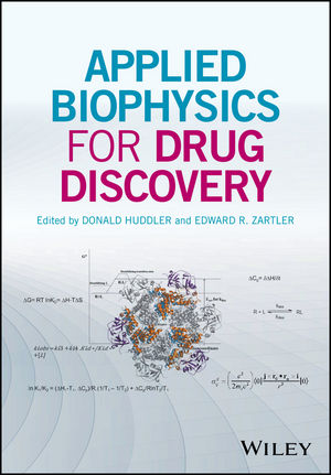
There are many great testaments to humanity's perseverance.   The Pyramids at Gizahttps://www.ancient.eu/uploads/images/display-5687.jpg, the Cathedral of Notre Dame https://upload.wikimedia.org/wikipedia/commons/thumb/b/be/Notre_Dame_de_Paris%2C_East_View_140207_1.jpg/220px-Notre_Dame_de_Paris%2C_East_View_140207_1.jpg, the Great Wall of Chinahttp://vizts.com/wp-content/uploads/2016/02/great-wall-of-china-view.jpg, and the most recent Applied Biophysics for Drug Discovery .   This blog typically reviews the content of the book, but I thought it would be interesting to describe how the book came together.

It is said that if you see inside a hotdog factory you'd never eat a hotdog again.  So, how did this hotdog get made?  Flash back 3 years, and Don Huddler (at that time at GSK) reaches out to me and says, "Hey do you have a contact at Wiley.  I have a great idea for a book." "Sure Don, here you go."
A week or so later, Don invites me to lunch so we go to greatest restaurant ever!!!  Over lunch , and maybe my second beer, Don says, lets write this together.  After almost choking on my Cajun Meatloaf GC, I said NO WAY!!!  There may have been at least one more beer involved but I agreed after Don said he would do all the heavy lifting.

Flash forward a few weeks, and Wiley accepted our book proposal, with 30 listed chapters, with 5 sections.  Part of the fun part is that they send your proposal out for review, so just like a paper, you get reviewer comments.   You then sign a contract with the Publisher that says you will deliver so many pages: 556 in our case by 31Jan2016 (14 months after the contract was signed).  Don had already defined many of the authors who he had thought would be appropriate.  So, at this point, you send out letters to authors and ask them to contribute.  So, "You would be perfect to write a chapter for our upcoming book on [insert what you want them to write on]."  Then you wait and hope that they actually respond.  Many authors responded right away, and it is not personal when they say no.  It is a pretty significant amount of work to contribute to a chapter.  Then you work with the authors to define the time frame of when drafts are due, and so forth.

You then wait and hope the authors deliver on time.  Some do, may don't.  So then you become Nagger-in-Chief.  "Where's the chapter draft?  Is it coming soon?"  As someone famous said, life is what happens while you are making other plans.  During the summer of 2015, GSK was re-orging, and Don decided to go to Law School full time.  So, here we are trying to get drafts done, edited and returned to authors and one editor makes a major career change.  Two months after that, I followed Don in changing careers, leaving the glamorous consulting life to join Pfizer.  As hard as we tried, our new careers required our focus and the book got short shrift.  

For some authors who had delivered on time, this was frustrating.  We had a given deadline and they delivered.  We dropped the ball for them.  For other authors who were less than timely in their contributions, they did not get nagged sufficiently, causing further delays.  2016 was by and large a horrible year if you were an author with a delivered chapter, and a fantastic year if you hadn't delivered a chapter yet.  And of course, as time drags on, other career changes happen.  One primary author retired, one changed jobs and stopped responding to emails, and so on.  So, our initial 20 chapters (from the 30 we wanted) was whittled down to 15 chapters.  

Its now 2017, and the book is a year late, and Don and I are still trying the best we can to manage new careers and editing the book.  Authors are angry with the delay and I didn't blame them.  We were actually able to finally get all the chapters together and to the publisher.  Phew, most of our work is done.  STOP!  One chapter got completely left out of the final submission.  So, the scramble was on to make sure that it was included.  You can see which one when you try to figure out the order of chapters.   

So, what is the role of the Publisher during this whole thing you ask?  I don't know.  My experience with the publisher on this book was VERY different from the first one.  I got the feeling that this book was a low priority for them.  Our emails took weeks sometimes to be responded to.  It was frustrating; but something we did not want to share this with the authors.  So, after this it is mostly on the publisher, but we had to design the cover.  To explain it, everything on the cover is from a chapter in the book: the structure and equations.  We get to galley proof stage and final publication, and Wiley tells us we need to index the book. This is a change from the first time.  Don and I take the option to have them do it for us and take it out of royalties (which is something on the order of 50$ a year).  So, finally in July of this year, it was finalized and given a publication date.  Project done.  So,  let me say to all the authors, thank you so much for being a part of this book.

So, what did I learn co-editing this book?  No matter how many beers I get fed, I will never EVER edit a book again.  There is actually a word ambit. I have a reputation for being "provocative" (from a reviewer); this probably didn't help.  The quality of the contact at the publisher makes a huge difference.  I hope our contact at Wiley was new and inexperienced because this experience was far worse than the first time I did the book.

So, what is inside the book?  291 pages (out of a promised 556).   There are 14 chapters of awesomeness, featuring people featured on this site previously.  This book is focused on biophysical methods and how they are used to triage and advance leads.  Many of these topics have been covered in depth on this site: thermodynamics, protein-protein interactions, HDX, MST, SPR, WAC, 1D NMR, Protein NMR, and how to use them in terms of residence time.  There are two case studies, one from Pfizer and one from FOB (Friend of the Blog) Michelle Arkin.  Lastly, and you can figure this out, there is a chapter from Martin Scanlon on fragment libraries.  I won't go into actually reviewing the book; that would be a major conflict of interest (I do have a financial interest in it and Don and I need to pay for that indexing!).

I would appreciate comments on it here, and of course any other questions I would be happy to answer.

23 October 2017

Poll results: does your primary fragment library contain racemates?

Our latest poll asked just this question. We received 72 responses, and the results are shown here.

Almost half of respondents said they include racemates in their library, as recommended by Claudio Dalvit and Stefan Knapp in the paper that inspired this poll. Another 40% said they had some racemates and some pure enantiomers in their library, which presumably reflects the fact that some enantiomers are more readily available than others.

Only about 10% of respondents said that all chiral fragments in their library are pure enantiomers.

And perhaps most surprisingly, only a single respondent said he or she doesn’t screen chiral fragments at all.

Personally I like racemates because they present an easy follow up experiment: if the two enantiomers have different activity, you are more likely looking at genuine activity as opposed to some sort of artifact

Of course, these poll results don’t tell how many chiral compounds are in the typical library. One source told me that his organization's 5000 molecule collection does contain chiral fragments - but only about 20 of them. It will be interesting to see whether we start to see more chiral fragments appear in fragment success stories.

16 October 2017

Docking for finding and optimizing fragments

Docking can sometimes seem like the Rodney Dangerfield of FBDD: it don’t get no respect. In last year’s poll of fragment finding methods, computational approaches ranked in seventh place. This partly reflects the largely biophysical origins of FBDD, but it is also true that ranking low affinity fragments is inherently challenging. Still, the continuing rise in computational power means that methods are rapidly improving. A recent paper in J. Med. Chem. by Jens Carlsson and collaborators at Uppsala University, the Karolinska Institute, and Stockholm University illustrates just how far they can take you.

The researchers were interested in the enzyme MTH1, whose role in DNA repair makes it a potential anti-cancer target. The crystal structure of the protein bound to an inhibitor had previously been reported, and this was used for a virtual screen (using DOCK3.6) of 300,000 commercially available molecules, all with < 15 non-hydrogen atoms, from the ZINC database.

Finding fragments is one thing, but one really wants slightly larger, more potent compounds to begin lead optimization. Thus, the top 5000 fragments were analyzed to look for analogs with up to 6 additional non-hydrogen atoms among the 4.4 million commercial possibilities. This led to 118,421 compounds, each of which was then virtually screened against MTH1. Of the initial 5000 fragments, the top 1000 that had at least 5 analogs with (predicted) higher affinity were manually inspected. Of 22 fragments purchased and tested in an enzymatic assay, 12 showed some activity, with the 5 most active showing IC50 values between 5.6 and 79 µM and good ligand efficiencies.

Since each of these fragments had commercially available larger analogs, the researchers purchased several to see if these did indeed have better affinities. Impressively, this turned out to be the case: both compounds 1a and 4a bound more than two orders of magnitude more tightly than their fragments. Interestingly, while the researchers were unable to obtain crystal structures of fragments 1 and 4 bound to MTH1, they were able to obtain crystal structures of 1a and a close analog of 4a, and these bound as predicted.


Of course, not everything worked: in the case of one fragment, among 19 commercial analogs purchased, the best was only 7-fold better. The crystal structure of this initial fragment bound to MTH1 was eventually solved, revealing that it bound in a different manner than predicted, thus explaining the modest results. In another case the most interesting commercial analogs turned out not to be available after all, but during the course of the study a different research group published a low nanomolar inhibitor with the same scaffold.

One notable aspect of this work is going from fragments to more potent leads without using experimentally determined structural information, something the majority of respondents in our poll earlier this year said they would not attempt. Although such advancement is not unprecedented, published examples are still rare.

In some ways this work is similar to the Fragment Network approach we highlighted last month, the key difference being that while Fragment Network was focused on looking for other fragments, this is focused on finding larger molecules. But how general is it? The researchers found that, while there are a median of just 3 commercial analogs in which a fragment is an exact substructure of a larger molecule, this increases to 700 when the criterion is relaxed to similarity (for example compound 1 and 1a). These numbers undoubtedly become even more favorable for organizations with large internal screening decks.

Eight years ago I ended a post about another successful computational screen with the statement that “the computational tools are ready, as long as they are applied to appropriate systems.” This new paper demonstrates that the tools have continued to improve. I expect we will see computational fragment finding and optimization methods move increasingly to the fore.

09 October 2017

Fragments vs ketohexokinase (KHK) deliver a chemical probe

Anyone who has paid any attention to health news will be aware of concerns over high fructose corn syrup. Just a few years ago the stuff was ubiquitous. Today, due to consumer backlash, it is less common, though still widely used as a cheap sweetener in foods and beverages.

In humans fructose metabolism, unlike glucose metabolism, is not regulated by feedback inhibition, so the sugar is metabolized preferentially. Overconsumption of fructose has been correlated with all sorts of metabolic disorders, from insulin resistance to obesity. But even if you avoid consuming any fructose, your body can still convert glucose into fructose.

The rate-determining step in fructose metabolism is the enzyme ketohexokinase (KHK). Mice lacking this enzyme are healthy and resistant to metabolic diseases. Could a pill do the same thing? Although previous KHK inhibitors have been reported – one starting from fragments discussed here – these did not seem suitable for in vivo studies, not least because they are considerably less potent on rat KHK than human KHK. In a recent J. Med. Chem. paper, Kim Huard and her colleagues at Pfizer describe a chemical probe for KHK.

The researchers used STD NMR to screen their 2592-fragment library in pools of 4 or 10 compounds, with each at 240 µM. This resulted in a formidable 451 hits, of which 448 were screened in full dose response curves using SPR. Of these, 179 confirmed, and 114 had affinities better than 100 µM. All of the SPR-validated hits were tested in an enzymatic assay, leading to 23 fragments with IC50 values from 46 to 439 µM. All 23 of these were soaked into crystals of KHK, and all of them yielded structures showing them bound in the ATP-binding pocket. (Incidentally, this is a lovely example of a successful screening cascade using multiple orthogonal methods, though it would be interesting to know what the outcome would have been had the researchers jumped directly to the X-ray screen.)

But what do you do with 23 fragment hits, all with decent ligand efficiencies and experimentally determined binding modes? Rather than focusing on a single fragment, the researchers noticed that many shared common features, for instance a central heterocycle surrounded by various lipophilic substituents, as in compounds 4 and 5. Many, such as compound 4, also contained a nitrile that made a hydrogen bond to a conserved water molecule.


Next, the researchers combed the full Pfizer screening library for compounds that merged common elements of the fragment hits. This led to more potent inhibitors, such as compound 9 (which was present in the library as a racemate – make sure to vote in the poll on the right!). Parallel chemistry around analogs of this and another hit led to compound 12. In contrast to previously reported molecules, this compound is equipotent on rat and human KHK. It also has decent pharmacokinetics, is orally bioavailable, and is quite selective against a broad panel of off-targets. Rat experiments revealed that the compound inhibits fructose metabolism in vivo.

This story is a nice illustration of how lots of different crystal structures can enable fragment merging. There is still some way to go – the potency in particular could be improved. Also, there are actually two human isoforms of KHK, and compound 12 hits both equally – which may or may not be desirable. Nonetheless, this chemical probe should help further elucidate KHK biology, and help to address whether the enzyme is druggable, or merely ligandable.

02 October 2017

Dynamic combinatorial chemistry revisited: why it’s so difficult

Last year we discussed the application of dynamic combinatorial chemistry (DCC) to fragment linking. The idea is that a protein will shift the equilibrium of a reversible reaction, selecting the tightest binder. Over the past twenty years practitioners of DCC have generated plenty of papers, some quite nice, but I do not recall seeing examples of the technique generating novel and attractive chemical leads. A new paper in Chem. Eur. J. by Beat Ernst and colleagues at the University of Basel explains why it is so difficult.

The researchers were interested in the bacterial protein FimH, which helps microbes colonize the urinary tract by adhering to human proteins that are decorated with mannose. The chemistry the researchers decided to explore for DCC was the reaction of aldehydes with hydrazides to form acylhydrazones. This reaction is slowly reversible at pH 7, allowing exchange between library members to occur, but it can be essentially frozen by raising the pH.

To try to understand every aspect of their system, the researchers focused on a tiny library. Two aldehydes were chosen, one based on mannose, the other based on glucose. Four (quite similar) commercially available hydrazides were purchased.

The researchers made and tested the affinity of each of the eight possible library members using surface plasmon resonance (SPR). The four acylhydrazones based on mannose had dissociation constants (KD) ranging from 0.33 to 0.76 µM, while the mannose aldehyde came in at 3.2 µM. In contrast, the four acylhydrazones based on glucose had KD values between 152 and 735 µM, comparable to the glucose aldehyde itself (194 µM). Since mannose is the natural ligand for FimH while glucose is not, this was expected.

One challenge of DCC is separating library members from the protein for analysis; releasing bound ligands can be particularly challenging if they bind tightly to the protein. A variety of methods were tested, including microfiltration, but this gave “massive alterations in composition.” Various attempts at protein denaturation and precipitation using organic solvents or heat also failed. The fact that this step was so difficult, even for closely related ligands (the difference between mannose and glucose is the stereochemistry around a single hydroxyl group) underscores the challenge of analyzing DCC mixtures.

The problem was finally solved by using a biotinylated version of FimH which could be captured using commercial streptavidin agarose beads.

The most general approach works as follows.

1. Incubate 100 µM FimH protein with library (with each aldehyde and hydrazide at 50-200 µM) at pH 7 for 3 days in the presence of 10 mM aniline, which catalyzes the acylhydrazone exchange.

2. Raise the pH to 8.5 to stop the reaction, add streptavidin agarose, centrifuge, and discard the supernatant containing the unbound molecules.

3. Resuspend the agarose beads containing the protein, add a competitor to release bound ligands, increase the pH to 12 to ensure release, and analyze the product ratios using HPLC.

Although cumbersome, this protocol does work: mannose-derived compounds were enriched relative to glucose-derived compounds, as expected due to their higher affinities, and the most potent compound was enriched over the less potent ones. That said, the robustness of the results were dependent on the ratios of library components.

So will DCC ever be practical? I’m not so sure. But, as the researchers end hopefully but not hypefully, their work “is a contribution to this challenge.”

25 September 2017

Flipping fragments in CDK8

The cyclin-dependent kinases (CDKs) are targets for a variety of diseases, particularly cancers. One of the earliest posts at Practical Fragments discussed the clinical-stage AT7519, which inhibits several CDKs. A new paper in Bioorg. Med. Chem. Lett. by Xingchun Han, Song Yang, and their colleagues at Roche Innovation Center Shanghai describes the discovery of a selective CDK8 inhibitor.

The researchers started with a biochemical screen (at 100 µM) of ~6500 fragments, all with less than 19 non-hydrogen atoms. A whopping 403 compounds showed >70% inhibition, and of 227 tested in full dose-response curves, 48 had IC50 < 50 µM with ligand efficiency > 0.3 kcal/mol/atom. Compound 1 was both potent and structurally interesting.


SAR by catalog led to several more active analogs, including compound 4, which was crystallographically characterized bound to CDK8 (blue). The pyridine nitrogen makes a hydrogen bond with the hinge-region of the kinase, while the pyrrole nitrogen makes a water-mediated bond to the protein. Interestingly though, benzylation of the pyrrole slightly improved affinity, suggesting that the molecule can bind in a flipped orientation, with the pyrrole nitrogen pointing out towards solvent. This binding mode would provide easy access to a small hydrophobic pocket, a hypothesis that was supported when compound 17 showed a dramatic increase in affinity. A crystal structure of compound 17 bound to CDK8 confirmed the flipped binding mode.

A closely related molecule (replacing the chlorine atom with a trifluoromethyl group) showed oral bioavailability and good pharmacokinetics in mice. And another closely related compound (methyl instead of chlorine) showed excellent selectivity against a panel of 43 kinases.

There are several practical lessons in this brief paper. First, very minor changes can lead to massive improvements in affinity. Indeed, compound 17 has the same number of non-hydrogen atoms as the initial fragment, yet binds almost 1000-fold more tightly. Second, it is possible to discover selective kinase inhibitors while staying well within the ATP-binding pocket, and doing so may cut down on molecular obesity too (compare this paper with the CK2 story highlighted last week.) And finally, while structural information can be enabling, it is always important to remember that molecules – even reasonably potent ones – can dramatically change binding modes with the slightest modification. Remaining alert to this possibility can open new opportunities.

18 September 2017

Fragment linking to a selective CK2 inhibitor

The kinase CK2 is an intriguing anti-cancer target, but most of the reported inhibitors bind in the conserved hinge region and so also hit other kinases, complicating interpretation of the biology. A team based at the University of Cambridge has taken a fragment-linking approach to discover more selective inhibitors. The first report was published last year by Marko Hyvönen, David Spring, and colleagues in Chem. Sci., and they have now published a more complete account in Bioorg. Med. Chem.

A crystallographic screen identified compound 1, which bound to six different sites! One of these sites was particularly interesting as it appeared to be a previously undiscovered “αD” pocket near the ATP-binding site. A couple cycles of SAR by catalog, informed by computational screening, led to compound 7, which binds in the desired pocket but not at other sites.

Although compound 7 has measureable affinity for CK2α as judged by ITC, it does not inhibit the enzyme, which is not surprising because it does not bind in the ATP-binding site. Thus, the researchers screened 352 fragments from Zenobia in cocktails of 4, each at 5 mM, and found 23 that bound in the ATP site. Reasoning that the hinge region is the most conserved portion of the ATP-binding site, the researchers avoided fragments that bound there. This led them to focus on compound 8, which has a synthetic handle pointing towards the αD pocket.


Next, modeling was used to generate a series of appendages from compound 7 to try to reach compound 8. Compound 19 looked like it could bridge the gap, a hypothesis which was confirmed when linking led to a low micromolar binder. Tweaking the linker led to CAM4066, which showed nanomolar binding as well as inhibition of CK2. Crystallography revealed that the linked molecule bound as expected.

CAM4066 was tested against 52 other kinases at 2 µM and showed at most only 20% inhibition, suggesting that it is indeed quite selective for CK2. Unfortunately, perhaps because of its carboxylic acid, it did not show any cell activity. This was addressed by making a methyl ester prodrug – a strategy that was also taken for another fragment linking campaign on a very different target.

As the researchers point out, CAM4066 follows the Evotec model of a largely lipophilic fragment linked to a more polar fragment. There is still much more to do: no pharmacokinetic data are provided, and the potency still falls short of what is needed for a chemical probe. Still, this is a nice illustration of the power of fragment linking, guided by both modeling and crystallography, to generate molecules with interesting properties.