Showing posts with label deconstruction. Show all posts

14 October 2024

Fragments vs KAT6A: ligand efficiency in action

Epigenetic writers such as histone acetyltransferases (HATs) control gene expression, which often goes awry in cancer. The gene encoding the lysine acetyltransferase KAT6A, for example, is amplified in multiple types of cancer, and its overexpression is associated with poor clinical outcomes for patients with ER+ breast cancer. A recent paper in Bioorg. Med. Chem. Lett. from Andrew Buesking and colleagues at Prelude Therapeutics reports a new series of inhibitors.
 
The researchers started by considering previously reported molecules such as PF-9363. Recognizing the importance of the central sulfonamide, they generated a library of 150 fragments containing this core and screened them in a biochemical assay. Taking a similar approach as other researchers reporting on a different epigenetic target we discussed last year, the Prelude team explicitly used ligand efficiency to call hits, setting the cutoff at > 0.3 kcal per mol per heavy atom.
 

A direct deconstruction of PF-9363 led to compound 6, but, seeking something novel, the researchers were drawn to compound 8, with similar ligand efficiency. Modifications were made to both of the terminal aromatic groups, leading to compound 13, which was co-crystallized with KAT6A. This led to further structure-guided modifications, with compound 25 being the most potent.
 
Unfortunately, further improvements in potency were not forthcoming, and the ligand efficiency had dropped steadily from both the initial fragment as well as PF-9363. The researchers conclude by stating that they “decided to pursue alternative approaches.” Still, this paper is a nice, concise example of using metrics both for pursuing a chemical series and, ultimately, discontinuing it.

19 April 2021

Fragments vs KEAP1: deconstruction and merging

One of the more challenging protein-protein interactions targeted by drug hunters is the interface between the transcription factor NRF2 and its repressor KEAP1. This is part of the cellular defense against reactive oxygen species; increasing NRF2 activity may be useful for treating a variety of diseases. Unfortunately, the binding site on KEAP1 that interacts with NRF2 is large and has a predilection for carboxylic acids. Thus, many of the molecules reported as inhibitors tend not to be druglike. Anders Bach (University of Copenhagen) and a multinational team of collaborators sought to do better, and have just published some of their journey in J. Med. Chem.
 
The researchers had previously tested 19 reported small-molecule KEAP1 inhibitors, of which only nine confirmed. (This is a salutary reminder to take any individual publication with a large grain of salt.) The nine fell into six chemical series (two shown below), and the researchers decided to fragment some of these molecules into 77 fragments. The fragments were then tested in four assays: fluorescence polarization (FP), a thermal shift assay (TSA), saturation transfer difference (STD) NMR, and surface plasmon resonance (SPR).
 
Primary hit rates were generally high, from 25%-64%, but long-time readers will not be surprised that the overlap was not great: no fragments hit in all four assays, and only eight hit in three. As the researchers point out, this could reflect differences in sensitivity, conditions (from 3-8% DMSO and from 0.5 to 8 mM fragment), and different types of false positives and false negatives. Interestingly, and in contrast to previous work, overlap was good between STD NMR and SPR.
 
Crystal structures of seven hits were solved bound to the protein, and compounds 4c and 1m (from different precursor molecules) were merged to provide compound 8, with low micromolar affinity. Compound 8 was the subject of considerable medicinal chemistry, with five different vectors chosen for growing. Despite being structurally enabled, the researchers struggled; changes that improved affinity in one context did not do so in another. After considerable effort, the researchers obtained compound 77o, with mid-nanomolar activity.
 

Compound 77o is stable in human plasma and mouse liver microsomes. Unfortunately, and unsurprisingly given the two carboxylic acids, it has poor permeability. Indeed, a fragment-derived KEAP1 inhibitor we described previously has only a single carboxylic acid, as does precursor compound 7. As the researchers themselves acknowledge, “the physicochemical properties of our compounds are not favorable for membrane permeability.”
 
Nonetheless, this paper is a lovely example of fragment-based deconstruction reconstruction (FBDR) and is well worth studying for the thorough descriptions of fragment screening in orthogonal assays and structure-based design. Another lesson may be that despite considerable effort, the final molecule is far from a chemical probe, let alone a drug. Perhaps some targets truly are undruggable. Or maybe – as for other seemingly undruggable targets – a change in strategy is needed.

16 November 2020

Kinase fragments galore: a free virtual collection

Kinases hold a special place in fragment-based drug discovery. Vemurafenib, the first approved FBDD-derived drug, targets a kinase, as do more than a third of fragment-derived drugs to enter the clinic. These efforts have produced a wealth of knowledge, and in a new paper in J. Chem Inf. Mod. Andrea Volkamer and collaborators at Universitätsmedizin Berlin and Bayer have extracted thousands of virtual fragments and made them freely available in a database called KinFragLib.
 
The researchers started with a prior database called KLIFS, which compiles thousands of crystal structures of kinases bound to small molecules. Kinase inhibitor binding modes are classified into several  types, and to keep things simple the focus here was on Type I and Type I1/2. Both bind to the active, DFG-in form of the kinase, the only difference being that Type I1/2 binders extend into a back pocket. A total of 2801 crystal structures were selected for analysis.
 
Next, the ligands were computationally fragmented using a methodology called BRICS (Breaking of Retrosynthetically Interesting Chemical Substructures). Molecules such as ATP and other substrate analogs were discarded to keep the focus on drug-like compounds, and some particularly large, complex molecules such as staurosporine could not be fragmented. The researchers were particularly interested in molecules that bind to the so-called hinge region, where the adenine moiety of ATP binds, so the few ligands that did not bind here were also removed. This reduced the total number of structures to 2553, which yielded 7486 fragments.
 
The kinase active site was divided into six sub-sites: the adenine pocket, solvent-exposed pocket, front pocket, gate area, and two back pockets. Each of the fragments was then assigned to one sub-pocket. More than 80% of the original (unfragmented) ligands bound to two or three sub-pockets, while another 13% bound to four sub-pockets. Just 5% of the original ligands bound only to the adenine sub-site, but these 127 ligands – with an average of 15 non-hydrogen atoms – could be quite interesting as crystallographically validated fragments.
 
Various analyses of the fragments binding in each of the sub-pockets reveal trends. Those binding in the adenine sub-pocket tend towards more hydrogen bond donors and acceptors than those in other pockets, as expected. The shapeliness of fragments binding in the various sub-pockets is not quantitatively analyzed, though the interested reader could run these calculations. The 50 most common fragments for each sub-pocket are presented as figures in the supporting information.
 
Aside from extracting interesting cheminformatic trends, what else can you do with these fragments? The researchers took a subset of 624 rule-of-three compliant fragments and recombined them to generate 6,720,637 distinct molecules. The vast majority of these appear to be novel, and among the 218 that had previously been reported in ChEMBL, more than 20% were potent (IC50 ≤ 500 nM) kinase inhibitors.
 
With the inexorable increase in docking speeds, this virtual collection of fragments will be useful for building even larger libraries and using them to find ligands for new kinases. And, as the researchers point out, the collection could be useful for fragment growing or merging to new experimentally identified fragments. This is a resource that should be broadly useful for the community.

08 June 2020

Deconstructing an HTS hit for GyrB inhibitors

COVID-19 is deservedly engaging most of our collective mindspace when it comes to infectious diseases. Unfortunately, plenty other threats are out there, including antibiotic-resistant bacteria. A paper recently published in ACS Omega by Fumihito Ushiyama and colleagues at Taisho reports progress in this area.

The researchers were specifically interested in the protein DNA Gyrase B (GyrB), which is essential for bacterial replication (see here for previous work on the same target). A high-throughput screen against the E. coli protein led to a few dozen hits that were validated using a variety of biophysical methods including SPR, isothermal titration calorimetry (ITC), and crystallography. Compound 1 binds in the ATP-binding site, which is also where the natural product inhibitor novobiocin binds. The latter molecule makes an interaction with an arginine residue in the protein, but introducing a carboxylic acid moiety onto compound 1 to make a similar interaction was not successful (compound 8e).


Taking a step back, the researchers stripped compound 1 down to the core fragment 2a, which makes both polar and hydrophobic interactions with GyrB. Unfortunately, this fragment was too weak to show any affinity by ITC, as were 120 related fragments.

Looking closer at the structure of compound 1 bound to the protein revealed a small unfilled hydrophobic pocket near the 2-quinolinone fragment. Making appropriately substituted fragments was “relatively complicated,” and most of them were inactive. However, compound 2d showed binding by ITC as well as excellent ligand efficiency. Growing from this fragment ultimately led to compound 13e, with low nanomolar affinity. In addition to binding, compound 13e is a potent inhibitor of GyrB and is selective against a panel of 96 human kinases. Unfortunately though, it displays only modest antibacterial activity, likely due to efflux.

Nonetheless, this is a nice example of thoughtful structure-based design. In particular, the dramatic boost in potency gained by filling a small pocket (nearly 400-fold from compound 8e to 13e) validates the willingness to explore difficult chemistry rather than sticking with available analogs. The paper ends by noting that optimization is continuing, and I wish them well. By my count only a single fragment-derived antibacterial agent has entered clinical development, and that program is no longer active. We could use more.

21 January 2019

Fragments vs PI3Kδ via deconstruction and regrowth

Ligand deconstruction, in which a larger molecule is dissected into component fragments that are subsequently optimized, can be useful for developing new chemical series. This is nicely illustrated in a paper recently published in J. Med. Chem. by Kenneth Down and colleagues at GlaxoSmithKline.

The researchers were interested in phosphoinositide 3-kinase δ (PI3Kδ), a popular target for a variety of indications from oncology to inflammation. They had already developed GSK2292767 as a clinical candidate, but they wanted a backup with a different chemotype. Crystallography revealed that the indazole moiety was interacting with the hinge region of the protein. Trimming off the top of the molecule (compound 4) led to a loss of both potency and specificity against three related members of the lipid kinase family, not surprising given the fact that indazole is a privileged fragment for kinases in general.


To generate a new series, the researchers sought to replace the indazole hinge binder using modeling and previously published information. Starting with a selection of more than 30 possible hinge binders, they synthesized 324 molecules and found that compound 11 was more potent and ligand efficient than compound 4, as well as reasonably selective against other PI3K isoforms. Growing this fragment-sized molecule led to compound 16, with low nanomolar potency against PI3Kδ, greater than 100-fold selectivity against three related PI3K isoforms and 29 additional kinases, good permeability, and activity in a cellular assay.

The careful observer will note that the dihydropyran hinge binder in compound 11 is shorter than the indazole in compound 4, and indeed crystal structures of compounds 11 and 16 complexed to PI3Kδ revealed that the pyridine sulfonamide fragment is shifted in the active site compared to the original drug molecule, accommodated by various conformational shifts in the protein.

This paper is a good illustration of what has been called fragment-assisted drug discovery. Nowhere in the article do the researchers use the phrase “fragment-based,” though they do refer to the pyridine sulfonamide as a “privileged fragment.” In the end, the proof of practicality is in the chemical matter, so we’ll need to wait until more is revealed about this series.

17 April 2017

Fragments vs PRC2 revisited: a chemical probe

Earlier this year we highlighted a paper from Novartis in which ligand deconstruction was used to deconstruct an HTS hit against the epigenetic target methyltransferase polychrome repressive complex 2 (PRC2). In a new J. Med. Chem. paper, Ying Huang and Novartis colleagues report a similar approach on a different HTS hit, ultimately yielding a promising chemical probe.

Compound 7 was identified as one of about 1400 hits from a high-throughput biochemical screen of 1.4 million molecules (described here at PLOS ONE). Crystallographic studies revealed that, like the previous molecule, this one also binds in the site on the EED subunit of PRC2 that normally recognizes trimethylated lysine 27 on histone H3.


Crystallography also suggested that the left half of the molecule didn’t seem to be making productive interactions with the protein. Lopping this off actually increased the activity and dramatically improved the ligand efficiency. Fragment-sized compound 8 was then subjected to extensive medicinal chemistry, ultimately resulting in EED226. Crystallography revealed that the binding modes of the initial hit and the final molecule are quite similar.

In addition to good biochemical activity, EED226 also shows good cell potency and impressive selectivity against other histone methyltransferases, kinases, and unrelated safety targets. It also shows excellent oral bioavailability in mice and acceptable pharmacokinetic properties. The compound caused complete tumor regression in a mouse xenograft model.

A separate paper in Nat. Chem. Biol. further characterizes EED226. A chemoproteomics study of a labeled version of EED226 revealed that it is remarkably selective for the PRC2 complex in human cell lysates. Also, EED226 is active against mutant cell lines that are resistant to other PRC2 inhibitors currently in the clinic, which are competitive with the cofactor rather than the trimethylated lysine residue. In fact, EED226 can bind to the PRC2 complex simultaneously with these other inhibitors, so dosing both together could give improved efficacy and slow the emergence of resistance.

As with the previous post on this target, the discovery of EED226 is a nice example of fragment-assisted drug discovery (FADD). Unlike that case, in which fragmentation led to an initial loss in potency, here trimming back the molecule paid immediate dividends.

Artists often talk about finding a sculpture within a stone by cutting away excess material. It is rewarding to see that chemists can use the same strategy.

10 April 2017

Unexplored but promising fragments

Sir James Black famously said that the best way to find a new drug is to start with an existing one. A drug not only has to bind to a target with reasonable affinity, it also has to survive an onslaught of metabolic insults – and avoid doing too much collateral damage. Compound libraries are often populated with derivatives of known drugs, but as Richard Taylor and collaborators at UCB and Bohicket Pharma Consulting show in a recent J. Med. Chem. paper, there is plenty of untapped chemical real estate out there.

The researchers started by deconstructing all FDA-approved drugs into component rings. As they’ve previously shown (and presented), this gives a surprisingly small set: just 95 monocyclic rings (such as benzene and succinimide), 124 bicycles (purine and quinazoline), and 58 tricycles.

Next, they computationally combined these rings with one another in various ways, focusing on monocycles and bicycles to maintain low molecular weights. For example, one set contained all combinations of drug-derived monocycles connected either to another monocycle or to a bicycle by linkers containing up to four bonds. That provides about 14.4 million possibilities. Among commercially available molecules, about 1.6 million are monocycles connected to another monocycle or a bicycle by up to four bonds, but many of these monocycles and bicycles have never appeared in a drug. Remarkably, the overlap among the computed and commercial sets is less than 58,000 compounds: only about 3% of relevant commercial compounds contain two rings which have both appeared in a drug.

Of course, chemical space is large; how do things fare among fragments? The researchers examined a subset of theoretical molecules having two monocycles or a monocycle and bicycle connected by just two bonds and with molecular weights less than 280 Da. They also allowed “decoration” with a fluorine atom or a methyl, amino, or hydroxyl group. This provided 421,929 molecules – a sizable number but, as the researchers note, a small enough set to be tractable with computational docking approaches.

Even with this fragment set the commercial availability is less than 1%. In fact, less than half of the decorated monocycles and less than 40% of the decorated bicycles are for sale. This seems like a ripe business opportunity for enterprising vendors of fragments. Unfortunately the researchers do not provide a comprehensive list of structures, but the analysis would be relatively straightforward to repeat.

This paper draws similar conclusions to one we highlighted a couple years ago focused on kinase inhibitors. Some chemists enjoy the challenge of making entirely novel molecules, but it may be worth taking another look at more conventional pharmacophores, particularly when they are connected in new ways.

30 January 2017

Fragments vs PRC2: ligand deconstruction

Ligand deconstruction is a strategy for early-stage drug discovery in which a known hit is dissected into component fragments and one or more of them is optimized. When successful, it can lead to new and improved chemical series. One such example was just published in J. Med. Chem. by Andreas Lingel and colleagues at Novartis.

The researchers were interested in finding inhibitors of the protein methyltransferase polychrome repressive complex 2 (PRC2). Although some drugs have entered the clinic against this anticancer target, all of these are competitive with the cofactor S-adenosylmethionine (SAM), and resistant mutants are already being detected. Thus, the team sought a molecule that would act through a different mechanism.

PRC2 is actually a complex of four different proteins. The SET domain of the protein EZH2 contains the catalytic machinery, but a protein called EED stabilizes the protein complex and is necessary for activity. EED also recognizes trimethylated lysine residues on histone substrates, allosterically activating methyltransferase activity.

A high-throughput biochemical screen identified compound 1, which has low micromolar activity and is noncompetitive with the SAM cofactor and substrate peptide. Subsequent NMR and crystallography experiments revealed that compound 1 binds to EED, with the tertiary amine binding in the same pocket that normally recognizes trimethyllysine. However, compound 1 is quite complex, with three stereocenters. Thus, the researchers sought to deconstruct it to something simpler. They began by chopping off two of the rings – an unconventional disconnection but one supported by crystallography, which revealed that the terminal rings were not closely associated with the protein.


The resulting fragment 2 was down more than an order of magnitude in potency but had improved ligand efficiency. Crystallography confirmed that it binds in a very similar fashion to compound 1. Initial SAR was conducted around the methoxybenzyl moiety, which is buried within the enzyme. Most changes were not tolerated, but compound 9 did show somewhat improved activity.

Next, the researchers sought to optimize the positively charged portion of the molecule. Replacing the amine with a guanidine improved the affinity but at a cost to cell permeability. This led to a search for less conventional replacements, ultimately yielding the 2-aminoimidazole moiety in compound 16. Not only did this regain the activity of the initial molecule, it also shows good permeability and cell activity. Crystallography revealed that it too binds in a similar fashion to the original hit.

This is a nice example of fragment-assisted drug discovery (FADD), in which concepts from FBDD were used to simplify and optimize a hit from HTS. There is of course much more to do with this series, not the least of which is figuring out exactly how the molecules actually inhibit PRC2. Trimethyllysine-containing peptides that bind to EED normally activate the enzyme, yet the small molecules that bind to the same site somehow allosterically inhibit activity. Despite multiple crystal structures, the researchers frankly acknowledge that they were “not able to decipher the molecular basis for this phenomenon.” A number of conformational changes occur when EED binds to ligands, and perhaps these propagate through the protein complex. A picture may be worth 1000 words, but we may have to wait for the movie to learn the full story. 

26 September 2016

Fragments vs DOT1L, two ways

This past July Practical Fragments was devoted almost entirely to bromodomains, an important type of epigenetic protein. Protein lysine methyltransferases (PKMTs) are another significant class: 51 human enzymes that transfer a methyl group from the cofactor S-adenosylmethionine (SAM) to the side chain amine of lysines, typically in histones. In two recent papers in ACS Med. Chem. Lett., researchers from Novartis describe how they discovered inhibitors of DOT1L, a target for certain leukemias.

The first paper, by Frédéric Stauffer and colleagues, started with a fragment screen of the DOT1L catalytic domain using surface plasmon resonance (SPR). This led to the discovery of compound 1, which is teetering on the edge of molecular obesity (at least for a fragment) but did show activity in a functional assay as well as binding by NMR. Moreover, a co-crystal structure revealed that it binds in a new pocket near the SAM binding site, primarily through hydrophobic and stacking interactions.


Replacing the potentially unstable pyrrole with a quinoline led to compound 3, and subsequent structure-based design led to compound 5. Interestingly, while the methoxy substituent on compound 5 was installed to form a hydrogen bond with the protein, this instead caused a shift in binding mode – a reminder that fragments don’t always retain their original orientations during optimization. This new binding mode provided a vector to grow through a narrow channel into another pocket, ultimately resulting in compound 8, with low nanomolar activity.

The second paper, by Christoph Gaul and colleagues, started with a high-throughput screen (HTS). One low micromolar hit turned out to be a (felicitous) regioisomeric impurity from a commercial supplier. Crystallography revealed that this binds in the same pocket as the fragment in the previous paper, and subsequent medicinal chemistry led to low nanomolar inhibitors such as compound 3’. Unfortunately these turned out to have low permeability, probably due to the high number of hydrogen bond donors and acceptors. Fragmenting compound 3’ led to compound 4’, with a dramatic loss in potency, but structure-based design ultimately led to potent molecules such as compound 12’. This compound is also selective against other PKMTs, cell active, and orally bioavailable in rats.

These two papers provide a nice window into the complexity of lead discovery. In contrast to other examples, the fragment made largely hydrophobic interactions, while the HTS hit made numerous hydrogen bonds. Both hits bound in a new pocket, a reminder that secondary ligand binding sites are common. And in both cases, extensive medicinal chemistry was necessary and led to molecules that scarcely resemble their starting points. Interestingly, a previously described clinical candidate against this target, EPZ-5676, was identified by yet another approach: structure-based design starting from the cofactor SAM. All of which is to say that there are lots of ways to find inhibitors, and they don’t always fall into neat categories.

22 February 2016

Fragments vs PRMT6

Epigenetics involves turning genes on or off without changing their sequence. This often relies on modifications to proteins or DNA that are recognized by other proteins. As Teddy pithily observed, this is a big field. However, in the realm of fragments, most of the attention has been on bromodomains; other classes of proteins, such as methyltransferases, have been largely neglected. A new paper in J. Med. Chem. by Masoud Vedadi and Matthieu Schapira and collaborators at the University of Toronto and Bayer suggests fragments are promising for these targets as well.

The researchers were specifically interested in protein arginine methyltransferases (PRMTs), which transfer a methyl group to one of the terminal side chain nitrogen atoms on specific arginine residues. PRMT6 in particular targets histone proteins to modulate transcription and has been implicated in cancer as well as neurodegenerative diseases. A few potent inhibitors have previously been reported for PRMTs, and the team started by deconstructing them to hunt for active fragments.

Ligand deconstruction involves chopping a known ligand into fragments to see whether any of these pieces will still bind. In this particular case, EPZ020411 had previously been characterized crystallographically bound to PRMT6 with the basic amine-containing “tail” in the substrate arginine-binding groove. Testing this fragment 6 by itself revealed a low micromolar inhibitor with a ridiculously high ligand efficiency.
Thus encouraged, the researchers ran a functional screen of 2040 diverse fragments (about half from Maybridge) at 1 mM concentration and retested hits at 0.5 mM. About half the resulting hits were false positives or other uglies, leaving the researchers with 14 fragments with IC50 values from 0.3 – 400 µM. As might be expected given the cationic nature of the substrate, 12 of these have basic nitrogens.

Compound 7 was particularly interesting: at 300 nM this is one potent fragment! ITC revealed a dissociation constant of 970 nM, with a favorable enthalpy and unfavorable entropy of binding. It did hit other PRMTs, but was remarkably selective against a panel of 25 other human methyltransferases.

The researchers also determined the crystal structure of compound 7 bound to PRMT6, which revealed it binding, as expected, in the arginine site, making hydrogen bonds with a conserved catalytic glutamic acid. Weirdly though, it seems to be a noncompetitive inhibitor: increasing concentrations of substrate peptide or cofactor had no effect on inhibition. The team speculates that the noncompetitive behavior could be because the substrate makes strong interactions with the protein outside the arginine-binding site. Nonetheless, the fragment did inhibit PRMT6 activity in a cell assay with IC50 = 21 µM.

Overall then it seems that the PRMTs are amenable to FBLD. They are interesting drug targets, and at the very least having more probes will help to unravel the biology.

09 November 2015

Group efficiency

Ligand efficiency (LE) is one of the more controversial topics we cover at Practical Fragments. One critic asserted – incorrectly – that it is mathematically invalid. Another has stated that it is “not even wrong,” because the metric is predicated on standard state conditions and thus "arbitrary". (As he acknowledges, this also applies to the value and even the sign of the Gibbs free energy for a reaction.) A related metric that has received less attention is group efficiency (GE). In a paper just published in ChemMedChem, Chris Abell and colleagues at the University of Cambridge use this to help them optimize pantothenate synthetase (Pts) inhibitors.

Ligand efficiency is defined simply as the free energy of binding divided by the number of non-hydrogen, or “heavy” atoms (often abbreviated as HAC for heavy atom count) in the ligand. (Geek notes: although the binding energy is negative, LE is expressed as a positive number, so LE = - ΔG / HAC. Also, on Practical Fragments, units are assumed to be kcal mol-1 per heavy atom unless otherwise stated.)

Instead of focusing on a single ligand, group efficiency compares two ligands that differ by the presence or absence of a given group of atoms. To calculate GE, you simply subtract the ΔG values for the two ligands and divide by the number of heavy atoms in the group. For example, if you add a methyl group to your molecule and are lucky enough to get a 100-fold pop in potency, the methyl group has a group efficiency of 2.7 kcal mol-1 per heavy atom.

The current paper chronicles lead discovery for Pts, a potential target for tuberculosis. Previous screening efforts followed by fragment growing and fragment linking had generated low micromolar and high nanomolar inhibitors. The researchers turned to group efficiency to improve their molecules further.

As expected from ligand deconstruction studies (see for example here, here, and here), different portions of a molecule are likely to have vastly different group efficiencies. Indeed, this turned out to be the case here: the acetate moiety had high group efficiency, whereas the pyridyl moiety had lower group efficiency. Thus, the researchers set out to replace the pyridyl with ten diverse substituents. Happily, one of these improved the dissociation constant to 200 nM as assessed by isothermal titration calorimetry of the fully elaborated molecule. Compound 11 also showed reasonable enzyme inhibition in a functional assay.

One potential problem with group efficiency is that it assumes the molecules being compared bind in a similar fashion, which is not always a safe assumption. In this case, the researchers obtained a crystal structure of compound 11 bound to the enzyme, which not only revealed that it binds similarly to compound 5, but also suggested that inserting a methylene may improve binding. The resulting compound 20 showed better activity in the inhibition assay, as well as activity against M. tuberculosis in a cell assay (though unfortunately the dissociation constant was not reported).

This paper offers a clear illustration of how group efficiency can be useful for prioritizing which portions of a molecule to change. In some cases, such as the example here, it makes sense to try to replace groups with low group efficiency. On the other hand, the core fragment may bind in a hot spot, and so just a slight tweak can dramatically boost potency. As with lead optimization in general, there are many paths – both to enlightenment and to perdition.

25 May 2015

Charting new chemical space for kinase inhibitors

Since the advent of imatinib, kinase inhibitors have become a thing in drug discovery, with more than two dozen already approved. Indeed, kinases are the targets of more than a third of reported fragment-derived compounds to reach the clinic. Given that all 500+ human kinases bind ATP, you would think that the chemical space would be pretty well picked over by now. As Hongtao Zhao and Amedeo Caflisch at the University of Zurich show in a recent Bioorg. Med. Chem. Lett. paper, this is not the case.

The researchers started by extracting all 26,668 kinase inhibitors with MW < 600 Da and IC50 or Ki < 10 µM from the ChEMBL database; three quarters of these were better than 1 µM. These have been tested in aggregate against 367 kinases, of which 88 have more than 100 reported inhibitors!

The molecules were then deconstructed into 10,302 ring-containing fragments, such as benzene (7.1% of kinase inhibitors), 2-methylaminopyrimidine (3.5%) and N-methylmorpholine (2.3%), as well as more obscure structures. In fact, more than half (53%) of these fragments were not found within 7.5 million commercial compounds in the ZINC database. In other words, many fragments that form a part of known kinase inhibitors are not represented among commercial compounds, despite many vendors offering “kinase inhibitor libraries”.

What about the reverse question, analyzing commercial molecules for new kinase inhibitors? The researchers focused on possible “hinge-binding” fragments – those that have at least one hydrogen bond donor and one acceptor in close proximity to one another so as to be able to interact with a conserved region of kinases. Not surprisingly, more than half of the fragments (5681) found by deconstructing the kinase inhibitors fit this description. More interestingly, 196,904 potential hinge binders resulted from deconstructing the ZINC compounds, of which only 1% had been reported as kinase inhibitors.

Digging into the data more deeply, the researchers classified hinge binders as monocyclic, bicyclic, and multicyclic. This analysis revealed that the overlap between kinase inhibitors and commercial compounds was particularly low for multicyclic fragments. This intuitively makes sense: medicinal chemists often turn to ring construction to fix all manner of problems, both pharmaceutical and IP-related, so the under-representation in commercial compounds is likely because medicinal chemists introduce rings into simpler starting molecules. Also, from a molecular complexity standpoint, multicyclic ring systems may be less likely to bind to a wide variety of proteins than simpler monocyclic fragments.

More than five years ago Practical Fragments highlighted a paper from Abbott describing their efforts to generate novel hinge binders. As this and related analyses show, there is still plenty of chemical space left to explore and exploit.

18 May 2015

Predicting protein ligandability and conservation of fragment binding modes

Say you have a protein target, and you want to know whether you will be able to find small molecules that bind to it. A fragment screen can give you a good idea as to the likelihood of success: if you find lots of different fragments with high affinities (say, better than < 0.1 mM), your protein is likely to be highly “ligandable.” On the other hand, if you get very few fragments, and most of them are weak (> 1mM), be prepared for a slog.

Of course, it would be even better if you didn’t have to do a physical screen at all, and two recent papers show how a computational approach may be sufficient. The first, by Dima Kozakov, Sandor Vajda, and their collaborators at Boston University and Acpharis is a detailed how-to guide in Nature Protocols. The second, in Proc. Nat. Acad. Sci. USA by Dima Kozakov, Adrian Whitty, and Sandor Vajda and their collaborators at Boston University, Northeastern University, and Acpharis, addresses some interesting questions about fragment binding.

The main program is called FTMap (also highlighted here); it and several related programs are accessible through a free web server. It is remarkably easy to use: just provide a protein data bank (PDB) ID or upload your own structure and away it goes.

The program works by docking a set of 16 virtual probes (such as ethanol, acetonitrile, acetamide – the largest molecule is benzaldehyde) against a protein and looking for “hot spots” where many fragments cluster. Previously the researchers demonstrated that known ligand-binding sites in proteins tend to be computational hot spots, where at least 16 probes bind. (Note that due to their small size, multiple probes of the same type – acetone, for example – can bind within the same hot spot simultaneously.) In other words,

The strongest hot spot tends to bind many different fragment structures, acting as a general “attractor.”

On the other hand, a hot spot with fewer probe molecules is unlikely to have enough inherent binding affinity to bind to ligands with low micromolar or better affinity.

A related program is called FTSite, which focuses on more thoroughly characterizing the best binding sites. Other programs allow for protein side chain flexibility, docking custom probes, or docking against ensembles of protein models such as generated by NMR structural methods.

The PNAS paper goes further to ask about ligand deconstruction. Specifically, why is it that when a larger ligand is dissected into component fragments, sometimes the fragments recapitulate the binding modes seen in the larger molecule, and sometimes they do not? The answer:

Because a substantial fraction of the binding free energy is due to protein-ligand interactions within the main hot spot, a fragment that overlaps well with this hot spot and retains the interacting functional groups will retain its binding mode when the rest of the ligand is removed.

The researchers support this assertion by examining eight literature examples in which structural information was available for fragments and larger ligands (some of which we’ve covered here, here, and here). In cases where the isolated fragments overlapped with 80% of atoms in probe molecules within a given hot spot, the fragment binding mode remained conserved. Also, these fragments tended to have high ligand efficiency values.

This is neat stuff, and it will be fun to see how general it is. I’m especially happy to see that all of the software is free and open access. Even though I’m hardly a computational chemist, I tried playing around with it and found it remarkably fast and easy to use. So if you have a protein with no known ligands, FTMap can find hot spots, and if they’re particularly promising, this should embolden experimental work.

16 February 2015

MELK part 2: fragment deconstruction

Last week we highlighted a paper from Chris Johnson and collaborators at Astex and Janssen in which they used fragment-growing to develop a selective inhibitor of maternal embryonic leucine zipper kinase (MELK). The paper immediately following in ACS Med. Chem. Lett. also describes the team’s efforts to discover MELK inhibitors, but using a very different approach.

In fact, the second paper doesn’t really start from fragments. The researchers were interested in designing compounds that bind MELK in a particular fashion: type II inhibitors fit into the hinge region but also insert themselves deep into a back pocket which is accessible when the so-called DFG-loop swings open (DFG-out). Since Type II inhibitors make more interactions with the kinase, they have the potential to be more selective.

The problem was that all previous crystal structures of MELK were “DFG-in”, so the researchers couldn’t use crystal soaking. Instead, they turned to structure-based design for molecules that would be able to span the hinge region and the back pocket. Happily, they succeeded with compound 2, and further optimization led to the low nanomolar compound 7. Co-crystallization experiments using a related molecule revealed that the compound binds as expected with MELK in the DFG-out conformation.


Compound 7 was tested in a panel of 243 kinases and inhibited 31 of them >50% at 1 µM; besides MELK, six other kinases were inhibited with IC50 values < 100 nM. This isn’t terrible, but it is far from the selectivity seen with MELK-T1, the Type I inhibitor discussed last week. Thus, one can’t assume that Type II binders will necessarily be more selective than Type I binders.

Fragments enter the picture at the very end of the paper, when the researchers “deconstructed” their molecules. Simply removing the aminomethyl group from compound 2 to give compound 8 reduced affinity by more than ten-fold. This was expected because crystallography had already revealed that this moiety makes electrostatic interactions with aspartate and glutamate residues in the protein.

More surprisingly, removing the phenyl group from compound 8 produced a molecule with greater affinity and ligand efficiency than the initial compound 2! The researchers determined the crystal structure of this (compound 9) bound to MELK and found that, in contrast to the other molecules, it binds in the DFG-in conformation. The isoquinoline hinge binder actually binds in a similar manner as it does for its DFG-out binding cousins, it’s just the back pocket that is cut off. The researchers speculate that the DFG-in conformation of the protein may be lower energy, giving the edge to compounds that bind to this state. Whether or not this is the case, it is certainly another reminder of the remarkable plasticity of proteins.

07 January 2015

Spinach affects the Water

People often ask what a fragment is.  I like to paraphrase Justice Potter and say that it is like pornography; it is in the eye of the beholder.  I am not one for hard and fast rules as to what a fragment should be.  But, I also have a definite opinion what a fragment is NOT.  To me, what a fragment should be is easily described: relatively unadorned molecules.  I have a whole set of rules as to what the substituents should look like (coined the Zartler Optical Filter or ZOF by a cheeky comp chem friend).  In this paper, a group from Merck Serono decide to probe exactly what role the spinach on fragments play.  

Specifically, they deconstructed a TIE2 inhibitor (Figure 1) into its core hinge binding motif (Figure 2). 
Figure 1.  Crystal Structure of the Intact Inhibitor
This hinge binding motif has the advantage in that "decoration" can be introduced at the 4 or 8 position (Figure 2) as well as giving three donor/acceptor moieties. 
Figure 2. 4-Amino-8H-pyrido[2,3-d] pyrimidin-5-one (compound 1)
as core hinge binding motif.
They determined crystal structures for this molecule and four related fragments (Figure 3)
Figure 3.  Fragments for this study.
and then went to town on them with in silico methods to study the roles of water.  In one of those "gotta love it" moments, they classified the waters as "happy" or "unhappy", depending on whether they have positive or negative free energy, respectively.

So, what do we learn?  First, changes in the decoration leads to different binding modes.  In this case, they conclude that replacement of different water molecules leads to differences in binding modes.  Well, not surprising.  But, I think this is part of a trend, studying water and how fragments affect them, and vice versa.  In fact, the authors suggest using WaterMap could help to rationalize the roles of waters.  So, are we entering a brave new world of experimental verification of in silico predictions?

29 December 2014

Review of 2014 reviews

The year is spinning to an end, and as we did in 2013 and 2012, Practical Fragments is looking back on notable events as well as reviews we didn’t cover previously.

2014 was full of conferences, starting with the CHI meeting in San Diego (here and here), moving to the Zing conference in the Dominican Republic, on to the Fall ACS meeting in beautiful San Francisco, and ending with FBLD 2014 in Basel.

In terms of reviews relevant to the fragment community, John Christopher and colleagues at Heptares published an extensive analysis of “Structure-based and fragment-based GPCR drug discovery” in ChemMedChem early in 2014. The last few years have seen an efflorescence of new structural information on G protein-coupled receptors, and this paper provides a thorough compilation of crystal structures and small molecule ligands. The review also discusses methods that have been used to discover fragments that bind to GPCRs, including TINS, SPR, CEfrag, radioligand binding, and fluorescence assays, and ends with case studies on A2A antagonists and β1AR ligands.

In contrast to GPCRs, kinases represent a well-established target class for fragment-based drug discovery, as exemplified by the first approved drug, vemurafenib. Structural biology has played a major role in this success; more than 200 of the 518 human kinases have had their X-ray crystal structures determined, and more than 3000 protein kinase structures have been deposited in the protein data bank. Astex has put several kinase inhibitors into the clinic, and in Methods in Enzymology Paul Mortenson and colleagues from the company discuss the state of the art. This is a clear and concise review of fragment-based drug discovery in general and as specifically applied to kinases. It serves as an excellent introduction to the topic.

Any chemist who has worked on kinases will be familiar with azaindoles, and in Molecules, Sylvain Routier and colleagues at Université d’Orléans discuss “the azaindole framework in the design of kinase inhibitors.” This provides a thorough compilation of azaindole inhibitors against ALK, Aurora, Cdc7, CHK1, C-Met, DYRK1A, FAK, IKK2, JAK2, KIT/FMS, PAK1, p38α, PIM1, B-Raf, ROCK, m-TOR, and TrkA, replete with synthetic methods. The paper also includes a nice analysis of binding modes. Of the 58 crystal structures of azaindoles bound to kinases in the protein data bank, the majority (48) are with 7-azaindole rather than the three other positional isomers. This isomer (found in vemurafenib) is also over-represented in the patent literature and among commercial compounds.

Another target that has yielded to FBLD is BACE1, a hot but still controversial target for Alzheimer’s disease, and in Bioorg. Med. Chem. Lett. Daniel Oehlrich and colleagues at Janssen review “the evolution of amidine-based brain penetrant BACE1 inhibitors”. This is very much a medicinal chemist’s review, with over 100 chemical structures, including a nice summary of the various chemotypes used by different companies. The authors do an excellent job synthesizing a tremendous amount of data, much of it reported only in the patent literature, and engage in some intriguing chemical sleuthing to guess at the identity of clinical candidates whose structures have not been publicly disclosed, such as MK-8931.

Jia Zhou and collaborators at the University of Texas Galveston and Fuzhou University discuss “Evolutions in fragment-based drug design: the deconstruction-reconstruction approach” in Drug Discovery Today. After briefly describing fragment-finding methods and library design, the review focuses on deconstruction of known ligands to generate “privileged” fragments that are then reassembled into new molecules. Although this approach can be productive, if one doesn’t exclude PAINS the result can be garbage-in, garbage-out.

Finally, in Methods in Enzymology, Katherine Warner (National Heart, Lung and Blood Institute) and Adrian Ferré-D’Amaré (University of Cambridge) review the crystallographic analysis of fragments binding to the TPP riboswitch. This is a concise how-to guide, and the methodology could be applicable to other RNA targets.

And with that, Practical Fragments says farewell to 2014. Thanks for reading, and may the New Year bring wonderful new discoveries!

17 November 2014

Deconstruction, superadditivity, and selectivity

One of the more exciting phenomena in fragment-based approaches is synergy (or superadditivity), in which the binding energy of linked fragments is greater than the sum of the binding energies of the individual fragments. Extreme cases are relatively rare, and the underlying thermodynamics can be counterintuitive, so it is always fun to see new examples. Cosimo Altomare and collaborators at the University of Bari and Consiglio Nazionale delle Ricerche (Italy) describe one in a recent paper in J. Med. Chem.

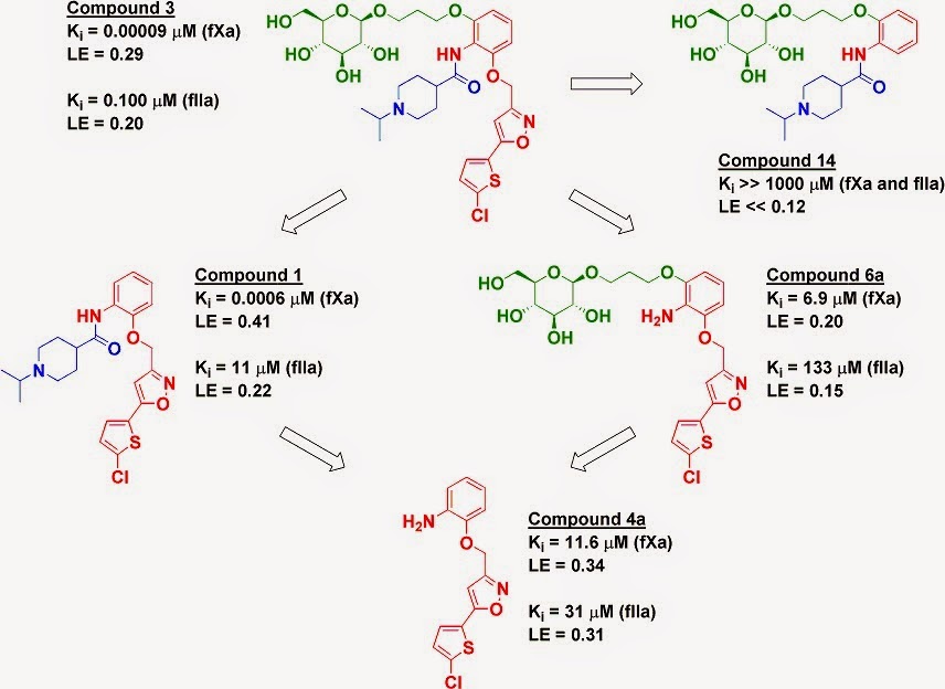
The proteases factor Xa (fXa) and thrombin (fIIa) are two heavily-studied anticoagulant targets. The paper characterizes a previously described molecule (compound 3) that is selective for fXa but still potent against fIIa, leading to good anticoagulant activity in human plasma as well as profibrinolytic activity. The researchers took a fragment deconstruction approach to better understand the binding to both targets.

As seen previously for fXa, the chlorothiophene moiety (red) is essential for binding, and removing it (compound 14) obliterates any detectable activity on both enzymes. However, while removing the glucose moiety (green) to give compound 1 reduced affinity for fXa by less than ten-fold, it reduced affinity for fIIa by more than two orders of magnitude. In contrast, removing the piperidine moiety (blue) to give compound 6a reduced affinity to both enzymes by several orders of magnitude.


However, these results are context-dependent. Removing both the piperidine moiety and the glucose moiety gives compound 4a, which has similar activity against fIIa as compounds 1 and 6a, where only a single moiety has been removed. In fact, compound 4a (without the glucose) is actually slightly more potent than compound 6a (with the glucose) against fIIa. But, as mentioned above, adding the glucose to compound 1 gives an impressive 110-fold boost in affinity for fIIa. In comparison, a famous early example of cooperativity in an NMR by SAR study gave only a 14-fold boost.

The researchers solved the crystal structure of compound 3 bound to fIIa, which reveals several hydrogen bond interactions between the glucose moiety and amino acid residues that have been previously implicated in allosteric activation of the protein. Perhaps compound 3 is exploiting this allosteric mechanism to bind more tightly.

This is a careful, thorough study and serves as a useful reminder that cooperativity can be huge, but it is still difficult to explain, much less predict.

27 May 2014

From substrates to fragments – or not

Recently we highlighted a paper in which enzyme substrates were deconstructed into component fragments and tested against an enzyme with unknown specificity. In a new paper in J. Am. Chem. Soc. a collaboration led by Karen Allen (Boston University), Frank Raushel (Texas A&M), and Brian Shoichet (UCSF) has performed a similar experiment to ask whether fragments could be used to identify substrates.

The researchers chose six enzymes from three different classes and collected various-sized fragments based on known substrates. These were then tested in functional assays to see whether they could be substrates or inhibitors. Stunningly, in most cases the fragments showed no activity against the enzymes; when activity was detectable, it was usually at least 100,000-fold lower than the natural substrate. Even subtle tweaks, such as removing a hydroxyl group, were enough to mess things up, as illustrated for adenosine deaminase (compare compounds 1 and 4). Breaking the substrate in two was sometimes better: compound 8 was turned over slowly by the enzyme, though its complementary fragment 9 had no effect on activity – positive or negative – when added to the assay along with compound 8 or the natural substrate.


Of course, functional assays are less sensitive than biophysical assays, but in the one case where the researchers tried soaking fragments into crystals of the enzyme they found that the fragments bound in a different manner than the substrate – echoing previous work deconstructing synthetic inhibitors of protein-protein interactions.

As the authors note, the remarkably sharp structure-activity-relationships (SAR) observed here could reflect a fact of nature: most enzymes need to be highly selective for their substrates to avoid mucking up cellular metabolism.

Moreover, the notion that two fragments, when properly linked together, can bind more tightly than the sum of their individual binding energies has been a primary motivator behind fragment-based lead discovery for more than 30 years. In a sense, this paper illustrates this principle in reverse. Indeed, it is possible for the energy gained by linking two fragments to exceed the binding energy of an individual fragment.

This is a nice study from which we can draw two lessons, one pessimistic, the other optimistic. On the down side, we are unlikely to be able to use fragments to predict the natural substrates of uncharacterized enzymes, at least on a general basis. As noted previously, this is not surprising: the concept of molecular complexity predicts that fragments should be fairly promiscuous, and we’ve seen time and again that fragment selectivity is not necessarily maintained during optimization.

On the positive side, this study beautifully illustrates that it is possible to achieve massive enhancements in affinity with relatively small changes. Beyond just the magic methyl effect, we’ve got the magic hydroxyl effect, the magic thiophene effect – heck – the magic fragment effect. Of course, these are retrospective analyses, and it’s easier to break things than make them. That said, folks at Astex demonstrated that it is possible to improve the affinity of a millimolar fragment a million-fold by adding just six atoms. Perhaps such opportunities are more general than we have previously dared to dream.

03 February 2014

How weak is too weak for PPIs?

Ben Perry brought up an interesting question in a comment to a recent post about fragments that bind at a protein-protein interface: “At what level of binding potency does one accept that there may not be any functional consequence?” I suspect the answer will vary in part based on the difficulty and importance of the target, and many protein-protein interactions (PPIs) rank high on both counts. In a recent (and open-access!) paper in ACS Med. Chem. Lett., Alessio Ciulli and collaborators at the University of Dundee, the University of Cambridge, and the University of Coimbra (Portugal) ask how far NMR can be pushed to find weak fragments.

The researchers started with a low micromolar inhibitor of the interaction between the von Hippel-Lindau protein and the alpha subunit of hypoxia-inducible factor 1 (pVHL:HIF-1α), an interaction important in cellular oxygen sensing. The team had previously deconstructed this molecule into component fragments, but they were unable to detect binding of the smallest fragments.

In the new study, the researchers again deconstructed the inhibitor into differently sized fragments and used three ligand-detected NMR techniques (STD, CPMG, and WaterLOGSY) to try to identify binders. As before, under standard conditions of 1 mM ligand and 10 µM protein, none of the smallest fragments were detected. However, by maintaining ligand concentration and increasing the protein concentration to 40 µM (to increase the fraction of bound ligand) or increasing concentrations of both protein (to 30 µM) and ligand (to 3 mM), the researchers were able to detect binding of fragments that adhere to the rule of three.

Of course, at these high concentrations, the potential for artifacts also increases, but the researchers were able to verify binding by isothermal titration calorimetry (ITC) and competition with a high-affinity peptide. They were also able to use STD data to show which regions of fragments bind to the protein, suggesting that the fragments bind similarly on their own as they do in the parent molecule. (Note that this is in contrast to a deconstruction study on a different PPI.) Even more impressively for a large (42 kDa) protein, the researchers were able to use 2-dimensional NMR (1H-15N HSQC) to confirm the binding sites.

Last year we highlighted a study that deconstructed an inhibitor of the p53/MDM2 interaction. In that case, the researchers were only able to find super-sized fragments, and they argued that for PPIs the rule of three should be relaxed. The current paper is a nice illustration that very small, weak fragments can in fact be detected for PPIs, though you may need to push your biophysical techniques to the limit.

But back to the original question of how weak is too weak. With Kd values from 2.7-4.9 mM, these are truly feeble fragments. Nonetheless, they could in theory have been viable starting points had they been found prospectively. That assumes, though, that these fragments would have been recognized as useful and properly prioritized. The ligand efficiencies (LE) of all the fragments, while not great, are not beyond the pale for PPIs. Previous research had suggested that much of the overall binding affinity in compound 1 comes from the hydroxyproline fragment (compound 6, which was originally derived from the natural substrate). Not discussed in the paper, but perhaps more significantly, the LLE (LipE) and LLEAT values are best for compound 6, which despite having the lowest affinity is the only compound that could be crystallographically characterized bound to the protein. In the Great Debate over metrics, this suggests that LLE and LLEAT may be more useful than simple LE for comparing very weak fragments.

15 July 2013

Fragments vs MDM2 – retrospectively

Protein-protein interactions (PPIs) are some of the tougher targets in drug discovery: they often have relatively flat interfaces that lack obvious small-molecule binding sites. But despite large surface areas, these interfaces often have smaller hot spots, and fragment-based approaches have succeeded against targets where traditional screening approaches have failed. In a paper in this month's issue of ACS Med. Chem. Lett., David Fry and colleagues at Roche describe a retrospective fragment study against a classic PPI (this story was also presented at the CHI conference earlier this year).

Molecules that disrupt the interaction between p53 and MDM2 have been sought since the 1990s as potential anti-cancer agents. The first such inhibitor to enter the clinic was RG7112, one of the so-called nutlins developed at the Roche Nutley site, which is sadly in the long process of closing. Although this series of compounds originated from a high-throughput screen, the researchers were interested to know whether they could have been discovered from fragments. To do so, they deconstructed RG7112 into a series of smaller pieces.

RG7112 is a “tripod-like” molecule with three hydrophobic moieties (blue, red, and green, below) that bind to subpockets on MDM2, as well as a polar “cap” (pink) that projects into solvent but is nonetheless important for activity. The researchers were unable to detect the binding of any of these individual fragments by either SPR or two-dimensional (HSQC) NMR, suggesting that each moiety alone was too weak, so the researchers tried lopping off one or two pieces from RG7112. In all cases, pared-down molecules containing three out of the four moieties led to molecules with affinities in the 14 to 1000 micromolar range. The tipping-point seemed to be when they kept just two of the four component fragments: in two cases the resulting molecules showed no detectable binding, while in two other cases they did. Compound 5, in fact, was a decent hit by any measure, and crystallography revealed that this molecule binds in the same manner as RG7112.

Interestingly, compound 5 makes interactions with two adjacent subpockets, including a deep hole that seems to be critical for all MDM2 binders; p53 inserts the indole moiety of a tryptophan into this site.

However, compound 5 does teeter on the edge of what could be called a fragment: with a molecular weight of 305 it falls just outside the Rule of Three (sorry Teddy!), and with 20 non-hydrogen atoms it sits right on the border of what most people seem to include in their libraries. The researchers suggest that larger fragments may be necessary for tackling PPIs, though they recognize that doing so will increase the number of fragments necessary to sample chemical space.

This is a beautiful, thorough study, but I do worry about super-sizing fragments. There are now multiple success stories of finding and advancing fragments against PPIs, including such “teflon-targets” as Mcl-1. Perhaps the nutlins would not have been discovered starting from small fragments, but with hundreds of billions of possibilities there are certainly other, even more ligand-efficient leads out there waiting to be discovered.