Showing posts with label activator. Show all posts

17 August 2020

Merge and grow: Fragment-based activators of SOS1

The RAS family of proteins is implicated in roughly one third of cancers, and as such has been a long-standing target for drug discovery. Earlier this year we highlighted how covalent fragment-based approaches were instrumental in discovery of direct KRAS inhibitors. A recent paper in J. Med. Chem. by Stephen Fesik and colleagues at Vanderbilt University takes a more unusual approach.

RAS proteins are activated when guanine exchange factors (GEFs) such as Son of Sevenless 1 (SOS1) exchange GDP for GTP. Clinical compounds bind to a mutant form of KRAS and block this process. Previous high-throughput screening in Fesik’s group had found molecules that bind to and activate SOS1-mediated nucleotide exchange. While it might seem counterintuitive to activate a known oncogene, these molecules can actually block downstream RAS signaling by inducing a feedback mechanism. Here, the researchers used fragment screening to look for a new series.

The catalytic core of SOS1 is ~65 kD, relatively large for the protein-detected NMR methods beloved of the Fesik group, so they produced proteins in which the methyl groups of Ile, Val, Leu, and Met were 13C-labeled. Selective Ile to Ala mutations allowed them to assign the various methyl groups. An 1H-13C HMQC screen of nearly 14,000 fragments yielded 59 hits (~0.1%), all quite weak: only five had dissociation constants better than 1 mM. Crystal structures were obtained for 16, revealing that all of them bind in the same site previously identified (see also here for similar work from a different group).

Fragments F-4 and F-7 bound in similar positions as each other and also as the HTS-derived compounds, so the researchers merged them to yield molecules such as compound 1b, with improved affinity and ligand efficiency.

Crystallography suggested that a nearby aspartic acid residue could be engaged through fragment growing, leading to molecules such as compound 2d. In addition to low micromolar affinity, this molecule also activated SOS1-mediated nucleotide exchange. In a cell-based assay, the compound caused enhanced phosphorylation of downstream target ERK at low concentrations and decreased phosphorylation at high concentrations, similar to what had been seen for the earlier series of molecules. Presumably, the biphasic response is due to a negative feedback loop that ultimately clamps down RAS signaling.

This is a nice example of structurally enabled fragment-merging and growing, assisted by knowledge of other ligands. While the compounds are probably not sufficiently potent to serve as chemical probes, they could be useful starting points. Activating the RAS pathway may or may not be a good approach for treating cancer, and we need suitable chemical tools to answer this question.

08 July 2019

Stabilizing apolipoprotein E4 with fragments

Among fragment-derived drugs that have entered the clinic, BACE1 inhibitors are well-represented. Sadly, multiple drugs targeting this protein have failed to show efficacy against Alzheimer’s disease. That said, every drug that has been thrown at Alzheimer’s has failed to slow the disease, so perhaps we need to think more boldly. An example was published recently in J. Med. Chem. by Andrew Petros, Eric Mohler, and colleagues at AbbVie.

The researchers were interested in apolipoprotein E4 (apoE4), one of three isoforms found in humans. Folks who have two apoE4 alleles are at increased risk for Alzheimer’s, suggesting that the protein might make a good drug target. Unfortunately, although it is known to be a lipid carrier, its precise function is unclear. What is known is that apoE4 is less stable to thermal denaturation than apoE2 or apoE3, so the team set out to find molecules that would stabilize the protein. This being AbbVie, they used two-dimensional NMR to find fragments.

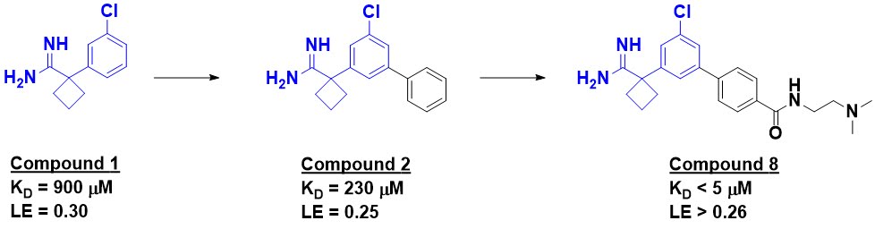
The methyl groups of all the isoleucine, leucine, methionine, and valine residues in apoE4 were 13C labeled, and the researchers looked for changes in the 13C-HSQC spectra upon addition of fragments; just over 4000 were screened in pools of 12, each at 1 mM. Of the dozen or so hits, compound 1 was among the best.


NMR titration studies revealed an affinity just under 1 mM, while SPR suggested slightly stronger binding. As hoped, compound 1 raised the melting temperature of apoE4. Adding the fragment also altered the kinetics of liposome breakdown, causing the protein to behave more like apoE2 and apoE3. Although this assay isn’t necessarily physiologically relevant, the reasoning is that causing apoE4 to behave more like the other isoforms may be useful.

A crystal structure of compound 1 bound to apoE4 revealed the fragment to be binding in a small pocket, and growing led to compound 2, with a slightly improved affinity. Introduction of polar substituents to interact with a nearby aspartic acid side chain led to compound 8, with low micromolar affinity (assessed by NMR). This molecule also stabilized apoE4 with respect to thermal denaturation.

As noted above, it is not entirely clear why apoE4 is associated with Alzheimer’s, but researchers had previously found that overexpression in a neuronal cell line caused release of the inflammatory cytokines IL-6 and IL-8. When human induced pluripotent stem cell (iPSC)-derived astrocytes carrying two copies of apoE4 were treated with compound 8, release of IL-6 and IL-8 cytokines was reduced to levels similar to those from iPSC-derived astrocytes carrying two copies of apoE3. The compound also showed no toxicity, even at relatively high concentrations (100 µM).

There is still a tremendous amount to do: affinity needs to be improved considerably, and permeability is also mentioned as an issue. Moreover, the highly polar nature of compound 8 will likely make transport across the blood-brain barrier challenging. Optimizing activity against a target whose function is poorly understood will present a host of problems. But if it were easy, Alzheimer’s disease would not be the scourge that it is. Practical Fragments salutes thinking outside the box, and wishes those involved the best of luck.

04 February 2019

Taking a step towards STEP activators

Most drugs – and small molecule modulators in general – inhibit something, often an enzyme. Enzyme activation, on the other hand, is rare; we’ve highlighted just a few cases on Practical Fragments over the past decade. A new example is described in J. Med. Chem. by Christofer Tautermann and collaborators at Boehringer Ingelheim and the Beckman Research Institute of the City of Hope.

The researchers were interested in the protein tyrosine phosphatase non-receptor type 5 (PTPN5), also known as striatal-enriched protein tyrosine phosphatase (STEP). As its name suggests, this enzyme is found in the brain, and has been implicated in multiple neuropsychiatric disorders. However, phosphatases are tough targets due to their small, polar active sites. The problem is exacerbated for CNS targets, because negatively charged molecules have a hard time crossing the blood-brain barrier. Thus, the researchers sought allosteric modulators.

They began with a screen of 3083 fragments using STD NMR, differential scanning fluorimetry, and microscale thermophoresis. Validation of the several hundred hits by 2-dimensional NMR confirmed just seven, and comparison of the protein chemical shifts with those caused by a non-specific active site binder (sodium vanadate) suggested that compound 2 bound outside the active site. Crystallography confirmed this, revealing that the compound binds on the “back side” of the protein, about 20 Å from the catalytic pocket. The affinity was extraordinarily weak, with no functional activity, so the researchers used NMR to drive the SAR. Ultimately this led to fragment-sized BI-0314, with measurable affinity by isothermal titration calorimetry (ITC). Crystallography revealed that it binds in the same pocket as compound 2.

Surprisingly, far from being an inhibitor, BI-0314 actually showed activation of the enzyme in functional assays, increasing the activity by up to 60% at 0.5 mM. Careful mechanistic analysis revealed that this was due to an increase of kcat, while the KM for substrate was mostly unchanged. Molecular dynamics simulations suggested that BI-0314 increases the rigidity of the enzyme, and also stabilizes the active conformation. As expected of an allosteric modulator, the molecule was selective for STEP, with no activity (activating or inhibitory) for a couple other phosphatases.

As it turns out, the researchers were actually interested in STEP inhibitors, so they didn’t pursue BI-0314 further. As they note, there is still much to be done to generate a useful chemical probe, in particular improving potency. Laudably, the researchers are making BI-0314 available to other researchers free of charge. Perhaps someone else will be able to take this forward, as we’ve seen for other published fragments. And indeed, as researchers at Novartis have shown with asciminib, the transition from an allosteric binder with no functional activity to an inhibitor is possible – perhaps the same will hold true for an activator. If you are interested in STEP, you now have a new site to explore, and even a well-characterized starting point.

30 July 2018

Dimerization: elegant but not essential

A special case of fragment linking is dimerization, in which two copies of the same fragment bind to adjacent sites in a protein and are subsequently linked together (see for example here, here, and here). A recent example was published in J. Med. Chem. by Bernard Pirotte, Julien Hanson (University of Liège), Lionel Pochet (University of Namur), Jette Kastrup (University of Copenhagen) and their collaborators.

The researchers have for some time been interested in AMPA receptors, critical components in neuronal synaptic transmission. Increasing their activity could be useful for treating diseases such as depression and schizophrenia, but increasing activity indiscriminately is known to be toxic. One approach has been to develop positive allosteric modulators (PAMs), which increase the activity only in the presence of the natural ligand glutamic acid, thus amplifying the normal biological signal.

AMPA receptors themselves are dimers of dimers. Many different PAMs have been reported for AMPA receptors, and some of these are in fact dimeric molecules that span two adjacent binding sites across the dimer interface. A crystal structure of a molecule closely related to compound 35 revealed that each molecule binds to two adjacent protein subunits, so the researchers designed compound 22, which pairs the molecules through a simple ethylene moiety. The strategy paid off with a low nanomolar activator, which crystallography confirmed binds as expected.


Interestingly, conceptually cleaving the bond connecting the two fragments generates a compound (33) which is slightly less active than the initial fragment 35; it is possible the methyl groups are too close to one another when two copies of compound 33 are bound.

As the researchers point out, compound 22 is one of the most potent AMPA receptor PAMs reported. However, it is also quite large, particularly since it needs to cross the blood-brain barrier. No animal data are reported, but a simple metric called the CNS MPO desirability score is reasonably predictive. This score is based on the molecular weight, lipophilicity, total polar surface area, number of hydrogen bond donors, and basicity; higher scores are better. By this measure, compound 22 is predicted not to have high brain penetration, though of course any metric needs to be taken with caution.

However, a separate J. Med. Chem. paper by many of the same researchers revealed that dimerizing the molecules is not essential: simply growing compound 35 could also generate a low nanomolar AMPA receptor PAM (compound 8). Crystallography revealed that the added phenyl group binds where the second molecule of compound 35 would normally bind. Moreover, compound 8 has a higher ligand efficiency as well as a higher CNS MPO desirability score than the dimeric compound 22, suggesting that it is more likely to be able to cross the blood-brain barrier.

In the absence of pharmacological or pharmacokinetic data, if forced to choose I would probably focus on compound 8 rather than compound 22. All of which is to say that although there is a certain elegance to dimerizing molecules, you might be able to replace one of them with a smaller, simpler moiety.

04 December 2017

Fragment activators of AMPK

Kinase inhibitors are common. Some 40% of the fragment-derived clinical compounds in our latest list target kinases. Kinase activators, on the other hand, are rare. It is easier to interfere with something than to enhance it, and of the 630+ posts on Practical Fragments, I believe only two discuss enzyme activators. A new paper (here) by Ping Lan, Iyassu Sebhat, and colleagues at Merck and Metabasis provides a third example.

The researchers were interested in adenosine monophosphate-activated protein kinase (AMPK), which plays a critical role in metabolism. As its name suggests, this kinase has naturally occurring activators, though these are nucleotides and thus not particularly useful as chemical probes. It also has fiendishly complex biology: the active enzyme is a heterotrimer of three distinct proteins, each of which comes in two or three flavors, leading to 12 different isoforms with their own unique tissue distributions – which vary among different animals.

After multiple HTS screens failed to produce anything of value, the researchers turned to fragments. Realizing that AMPK was a tough target, they assembled a library of 25,000 highly diverse fragments tending towards super-sized (all of them were greater than 200 Da, and they had up to 22 non-hydrogen atoms). A biochemical screen yielded just three hits, including compound 4.


Despite the generally larger size of fragments in the library, it is interesting that compound 4 follows the rule of three, with just 16 non-hydrogen atoms. Although only modestly active, the small size gave an impressive ligand efficiency, and the activation was nearly 80% that of the natural ligand AMP.

Rigidification of the linker between the benzimidazole and the acid moiety led to compounds such as 27, with low micromolar potency, while growing from the benzimidazole itself led to high nanomolar compounds such as compound 36. Combining these two modifications and further optimization for pharmacokinetic properties led to MK-3903, which was chosen as a development candidate.

MK-3903 activates 10 of the 12 AMPK isoforms and is fairly selective against a panel of off-targets. As predicted mechanistically, administering the compound to mice increases the phosphorylation of downstream substrates of AMPK. It also causes decreased fatty acid synthesis and increased insulin sensitivity. However, a related molecule causes cardiac hypertrophy in rats and monkeys.

In addition to the fact that MK-3903 is an enzyme activator, there are several other notable features about this story. First, despite the difficulty of the target, the team made rapid progress, moving from the initial screen to useful tool compounds in less than a year. Second, as near as I can tell, this optimization was done in the absence of direct structural information on how the compounds bind. (A publication by a separate team, who was closely monitoring the patent literature, describes the crystal structure and mechanistic analysis of a related molecule.) Third, all of this work stemmed from a single fragment: although more ligandable targets may produce lots of hits, in the end you only need one.

Finally, this paper illustrates the lag time that can occur between research and publication: several of the authors are from Metabasis, which was acquired by Ligand Pharmaceuticals way back in 2010. That was also when the patent publication describing these molecules was filed, suggesting the work could have been done a decade ago. That’s something to keep in mind when using the literature to guess who is working on fragments.

03 November 2014

Fragments as enzyme activators

About half of all approved drugs are small molecules that inhibit enzymes. This makes sense intuitively: an enzyme is like a complicated little machine, and there are lots of ways you can wreck a machine. But there are times when you might want to activate an enzyme, and this is conceptually more difficult. In a new paper in Angew. Chem. Int. Ed., Rod Hubbard and colleagues at the University of York show how fragments can help.

The researchers were interested in the enzyme O-GlcNAc hydrolase (OGA), which removes N-acetylglucosamine from proteins. They were looking for inhibitors of a bacterial version of this enzyme, so they screened it against 100 fragments using three ligand-observed NMR methods (STD NMR, PO-WaterLOGSY, and T1ρ-filter). This resulted in a very high hit rate: 22 fragments showed binding in all three assays, and 18 of these were competitive with a known substrate-like inhibitor, PUGNAc. Some of these were also active in an enzymatic assay.

More interestingly, the four hits that were not competitive with PUGNAc actually appeared to bind more strongly to the protein in the presence of that inhibitor. In this case, the inhibitor can be considered a stand-in for the natural substrate, and the results suggested that the fragments might enhance binding of the enzyme to its substrate. Indeed, when these fragments were tested in the enzymatic assay, one of them (compound 2) actually activated the enzyme, with an “AC50” value of 3.5 mM.

The activator was further characterized by several orthogonal methods. NMR revealed that, in the presence of PUGNAc, compound 2 bound with a dissociation constant of 3.1 mM, very close to its AC50 value. In the absence of PUGNAc, the dissociation constant was too weak to be determined. Isothermal titration calorimetry experiments revealed that compound 2 increased the affinity of the enzyme for PUGNAc by more than three-fold, while Michaelis-Menten kinetics revealed that, at a concentration of 8 mM, compound 2 nearly doubled the kcat/KM. A crystal structure of both compound 2 and PUGNAc bound to the enzyme revealed that the two molecules bind near one another in what appears to be a catalytically active conformation of the protein.

Next, the researchers took an “SAR by catalog” approach to find more potent molecules, leading to compound 4, with an AC50 value roughly ten-fold better than compound 2. Detailed enzymatic characterization revealed that the molecule acts as a “nonessential reversible activator,” and improves substrate binding roughly 7-fold while improving kcat by a factor of 1.7.

Interestingly, differential scanning fluorimetry (DSF) revealed that all the activators destabilize the enzyme, while PUGNAc stabilizes the enzyme. This is yet more evidence that compounds can decrease the melting temperature of a protein while still binding specifically.

This is an academic study in the best sense of the phrase. The immediate utility of these molecules is tenuous, as they do not work with human OGA (which has some important active site differences), though there may be industrial applications. The more important finding of this rigorous paper is that discovering enzyme activators might be easier than expected. There aren’t that many reported, but this may just be because people tend not to look for them: if you find an activator of an enzyme you are trying to shut down you probably won’t pursue it. That said, a few companies have been founded on enzyme activators. Perhaps fragments can help discover more.