Showing posts with label Tethering. Show all posts

08 May 2023

From reversible to irreversible fragments vs caspase-6

The caspase family of cysteine proteases is critical for initiating and executing programmed cell death, and some members also play a role in inflammation. Humans have a dozen caspases, all of which cleave substrate peptides immediately after an aspartic acid residue. Developing chemical probes for specific family members has been challenging for several reasons, not least because of similar active sites. In a newly published open-access J. Am. Chem. Soc. paper, Michelle Arkin, Adam Renslo, and colleagues at UCSF have done just that. (Michelle spoke about this work at the CHI DDC meeting last month.)
 
The researchers were particularly interested in caspase-6, which has been implicated in neuroinflammation. They recognized that, in addition to the active site cysteine, caspase-6 has another cysteine not found in other caspases. C264 sits at the far end of the active site, on the smaller of the two separate protein subunits that come together at the active site. They used Tethering (previously described here) to screen a library of 1500 disulfide-containing fragments. Since C264 is on the small subunit and the active site cysteine is on the large subunit, it was easy to determine specificity using mass spectrometry.
 

There were quite a few hits against the small subunit of caspase-6, including compound 1. The researchers replaced the disulfide moiety with an irreversible vinylsulfonamide warhead and made a few other modifications to the core to obtain compound 2a. This showed sub-micromolar inhibition of the enzyme after just 15 minutes, and crystallography revealed that it bound covalently to C264, as expected. The crystal structure also suggested where to grow the molecule, leading to compound 3a, which was a low nM inhibitor at 15 minutes. The researchers determined the kinact and Ki values, which are important parameters for irreversible inhibitors. The molecule was also quite selective, showing little to no inhibition of nine other human caspases at 10 µM in biochemical assays.
 
Compound 3a effectively blocked caspase-6 mediated cleavage of the substrate lamin A in a cellular assay. To assess cellular specificity, the researchers made an alkyne-containing version of compound 3a which could be used in pull-down experiments using click chemistry. This revealed a couple dozen protein targets, of which only eight could be competed by the parent compound 3a. Interestingly, all except caspase-6 were membrane-bound proteins, and none were proteases.
 
This is a nice, concise paper reminiscent of the early work that ultimately led to sotorasib. A key liability of the series is the vinylsulfonamide warhead, which is probably too intrinsically reactive for in vivo studies. The researchers did try replacing this with the clinically validated acrylamide warhead, but unfortunately the resulting molecule was not active. Nonetheless, compound 3a is worth considering as a chemical probe, and the researchers note that further optimization efforts are ongoing. I look forward to seeing the evolution of these molecules.

20 January 2020

Fragments in the clinic: AMG 510

Few cancer targets are as prominent as KRAS, a molecular switch that has been called “the beating heart of cancer.” Mutations that cause the switch to be stuck in the “on” state occur in roughly a quarter of human tumors. The protein’s role in driving cancer has been known for nearly forty years, but for most of that time it has been considered undruggable.

Until now.

When bound to GTP, KRAS is turned on, promoting cell proliferation until the GTP is hydrolyzed to GDP. Activating mutants impair this hydrolysis. An obvious approach to targeting KRAS would be to develop small molecules that bind in the nucleotide-binding pocket, as has been done successfully for kinases. Unfortunately, the extremely high affinity of KRAS for GTP, along with the high intracellular concentration of GTP, makes this impossible.

One activating mutant replaces a glycine with cysteine (G12C), providing a convenient handle for covalent inhibitors. In 2013, Kevan Shokat and colleagues at University of California San Francisco described in Nature how they used Tethering to identify fragments such as 6H05 that bound to this cysteine through a disulfide bond. Because disulfides are not stable in cells, they replaced this “warhead” with an acrylamide moiety and made a few other tweaks to arrive at compound 12. Extensive optimization described by Yi Liu and collaborators at Kura, Wellspring, and Janssen in a 2018 Cell paper led to molecules such as ARS-1620, which showed potent biochemical and cell-based activity and promising results in mouse xenograft models.


In 2014, Carmot Therapeutics began a collaboration with Amgen to discover covalent inhibitors of the G12C mutant of KRAS. Carmot’s technology, Chemotype Evolution, entails rapid synthesis and testing of large libraries around an existing molecule such as a fragment (see here for a nice animation). In this case, fragments chosen included simple acrylamides, the thought being that – unlike the disulfides in Tethering – these could be carried through into the final molecule.

Initial hit 2 was truncated and cyclized to compound 4, which had comparable activity but with fewer atoms and rotatable bonds. Additional iterations of Chemotype Evolution and medicinal chemistry ultimately led to compound 1, with nanomolar activity in cells containing KRASG12C. Crystallography revealed that the tetrahydroisoquinoline moiety bound in a previously cryptic pocket formed by movement of a histidine side chain. These interactions contributed to the high affinity of the molecule; more details are provided in a paper published last year in ACS Med. Chem. Lett. by Victor Cee, myself, and our collaborators at Amgen and Carmot.


Unfortunately, although compound 1 was more potent in cell assays than ARS-1620, it had low oral bioavailability and rapid clearance in mice and rats. However, as described in J. Med. Chem., Brian Lanman and colleagues at Amgen superimposed ARS-1620 with compound 1 and realized that it would be possible to access the cryptic pocket from the former compound. This strategy proved successful, ultimately leading to AMG510, which entered the clinic in August 2018. And although it is still early, a paper in Nature by Jude Canon, J. Russell Lipford, and collaborators at Amgen and elsewhere describes promising responses to the drug by a handful of patients with non-small-cell lung carcinoma.


Amgen is not the only company to have capitalized on the Tethering results: Mirati used the information to develop their clinical-phase MRTX849. And Janssen has also entered the clinic with JNJ-74699157.

There are multiple lessons here. First, as we’ve seen previously, a single fragment can lead to multiple clinical compounds. Second, progress often requires considerable changes to the initial fragment. This story is clearly a case of fragment-assisted drug discovery, and in the interest of space I’ve had to omit most of the lovely medicinal chemistry, not to mention biology and biophysics, detailed in these five papers.

Another lesson is that covalent fragments can enable lead discovery for targets not accessible through other means. But this enablement may require non-conventional molecules; the initial fragments violate the rule of three, and the disclosed clinical compounds have molecular weights in excess of 500 Da. Despite these challenges, remarkably rapid progress is possible: less than five years elapsed between the first Nature publication and the entry of AMG 510 into the clinic.

Most important, this research has led to possibly life-extending molecules – one of the responders described in the second Nature paper had been on drug for 42 weeks. Practical Fragments wishes her or him, and everyone else involved with the trials, the best of luck.

15 April 2019

Fourteenth Annual Fragment-based Drug Discovery Meeting

CHI’s Drug Discovery Chemistry (DDC) meeting took place last week in San Diego. I think this was the largest yet, with >825 attendees, a third from outside the US, and nearly 70% from industry. The initial DDC meeting in 2006 had just four tracks, of which FBDD is the only one that remains. This one had nine tracks and four one-day symposia, so it was obviously impossible to see everything. Like last year, I’ll just stick to broad themes.

Success Stories
As always, clinical compounds received deserved attention. Among two I’ve covered recently, Paul Sprengeler described eFFECTOR’s MNK1/2 inhibitor eFT508, while Wolfgang Jahnke discussed Novartis’s allosteric BCR-ABL1 inhibitor ABL001. As previously mentioned, ABL001 is a case study in persistence: the project started in stealth mode and was put on hold a couple times until seemingly intractable problems could be overcome.

Another story of persistence, albeit with a less happy outcome, was presented by Erik Hembre, who discussed Lilly’s BACE1 program. Teddy wrote about their first fragment-derived molecule to enter the clinic, LY2811376, back in 2011. Unfortunately this molecule showed retinal toxicity in three-month animal studies, so the researchers further optimized their molecule to LY2886721, which made it to phase 2 studies before dropping out due to elevated liver enzymes. Reasoning that a more potent molecule would require a lower dose and thus lower the risk of toxicity, the researchers used structure-based drug design to get to picomolar LY3202626, which also made it to phase 2 before being scuttled due to the apparent invalidation of BACE1 as an Alzheimer’s disease target.

Talks on BCL2 and MCL1 inhibitors from Vernalis, AstraZeneca, and Servier all involved fragments in some capacity, but unfortunately they were in the protein-protein interaction track which was held concurrently with the FBDD session I was chairing. Suffice it to say you can expect to hear more about the phase 1 compounds AZD5991 and S654315.

A few earlier-stage success stories included Till Maurer’s discussion of the Genentech USP7 program (see here), Santosh Neelamkavil on Merck’s Factor XIa inhibitors, and Rod Hubbard on Vernalis DYRK1A, PAK1, and LRRK2 inhibitors. We have previously written about how displacing “high-energy” water molecules can be useful, and this tactic was used by Sven Hoelder at the Institute of Cancer Research for their BCL6 inhibitors. Last week we highlighted halogen bonds, which proved important for transforming molecules that simply bind to MEK1 to molecules that bind and inhibit the protein, as described by AstraZeneca’s Paolo Di Fruscia.

Methods
The MEK1 story Paolo told began with a very weak (0.45 mM) fragment that the team was able to advance to 300 nM in the absence of structure, though they did eventually obtain a crystal structure that supported further optimization. On the topic of crystallography, Marc O’Reilly discussed the Astex MiniFrag approach, which we recently wrote about here. Only a couple of these fragments contain a bromine atom, but Marc did mention that, of the 10,051 X-ray complexes solved at Astex, a number show halogen bonds, including some to the hinge region in kinases.

At FBLD 2018 Astex’s Chris Murray showed the first cryo-EM structure of a fragment bound to a protein, and Marc confirmed that they have now obtained structures of fragments bound to two targets, with fragments as small as 120 Da and resolution as good as 2.3 Å. They are increasing automation, with turnaround times of less than 24 hours in some cases. Santosh also mentioned that Merck is applying cryo-EM to fragments.

Frank McCormick (UCSF) highlighted multiple fragment-finding methods used to discover inhibitors against RAS family proteins, which are responsible for more than a million cancer deaths each year. In addition to stalwarts such as crystallography and NMR, these include less common methods such as Tethering and the second harmonic generation (SHG) approach for detecting conformational changes used by Biodesy. RAS was reported as a cancer driver almost forty years ago, but only now are the first direct inhibitors entering the clinic – a testimony to both the challenging nature of the target and how far we’ve come.

SHG and Tethering were also highlighted elsewhere: Charles Wartchow described how SHG identified 392 hits from a collection of 2563 fragments against an E3 ligase bound to a target protein at Novartis, while Michelle Arkin described her use of Tethering at UCSF to find molecules that could stabilize a complex of 14-3-3 bound to a specific client protein (see here).

An effective sponsored talk was presented by Björn Walse of SARomics Biostructures and Red Glead Discovery, who described weak affinity chromatography (WAC). Once they saw the schedule for DDC, they looked for a target that would be presented shortly before their presentation, and chose the protein USP7 as a test case. Beginning in January, they screened a library of 1200 fragments to obtain 34 hits, of which 7 confirmed in a thermal shift assay. This led to an SAR-by-catalog experiment, and 11 of the 31 fragments tested showed activity, as did a Genentech positive control compound.

All methods can generate false positives and false negatives (see for example here and here), some of which were described in an excellent talk by Engi Hassaan of Philipps University. Engi discussed how improving the sensitivity of an STD assay by decreasing salt concentration identified more fragments that had previously been found by crystallographic screening. She also presented a case study of how introducing a tryptophan residue into a small protein to facilitate purification led to problems down the road when the tryptophan side chain blocked a key pocket in the crystal lattice. Gregg Siegal (ZoBio) also highlighted a case where a fragment bound to the dimer interface in a crystal structure, whereas in solution the fragment bound to the active site, as observed by NMR.

Finally, among computational methods, Pawel Sledz (University of Zurich) gave a nice overview of the SEED and AutoCouple methods, while Paul Hawkins (OpenEye) described rapid searching of more than 10 billion chemical structures using ROCS (rapid overlay of chemical features). SkyFragNet is looking closer with each passing year.

There is much more to say, so please feel free to comment. Several good events are still coming up this year, and mark your calendar for 2020, when DDC returns to San Diego April 13-17!

25 February 2019

Stabilizing protein-protein interactions

Despite the fact that the second FDA-approved fragment-derived drug targets a protein-protein interaction (PPI), these types of targets have a well-earned reputation for being difficult. Most researchers try to disrupt PPIs. An alternative is to stabilize PPIs. This is not as crazy as it sounds: rapamycin, tafamidis, and PROTACs all stabilize PPIs. In a paper just published in J. Am. Chem. Soc., Michelle Arkin, Christian Ottmann, and collaborators at UCSF, Eindhoven University of Technology, Novartis, and the University of Duisburg-Essen bring fragments to bear on the problem.

The researchers were interested in the protein 14-3-3δ, a “hub” protein that binds to more than 300 other proteins (not all at the same time). One of these is estrogen receptor α (ERα): binding prevents the transcription factor from dimerizing and binding to DNA. The natural product fusicoccin A (FC-A) binds at the interface of 14-3-3δ and ERα and stabilizes that interaction, thereby inhibiting the growth of breast cancer cells. Because FC-A is a structurally complex natural product, the researchers sought fragments that would have a similar effect. They used Tethering, in which reversible disulfide bond formation stabilizes a protein-ligand complex, allowing its identification (see here and here). Specifically, fragments that bind near a cysteine residue are resistant to reduction, and the extent of binding can be detected by mass spectrometry.

The 14-3-3δ protein conveniently contains a cysteine residue in the vicinity of the ERα binding groove; the researchers used this native protein and also created two additional mutant proteins in which the native C38 cysteine was removed and new cysteine residues were introduced nearby. These three proteins were then screened against a library of 1600 disulfide-containing fragments under mildly reducing conditions in the presence or absence of a phosphopeptide derived from ERα. Most of the hits against the native protein were weak, but several hits against the N42C mutant were both resistant to reduction and also bound preferentially to the 14-3-3δ/ERα peptide complex compared to 14-3-3δ alone. Thus, ERα could enhance the binding of fragments to 14-3-3δ.

Next, the researchers used a fluorescently labeled peptide derived from ERα to show that one fragment could improve the apparent dissociation constant for the peptide and 14-3-3δ about 40-fold, from 1.3 µM 32 nM. Crystallography revealed that the cooperative fragments bound at the PPI interface, as expected given the location of the cysteine residues. The cooperative fragments placed a phenyl group in close proximity to a valine residue from the ERα peptide.

The researchers then examined the selectivity of one of their stabilizing fragments for other 14-3-3δ client proteins. In the case of a phosphopeptide derived from TASK3, which has a similar sequence to that of the ERα peptide, the fragment also showed cooperative binding. However, two peptides from other client proteins competed with the fragment for binding, and crystal structures revealed that the binding modes would be incompatible.

This is a nice illustration of site-directed fragment discovery to identify fragments that can modulate protein function in a more sophisticated manner than simple inhibition. One of the nice features of Tethering is that – like crystallography – it is able to identify extraordinarily weak binders. Unfortunately, this sometimes makes the hits challenging to advance: NMR experiments do show binding between a non-disulfide-containing derivative of one of the fragments and the 14-3-3δ/ERα peptide complex, but at high concentrations. It will be interesting to see whether this can be built into a potent non-covalent binder, and/or whether other types of covalent modifiers will be able to produce useful chemical probes for this target.

22 September 2015

Crystallography as a primary screen: the case of HisRS

X-ray crystallography plays a starring role in fragment-based lead discovery. But, with a few exceptions, it is rarely the primary screen. One of these exceptions was reported recently in Acta Crystallogr. D by Wim Hol and coworkers at the University of Washington, Seattle.

The researchers were interested in the histidyl-tRNA synthetase (HisRS) from Trypanosoma cruzi, the parasite that causes Chagas disease. They had previously constructed a library of 680 fragments in pools of 10, designed such that the fragments within each pool had different shapes to facilitate crystallographic screening. Crystals of HisRS•His (ie, the enzyme in complex with its histidine substrate) were soaked with the pools, such that the final concentration of each fragment was 1.5 mM. Fifteen of these pools showed new electron density, and these in turn yielded 15 different fragment hits when the pools were deconvoluted. Two additional fragments from the deconvolution process showed weak density, though these were not further pursued.

Strikingly, all 15 fragments bound to the same site, a site not observed in the absence of the fragments. This is a narrow groove described by the researchers as a “document sleeve,” and in fact all the hits are single six-membered aromatics or double aromatics with few or no aliphatic substituents. Most of the fragments are also quite small, with the majority having just 8 or 9 non-hydrogen atoms. Although all the fragments bind in roughly the same plane, there is considerable variation in the positions of substituents, and some of the fragments appear to bind in multiple orientations.

Next, the researchers tested their hits in orthogonal assays. Only one fragment showed thermal stabilization of the enzyme, and only three showed any inhibition in a functional assay (at most 39% at 2 mM of fragment). Thus, these are very weak binders.

The fragment-binding pocket is a few Ångstroms away from the histidine substrate, making linking the two ligands feasible. In preparation for this step, the researchers acetylated the amino group of the most potent fragment. This caused little change in the functional activity, but crystallography revealed that the fragment’s binding orientation had flipped around, such that the acetamide group pointed away from the histidine and towards a cysteine residue. Attempting to turn lemons into lemonade, the researchers added electrophiles to try to interact with the cysteine residue. Some of these molecules had measurable IC50 values (on the order of 1 mM), and crystallography of one of these showed covalent bond formation between the fragment and the targeted cysteine. This cysteine residue is found in the T. cruzi enzyme but not the human version, and indeed these molecules appear to be more active against the parasitic HisRS.

This is a nice example of fragment screening by crystallography that illustrates one of its main challenges: crystallography is capable of detecting extremely weak binders that may prove difficult to advance. Still, the researchers have taken some promising initial steps, and it will be fun to see what they come up with.

20 April 2015

Tethering versus RNA

Last week we highlighted one of the less common fragment-finding methods, and today we turn to another. Tethering uses reversible disulfide exchange chemistry to trap thiol-containing fragments near binding sites. Back when we developed the technology at Sunesis we used cysteine residues in proteins. We occasionally discussed applying it to nucleic acids, but at the time it was hard to make a good business case. Now that microRNAs (miRNAs) have become hot, there is more interest in going after nucleic acid targets, and in a recent paper in Molecules Kiet Tran and Peter Beal (UC Davis) and Michelle Arkin (UC San Francisco) have done just that.

The researchers were interested in an RNA sequence that is cleaved in cells to generate miR-21, a potential cancer target. The idea is to find small molecules that bind to pre-miR-21 and prevent its processing to the mature miRNA. To perform Tethering, the researchers first introduced a thiol group into adenosine and incorporated this into RNA. They made two separate versions of pre-miR-21, with the modified adenosine at a different site in each, and also made a control RNA with a completely different sequence.

Next, they incubated the modified RNAs with 30 different disulfide-containing small molecules under partially reducing conditions and used mass spectrometry to identify those that covalently bound. As expected most showed minimal binding, but there were a couple hits. One of these, a 2-phenylquinoline, bound to both modified versions of the pre-mR-21 as well the control RNA, suggesting non-specific binding. In fact, 2-phenylquinoline is a known intercalator, so while its identification is not surprising, it does validate the ability of Tethering to identify binders. The other hit, however, appeared to be specific for one of the two pre-mR-21 sequences.

Of course, there is still a long way to go; it is unclear how much affinity the hit has for the RNA, or how specific it would prove if tested against a large panel of decoy RNAs. A key challenge for Tethering – as with many fragment-finding methods – is figuring out what to do with a hit. This is all the more true with RNA, about which we’ve written several times over the years. Still, one nice feature of Tethering is that it allows one to target a specific site of interest. Also, the covalent (disulfide) bond helps with both crystallography and modeling. It will be fun to watch this story develop.

16 June 2014

Irreversible fragments

Last year we highlighted a paper in which fragments were attached to a “warhead,” a chemical group that could form a reversible covalent bond with cysteine residues in proteins. These fragments were then screened against a kinase to identify nanomolar inhibitors. In a new paper in J. Med. Chem., Alexander Statsyuk and coworkers at Northwestern University describe a similar approach using irreversible fragments. (See here for In the Pipeline’s take.)

As we noted last year, one of the nice things about using reversible warheads is the fact that you can run experiments under thermodynamically-controlled conditions. Indeed, this was the idea behind Tethering (here and here), in which a library of disulfide-containing fragments is allowed to equilibrate with a cysteine-containing protein; if a fragment has inherent affinity for the protein, the disulfide bond will be stabilized towards reduction and can be identified using mass spectrometry. (Full disclosure: I am an inventor of Tethering, and my company, Carmot Therapeutics, Inc., has an exclusive license to the intellectual property covering this and other technologies.)

Irreversible warheads operate at least partly under kinetic, rather than thermodynamic, control. For example, if all the fragments are extremely reactive, the protein will react with whichever fragment it happens to encounter first, regardless of whether the fragment has any inherent affinity for the protein.

Acrylamide moieties are able to form irreversible bonds to cysteine residues and are even starting to be found in drugs. In 2012, researchers at Imperial College London tested an acrylamide-containing analog of one of the (disulfide) hits from the original Tethering paper. This successfully labeled the target protein thymidylate synthase (TS), while several other acrylamide-containing molecules did not, and the fragment was selective for TS over two other enzymes with active-site cysteine residues.

Regardless of whether your warhead is reversible or not, it is important that different members of the library have similar reactivities: if the inherent reactivities of the fragments are different, it will be difficult to distinguish inherent binding energies from chemical reactivities. One of the problems with acrylamides is that subtle changes in chemical structures can have dramatic effects on intrinsic reactivities. The new paper compares rate constants of two acrylamides reacting with a low molecular-weight thiol, and finds that one reacts 2,000-fold faster than the other. The researchers tested three other classes of electrophiles – vinylsulfonamides, aminomethyl methyl acrylates, and methyl vinylsulfones – and found that these had narrower ranges of reactivity. They chose acrylates and built a set of 100 acrylate-modified fragments. Happily, when 50 of these were tested for reactivity, there was only a 2.4-fold difference between the most reactive and the least reactive members.

These 100 fragments were tested in pools of ten against the classic cysteine protease papain using mass spectrometry, and three hits were identified. Enzymatic assays revealed that these three hits were also irreversible inhibitors of the protein with respectable activities (kinact/Ki = 0.46-1.2 M-1s-1), while a member of the library that did not label in the mass spectrometry assay was a weaker inhibitor (kinact/Ki = 0.037 M-1s-1). Moreover, the papain hits did not label three other enzymes containing active-site cysteines, though these enzymes could be modified with other fragments in the library.

In the case of Tethering, the disulfide linker was a means for finding fragments; when these fragments were developed further, the disulfide linker was replaced. However, given the renewed interest in covalent drugs, some warheads might be able to be retained. It will be fun to see how these types of strategies develop.

30 December 2013

Review of 2013 reviews

The year is coming to an end, and as we did last year, Practical Fragments is looking back at notable events as well as reviews that we haven’t previously highlighted.

The fragment calendar started in March in Oxfordshire, at the RSC Fragments 2013 conference, closely followed in April by CHI’s FBDD meeting in San Diego (here and here). Closing out the year for conferences that Teddy or I attended was the Novalix conference on Biophysics in Drug Discovery in Strasbourg (here, here, and here).

There weren’t any new books published (though the special issue of Aus. J. Chem. practically counts as one), but there were several notable reviews.

Stephen Fesik and colleagues at Vanderbilt University published “Fragment-based drug discovery using NMR spectroscopy” in J. Biomol. NMR. This is an excellent overview that covers library design, NMR screening methodologies, and compound optimization. The researchers make an interesting case for including multiple similar compounds and allowing for larger, more lipophilic fragments, while always being careful to avoid “bad actors”. They also do a good job of summarizing the various NMR techniques, including their strengths and limitations, in language accessible to a non-spectroscopist. Finally, the section on fragment linking discusses the theoretical gains in affinity, the practical challenges to achieving these, and strategies to overcome them.

Turning to the other high-resolution structural technique, Rocco Caliandro and colleagues at the CNR-Istituto di Cristallografia in Italy published “Protein crystallography and fragment-based drug design” in Future Med. Chem. This provides a fairly technical description of X-ray crystallography and its role in FBDD, along with a table summarizing around 30 examples, five of which are discussed in some detail.

Of course, it’s always best to use multiple techniques for finding fragments, so it’s well worth perusing “A three-stage biophysical screening cascade for fragment-based drug discovery,” published in Nature Protocols by Chris Abell and colleagues at the University of Cambridge. This expands on a gauntlet of biophysical assays (involving differential scanning fluorimetry (DSF), NMR, crystallography, and isothermal titration calorimetry (ITC)) that we discussed earlier this year. Nature Protocols are highly detailed, with lots of troubleshooting tips, so this is a great resource if you’re exploring any of these techniques.

Finally, Christopher Wilson and Michelle Arkin at the University of California San Francisco published “Probing structural adaptivity at PPI interfaces with small molecules” in Drug Discovery Today: Technologies. Protein-protein interactions are frequent targets for FBLD: see for example here, here, here, here, and here – and that’s just for 2013! The current review gives a nice overview of the technology called Tethering, focusing on the cytokine IL2 and an allosteric site on the kinase PDK1.

And with that, Practical Fragments thanks you for reading and says goodbye to 2013. May your 2014 be happy and fulfilling!

22 March 2011

Fragments vs. PDK1 again: into the adaptive pocket

PDK1 holds a certain appeal for fragment-based approaches. This kinase, an upstream member of the phosphatidylinositol-3 kinase (PI3K) signaling pathway, is an attractive anti-cancer target and has been successfully targeted using FBDD by researchers at Vernalis and GlaxoSmithKline (see here for a very recent publication). A series of allosteric activators was also discovered by researchers at Pfizer. The latest report on this target, an inhibitor which is particularly potent against the unphosphorylated form of PDK1, was just published online in Bioorg. Med. Chem. Lett.; it describes some of the work my colleagues at Sunesis and I did in collaboration with researchers at Biogen Idec.

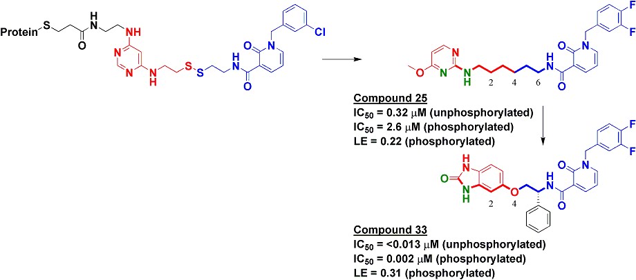
To find inhibitors against PDK1, we used Tethering with extenders. This approach starts by covalently modifying a protein of interest with an “extender,” which is a fragment designed to bind in a desired site on a protein and to capture other fragments; it contains a protein-reactive functionality as well as a masked thiol. In this case, we used a generic fragment known to interact with the purine-binding pocket of kinases, a diaminopyrimidine (red in upper left of figure). The protein-extender complex was then screened against a library of several thousand disulfide-containing fragments under partially reducing conditions; only fragments with some affinity for the protein will remain covalently bound to the protein and thus be detectable by mass-spectrometry. The pyridinone (blue in figure) was one of the strongest hits.


We synthesized several molecules containing purine-pocket binding elements connected to pyridinones by flexible linkers and found compound 25 to be one of the most potent. The SAR revealed the importance of a hydrogen-bond donor-acceptor pair (green atoms in figure) a specific distance from the pyridinone fragment, and further medicinal chemistry led ultimately to compound 33. In addition to being quite potent, compound 33 was remarkably selective for PDK1: in a panel of 241 kinases screened at 10 micromolar concentration of compound 33, only PDK1 and one other kinase were inhibited by 80% or more.

The selectivity of compound 33 was partially explained by the X-ray crystallographic structure, which revealed that the pyridinone fragment discovered through Tethering binds in the so-called adaptive pocket with the activation loop in the “DFG-out” conformation. I believe this is the only series of inhibitors for which this binding mode has been observed for PDK1. Consistent with the structure, these inhibitors are more potent against the unphosphorylated (inactive) form of PDK1 than against the phosphorylated (active) form.

Compound 33 was effective at preventing phosphorylation of the PDK1 substrate Akt both in cells as well as in a xenograft model. Though I may be biased in thinking so, this is a nice application of a fragment-based approach to discover a strikingly specific kinase inhibitor. I hope that people will use compound 33 to further probe the biology of the PI3K pathway. Indeed, at least one completely independent team of researchers seems to already be doing so.