Showing posts with label STD. Show all posts

28 August 2023

Affinity measurements in a single NMR tube?

Last week we highlighted a ligand-detected NMR method to measure affinities of protein-ligand interactions. That technique, R2KD, requires preparing multiple NMR samples with the ligand at different concentrations. In a new open-access paper published in J. Am. Chem. Soc., Serena Monaco and collaborators at University of East Anglia and Universidad de Sevilla describe a method that can be done in a single NMR tube.
 
The researchers have actually combined two methods, chemical shift imaging (CSI) and Saturation Transfer Difference (STD) NMR, to create imaging STD NMR. We’ve written previously about STD NMR, which relies on the transfer of magnetization from an irradiated protein to a bound ligand. In CSI, chemical shift information is recorded at multiple slices along the length of an NMR tube. Normally the solution in an NMR tube is homogenous and so the chemical shifts would be identical at the bottom and top of the NMR tube. Here, though, the researchers create concentration gradients by carefully pipetting a solution containing ligand on top of a solution containing protein and allowing the ligand to diffuse the length of the NMR tube.
 
Like all things NMR-related, the mathematics get a bit complicated. One important factor is the rate of diffusion for a given small molecule. This “diffusion coefficient” can be experimentally measured by creating a concentration gradient in the absence of protein and measuring the ligand concentration at various positions in an NMR tube after a given length of time (typically more than 12 hours). Diffusion is dependent on molecular weight, so it is also possible to calculate the diffusion coefficient, and in fact the researchers found that the calculated values matched the experimental values for three different small molecules.
 
Knowing the diffusion coefficient helps establish the maximum ligand concentration to use and the ideal diffusion time. The researchers examined three different protein-ligand pairs, all of which had weak affinities, with KD values from 0.2 to 2 mM. Measuring STD signals at different slices along the NMR tube effectively yields STD signals at different concentrations of ligand, and fitting this to an equation allows calculation of the dissociation constant. For the three model systems the affinities agreed with literature values, which had been determined using ITC or WAC.
 
One nice feature of imaging STD NMR is that it can identify non-specific binding. This is because STD signals vary depending in part on how close a proton on the ligand is to the protein, resulting in different STD signals for different protons for specific binders. If this “epitope pattern” is lost at higher concentrations, this suggests non-specific binding, where the ligand can bind in random orientations to multiple sites on the protein. The researchers demonstrated this for one of their model systems: tryptophan binds specifically to bovine serum albumin with a dissociation constant of 0.2 mM, but above 1 mM or so the epitope disappears, suggesting non-specific binding.
 
Imaging STD NMR does have some limitations. For one thing, it requires a high initial concentration of ligand: 30 mM in the case of tryptophan, and even higher for the other two ligands. Most small molecules are nowhere near this soluble in water. The researchers suggest that ligands could be dissolved in DMSO and placed on the bottom of the NMR tube, with the protein solution gently layered on top. They show that the concentration gradients develop in a similar manner as a fully aqueous system, but acknowledge that high DMSO concentrations may not play well with most proteins.
 
Also not stated is the sensitivity of the method for higher affinity binders. Last week’s R2KD could measure affinities as tight as 10 µM, but it is unclear how much below 200 µM imaging STD NMR can go.
 
Finally, as we noted in 2019, STD effects are remarkably complex and not well-correlated with affinity. In particular, binding kinetics can play a role in the strength of the signal. It would have been nice to see more than three protein-ligand pairs tested.
 
All that said, this is an intriguing approach. Laudably, the researchers provide extensive supporting information, including mathematical derivation of the fitting equations, a spreadsheet, NMR pulse sequences, and macros. I’ll be curious to see how it works for others.

24 April 2023

RSC Medicinal Chemistry special FBDD issue

The Royal Society of Chemistry puts out RSC Med. Chem., and last year they asked David Rees (Astex), Anna Hirsch (Helmholtz Institute for Pharmaceutical Research Saarland), and me whether a special themed issue on FBDD would be useful for the community. Naturally we said yes, and the results have now been published. You can read our introduction here.
 
Unlike olden days, when special issues were bound between covers, this is a virtual special issue, with papers published over a period of several months. Indeed, we already wrote about two of them last year: one on combining DNA-encoded libraries (DEL) with FBLD and one on inhibitors of PRMT5/MTA. (Both of these were also topics at the CHI FBDD meeting earlier this month.) In the next few paragraphs we highlight the rest.
 
AstraZeneca has been doing FBDD since 2002, and has gained hard-won wisdom, some of which was shared in a 2016 review we wrote about here. After years of screening, their fragment library had started to deteriorate, so they rebuilt it entirely, as described by Simon Lucas and colleagues. Some of the starting fragments came from their previous library, but they also considered molecules from their larger collection. Rather than focusing on the rule of three, they developed their own multiparameter optimization function, “FragScore,” which incorporates logD7.4, heavy atom count, number of rotatable bonds, and number of hydrogen bond donors. All compounds were inspected to make sure they would be synthetically tractable, and quality was assessed by SPR, NMR, redox activity, and solubility. The final set consists of 2741 fragments, with a subset of 1152 maximally diverse and attractive fragments for ligandability assessments or screening hard-to-make proteins. They also gathered 16,806 near neighbors for hit follow-up. So far the effort has paid off, with all four of the targets screened thus far yielding progressible hits. If you’re building or renovating a fragment library, you should read this paper.
 
Continuing on the theme of libraries, Bradley Doak, Martin Scanlon, and colleagues at Monash University describe their “MicroFrag” library, a set of 91 tiny (5-8 non-hydrogen atom) compounds similar to MiniFrags and FragLites. A crystallographic screen (at 1 M concentration!) of the MicroFrag library against the difficult E. coli target DsbA yielded a 52% hit rate, compared with a 2% hit rate with a conventional fragment library. Importantly, the MicroFrag screen identified the two main hot spots previously discovered from the conventional fragment library, along with ten others that may be less actionable. Interestingly, a crystallographic screen of 15 organic solvents at even higher concentrations (50-80%) was less informative: the primary hot spot did not distinguish itself from others. In the case of MicroFrags, not only did this hotspot bind the largest number of fragments, but all the molecular interactions seen for larger fragments were observed.
 
Fluorine NMR takes advantage of its own specialized library, the subject of a paper by Chojiro Kojima (Osaka University), Midori Takimoto-Kamimura (CBI Research Institute) and collaborators from several institutions. The researchers describe the construction of a 220-member library divided into pools of 10-21 compounds. This library was screened against four diverse proteins, yielding between 3 and 16 hits. The three hits against FKBP were characterized in more detail, including two-dimensional NMR and isothermal titration calorimetry. The researchers also discuss using 19F STD experiments to determine the binding mode of bound fragments.
 
Fluorine is not the only halogen of interest for library design. We’ve previously described the halogen-enriched fragment library (HEFLib, here and here), which consists of chlorine, bromine, and iodine-containing molecules. Frank Boeckler and collaborators at Eberhard Karls Universität Tübingen and the Max Planck Institute describe screening this library against the Y220C mutant of p53 in an expansion of work they first described back in 2012. Of 14 hits identified by thermal shift or STD NMR, ten confirmed by two-dimensional 1H-15N-HSQC NMR. Four of these bound in the cleft created by the Y220C oncogenic mutation. Two other fragments turned out to be covalent binders, though they reacted with more than one cysteine residue. Although all the fragments have low affinities, they could potentially serve as starting points for optimization.
 
An ongoing debate is whether there is an advantage to screening more “three dimensional” fragments as opposed to planar aromatic fragments. If your taste tends towards the former, the synthetic chemistry can get tricky. According to an analysis we highlighted last year, the piperidine ring is the third most common scaffold found in drugs. Now, Peter O’Brien (University of York) and an international group of collaborators report efficient synthetic routes to all 20 cis- and trans-piperidines substituted with a methyl group and a methyl ester. A virtual library of 80 compounds in which the secondary amine is capped with simple substituents such as methyl or acetyl groups was found to be quite shapely, particularly compared with the disubstituted pyridyl starting materials. Moreover, the fragments are still reasonably sized, with no more than 15 non-hydrogen atoms and ClogP values < 2.
 
Machine learning is gaining prominence everywhere, not least in drug discovery. In 2021 we highlighted an “autoencoder” designed for constructing fragment libraries biased towards “privileged” fragments more likely to generate hits. However, the method required considerable programming savvy. Now Angelo Pugliese (BioAscent) and collaborators at the Beatson Institute have implemented their model in the open-source KNIME platform, making it accessible to a wider range of researchers. As an example they use the method to construct a GPCR-focused fragment library, with the structures of all the members provided in the supporting information.
 
On the subject of fragment libraries, please make sure to vote in our 6-question poll on library design (right side of page; you may need to scroll up).
 
Not all the papers in this special issue involve library design. Marko Hyvönen, David Spring, and collaborators at University of Cambridge and National University of Singapore describe allosteric inhibitors of the kinase CK2α, which has been implicated in cancer cell survival. We highlighted some of their work against this target in 2017, in which they used fragment linking to find high nanomolar inhibitors of the enzyme. In the new paper, the researchers describe additional fragment binders at the so-called αD pocket, distant from the ATP-binding site. Virtual screening for analogs led to a fragment with mid-micromolar activity in biochemical and cell assays, and fragment merging led to low micromolar inhibitors.
 
This is a nice collection of papers, and for those of you without easy literature access make sure to check them out soon: for the next six months all of them are free to read after free RSC registration. Enjoy!

17 October 2022

Inter-ligand STD NMR: Better than ILOE?

Although our poll in 2019 suggested that crystallography has surpassed NMR in FBLD, not all proteins can be crystallized. Ligand-detected methods such as saturation transfer difference (STD) NMR can be particularly useful for quickly identifying individual fragment binders and getting some sense of how they bind. A new variation published (open access) in Pharmaceuticals by Jesus Angulo and collaborators at University of East Anglia and Universidad de Sevilla provides information on the relative binding modes of two ligands.
 
Long-time readers may remember the inter-ligand NOE method (ILOE) we wrote about in 2010, in which proximity of two ligands is assessed by measuring NOE signals between them. However, despite being described more than 20 years ago, the technique seems to be rarely used, with fewer than a dozen papers in Pubmed, perhaps because ILOE requires large amounts of both protein and NMR time.
 
The new method is called inter-ligand STD NMR (IL-STD NMR), and it was discovered serendipitously while studying the binding of the drug naproxen to bovine serum albumin (BSA). As Teddy discussed several years ago, STD NMR normally involves irradiating specific protons in a protein (for example, the hydrogen atoms on buried methyl groups) and then measuring the “transfer” of this magnetization to bound ligands. When the researchers instead irradiated protons on one end of naproxen, they found that while the STD effect fell along the length of the molecule as expected based on distance, the signal suddenly increased at the other end of the molecule. Naproxen is known to bind to three sites on BSA, and this increased signal was attributed to the proximity of two adjacently bound naproxen molecules.
 
Inspired by this observation, the researchers developed IL-STD NMR. The experiment requires two samples, with two NMR experiments on each. One sample contains the protein and ligand of interest, while the other sample also contains a “reporter ligand” with a known binding mode. For each sample, one NMR experiment is run with selective irradiation of protons on the protein, while the other is run using irradiation of the reporter ligand. Comparison of the spectra reveals which regions of the ligand of interest are near the reporter ligand. The researchers demonstrated that the method works using a model system they had previously studied, the cholera toxin subunit B (CTB), which binds two ligands at nearby sites.
 
Importantly, the time and amount of protein is considerably less than required for ILOE: in this case 2 hours of NMR time and 0.3 mg of protein compared with 88 hours (!) and 1.8 mg. Moreover, the experiment could be run on a 500 MHz NMR, which is a relatively common instrument.
 
IL-STD NMR does have limitations. The researchers note that it is important to avoid irradiating the ligand of interest while irradiating the reporter ligand. Also, the approach obviously only applies to proteins with two nearby pockets (or one larger pocket). Still, it does look interesting, and I could imagine it being used as part of a screening cascade to find candidate fragments for merging or linking. What do the NMR aficionados think?

26 April 2021

STD NMR on putative SARS-CoV-2 main protease ligands

Over the past sixteen months SARS-CoV-2 has infected more than 146 million people worldwide and killed over 3 million. Highly effective vaccines are now available, but not everywhere, and how long the vaccinations will last as new variants arise remains unknown. COVID-19 will likely be with us indefinitely, necessitating drugs as well as vaccines.
 
More than a year ago we highlighted the COVID Moonshot effort, which began with a crystallographic screen against the essential main protease (Mpro) to find starting points for drug discovery. In a new open-access J. Biomol. NMR paper, Ioannis Vakonakis and collaborators at University of Oxford and University of Patras have attempted to characterize some of the hits using saturation transfer difference NMR (STD NMR, see here for a brief description).
 
The researchers had access to a 950 MHz NMR (jealous much?). Samples were screened at 10 µM protein using irradiation of a Mpro methyl group with a chemical shift at 0.5 ppm. To try to minimize differences in relaxation parameters among different ligands, only the strongest STD signals in aromatic moieties were examined.
 
Of 39 non-covalent ligands discovered in the crystallographic screen, five either did not produce an NMR signal or the spectra were inconsistent with the expected structures, suggesting the ligands may be insoluble or unstable in aqueous buffer. The remaining 34 compounds were nominally screened at 0.8 mM each, but the reference spectra differed in intensity by as much as 15-fold, suggesting dramatic differences in concentration. Since the strength of an STD signal is related to both affinity and concentration, this could obviously complicate interpretation of results. As we’ve written previously, careful curation of your library is essential.
 
Of thirteen active site ligands, only four showed strong STD signals. Dose-response titrations between 0.05 and 4 mM revealed dissociation constants of 1.6-1.7 mM for two of them, with the other two being too weak to accurately measure. Molecular dynamics simulations starting with the known structures were consistent with these results, with the tighter binders tending to maintain their positions more than the weaker binders.
 
The researchers also characterized 650 elaborated molecules from the COVID Moonshot, some of which had been reported to be nanomolar inhibitors. Disturbingly, 35 gave no NMR signal and another 86 yielded weak signals. Among those remaining there was a weak correlation (R2=30%) between IC50 in an enzymatic assay and the STDratio (integrated signal intensity of peaks in the STD spectrum over reference spectrum). STD NMR is not appropriate for molecules with Kd < 10 µM, so the researchers also used a competition experiment in which four putative high-affinity molecules would compete a weaker “spy” fragment. This exercise confirmed two ligands but not two others, calling into question their mechanism.
 
STD NMR is often used as part of an assay cascade prior to attempting crystallography, but as crystallography throughput increases there is a case for starting with crystallography, as we argued five years ago. Results from the 39 crystallographic hits perhaps gives pause to that notion, or at least emphasize the need for confirmatory assays. It is easy to be seduced by a high-resolution structure, but because of its sensitivity crystallography may identify ligands so weak as to be unadvanceable. As for the 650 elaborated molecules, it’s too early to draw conclusions, though it’s good to always be on the lookout for false positives.
 
Hopefully the COVID Moonshot will ultimately lead to drugs against SARS-CoV-2. But even if it doesn’t, the intensive focus of multiple techniques on a few proteins is providing useful guidance and best practices that will be applicable to other targets.

19 April 2021

Fragments vs KEAP1: deconstruction and merging

One of the more challenging protein-protein interactions targeted by drug hunters is the interface between the transcription factor NRF2 and its repressor KEAP1. This is part of the cellular defense against reactive oxygen species; increasing NRF2 activity may be useful for treating a variety of diseases. Unfortunately, the binding site on KEAP1 that interacts with NRF2 is large and has a predilection for carboxylic acids. Thus, many of the molecules reported as inhibitors tend not to be druglike. Anders Bach (University of Copenhagen) and a multinational team of collaborators sought to do better, and have just published some of their journey in J. Med. Chem.
 
The researchers had previously tested 19 reported small-molecule KEAP1 inhibitors, of which only nine confirmed. (This is a salutary reminder to take any individual publication with a large grain of salt.) The nine fell into six chemical series (two shown below), and the researchers decided to fragment some of these molecules into 77 fragments. The fragments were then tested in four assays: fluorescence polarization (FP), a thermal shift assay (TSA), saturation transfer difference (STD) NMR, and surface plasmon resonance (SPR).
 
Primary hit rates were generally high, from 25%-64%, but long-time readers will not be surprised that the overlap was not great: no fragments hit in all four assays, and only eight hit in three. As the researchers point out, this could reflect differences in sensitivity, conditions (from 3-8% DMSO and from 0.5 to 8 mM fragment), and different types of false positives and false negatives. Interestingly, and in contrast to previous work, overlap was good between STD NMR and SPR.
 
Crystal structures of seven hits were solved bound to the protein, and compounds 4c and 1m (from different precursor molecules) were merged to provide compound 8, with low micromolar affinity. Compound 8 was the subject of considerable medicinal chemistry, with five different vectors chosen for growing. Despite being structurally enabled, the researchers struggled; changes that improved affinity in one context did not do so in another. After considerable effort, the researchers obtained compound 77o, with mid-nanomolar activity.
 

Compound 77o is stable in human plasma and mouse liver microsomes. Unfortunately, and unsurprisingly given the two carboxylic acids, it has poor permeability. Indeed, a fragment-derived KEAP1 inhibitor we described previously has only a single carboxylic acid, as does precursor compound 7. As the researchers themselves acknowledge, “the physicochemical properties of our compounds are not favorable for membrane permeability.”
 
Nonetheless, this paper is a lovely example of fragment-based deconstruction reconstruction (FBDR) and is well worth studying for the thorough descriptions of fragment screening in orthogonal assays and structure-based design. Another lesson may be that despite considerable effort, the final molecule is far from a chemical probe, let alone a drug. Perhaps some targets truly are undruggable. Or maybe – as for other seemingly undruggable targets – a change in strategy is needed.

26 August 2019

Biophysics beyond fragments: a case study with ATAD2

Three years ago we highlighted a paper from AstraZeneca arguing for close cooperation of biophysics with high-throughput screening (HTS) to effectively find genuine hits. A lovely case study just published in J. Med. Chem. shows just how beneficial this can be.

Paul Bamborough, Chun-wa Chung, and colleagues at GlaxoSmithKline and Cellzome were interested in the bromodomain ATAD2, which is implicated in cancer. (Chun-wa presented some of this story at the FragNet meeting last year.) Among epigenetic readers, bromodomains are usually quite ligandable, but ATAD2 is an exception, and when this work began there were no known ligands.

Recognizing that they might face challenges, the researchers started by carefully optimizing their protein construct to be stable and robust to assay conditions. This included screening 1408 diverse compounds, none of which were expected to bind. Disturbingly, a TR-FRET screen at 10 µM yielded a 4.1% hit rate, suggesting many false positives. Indeed, when an apparently 30 nM hit from this screen was tested by two-dimensional 15N-1H HSQC NMR, it showed no binding. The researchers thus made further refinements to the protein construct to improve stability and reduce the hit rate against this “robustness set.”

This exercise illustrates an important point: make sure your protein is the highest quality possible!

Having done this, the researchers screened 1.7 million compounds and obtained a relatively modest 0.6% hit rate. Of these 9441 molecules, 428 showed dose response curves and were tested using SPR and HSQC NMR. In the case of SPR, the researchers also tested a mutant form of the enzyme that was not expected to bind to the acetyl-lysine mimics being sought. Most of the hits did not reproduce in either the SPR or the NMR assays, and at the end of the process just 16 closely related molecules confirmed – a true hit rate of just 0.001%!

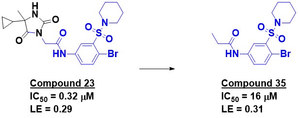
Compound 23 is the most potent molecule disclosed, but the researchers mention a manuscript in preparation that describes further optimization. The compound shows promising selectivity against other bromodomains; it certainly doesn’t look like a classic bromodomain binder. X-ray crystallography revealed that the binding mode is in fact different from other bromodomain ligands. Trimming down compound 23 produced compound 35, which shows reasonable activity and ligand efficiency.

This paper nicely demonstrates the power of biophysics to discern a still small signal in a sea of noise. As the researchers note, PAINS filters and computational approaches would not have worked due to the sheer diversity of the misbehaving compounds. (That said, if the library had been infested with PAINS, the false positive rate would have been even higher!)

The paper is also a good argument for FBLD. Compound 35 is probably too large to really qualify as a fragment, but perhaps related molecules could have led to this series. And GSK also discovered a different series of potent ATAD2 inhibitors from fragments, which Teddy wrote about.

20 May 2019

SAR by STD: NOT

As noted last week, Practical Fragments has been on something of a crystallography binge. But according to polling, NMR is the most common fragment-finding method. And, according to a different poll, saturation transfer difference (STD) is the most popular NMR technique. Familiarity breeds complacency, and widespread assumptions go untested. A new paper in Front. Chem. by Jonas Aretz and Christoph Rademacher (Max Planck Institute and Freie Universität Berlin) suggests that this is a mistake.

In STD NMR, a protein is saturated by specific electromagnetic pulses, and the resulting magnetization transfers to bound ligands. Assuming that the bound ligands are in rapid equilibrium with ligands free in solution, this “saturation transfer” results in a reduction of NMR signal for the small molecule in the presence of protein compared to no protein. High affinity ligands will remain bound to the protein and thus be missed by STD NMR, but this is usually not relevant in FBLD, where most fragments bind with dissociation constants weaker than 10 µM.

A common assumption with STD NMR is that the strength of an STD signal increases with the affinity of the ligand (again, in affinity ranges between about 10 µM and 10 mM). Indeed, when STD NMR is used as part of a screening cascade, molecules showing the strongest effect are generally prioritized as hits. But is this assumption correct?

To find out, the researchers retrospectively analyzed a fragment screen against langerin, a carbohydrate-binding protein we discussed last year. When they plotted the STD amplification factor against the affinity (measured by SPR) for several dozen fragments, the resulting scatter plot showed no correlation.

Recognizing that experimental errors could obscure a true correlation, the researchers ran virtual STD experiments using COmplete Relaxation and Conformational Exchange MAtrix (CORCEMA) theory. They used well-characterized fragments with published crystal structures and affinities for some dozen diverse proteins. As they conclude, “varying saturation time, receptor size, binding kinetics, and interaction site… there were no conditions in which the STD NMR amplification factor correlated unambiguously with affinity.”

But it gets worse. When the researchers explored the effects of binding kinetics, they found that ligands with slower on-rates or off-rates also had lower STD signals. Several groups have advocated prioritizing compounds with slower-off rates, yet these are the very compounds STD is most likely to miss.

All in all this paper could go some way toward explaining the sometimes poor correlation between different fragment-finding methods.

That said, I’m no NMR spectroscopist, so I’m certainly not as qualified to comment on the importance of this paper as someone like Teddy, who co-wrote this how-to guide for STD NMR. I’d be interested to hear what NMR folks think, and whether we should rethink use of STD. In any case, this work is a useful reminder that skepticism is a scientific virtue.

18 December 2017

New tools for NMR: 2017 edition

NMR was the first practical fragment-finding method, and continues to be popular. Just over the past year we’ve discussed several new techniques, (here, here, and here), and this post highlights three more.

In Angew. Chem. Int. Ed., Jesus Angulo and colleagues at the University of East Anglia describe differential epitope mapping by STD NMR (DEEP-STD NMR). STD NMR, the most popular of ligand-detected methods according to our poll, can provide some information as to which portions of a ligand are close to a protein, but doesn’t show where on a protein the ligand binds. In DEEP-STD NMR, two separate NMR experiments are conducted and the results compared to provide this information.

The researchers provide two implementation of the technique. In the first, the protein is “irradiated” at two different frequencies; for example, the aliphatic and aromatic regions. Protein residues that are directly irradiated will show a stronger STD to ligand protons than those that are indirectly irradiated, thus revealing whether one region of the ligand is closer to an aromatic or an aliphatic amino acid side chain. If the structure of the protein is known, this can then reveal the orientation of the ligand within the binding site. A similar experiment can be done using H2O vs D2O to determine whether a portion of a ligand is in close proximity to polar residues in the protein.

Water is the subject of the second paper, in J. Med. Chem., by Robert Konrat and colleagues at the University of Vienna and Boehringer Ingelheim. As we’ve previously noted, water often plays a critical role in protein-ligand interactions. The new method, called LOGSY titration, involves doing a series of WaterLOGSY experiments at different protein concentrations and plotting the signals for each proton in the ligand as a function of protein concentration; ligand protons close to the protein show steeper slopes. The researchers examine pairs of bromodomain ligands and demonstrate that LOGSY titration can confirm changes in binding mode previously seen by crystallography. The technique could also reveal what portions of the ligands make interactions with disordered water molecules, which are more difficult to detect in crystal structures.

Both of these techniques provide useful but incomplete information about ligand binding modes. A paper in J. Am. Chem. Soc. by Andreas Lingel and his Novartis colleagues describes how to generate more detailed models. The researchers used a deuterated protein in which all methyl groups (in methionine, isoleucine, leucine, valine, alanine, and threonine) were 13C-labeled. Multiple intermolecular NOEs between the protein and several previously characterized ligands were collected and the resulting distances fed into modeling software to produce good agreement with the known structures. More significantly, the researchers were able to use the method prospectively with two weak (0.9 and 2.8 mM) fragments. The binding models were sufficiently accurate to guide chemical optimization, resulting in molecules with 30-50 µM affinities. Subsequent crystal structures revealed that these bound as predicted. Impressively, this was done on a protein that forms 115 kD hexamers – larger than those typically tackled by NMR.

Teddy would normally close his NMR posts by stating – usually quite forcefully – whether he felt the technique was practical or not. I’m no NMR spectroscopist, so I’ll throw this question out to readers – do you plan to try any of these approaches?

23 January 2017

New tricks for old methods: STD NMR

According to our last poll, ligand-detected NMR is the most popular method for finding fragments. And among the several ligand-detected NMR techniques, the most popular appears to be saturation transfer difference (STD) NMR. The basic concept behind this approach is to selectively irradiate a protein, which then transfers its magnetization to any bound ligand, thus “saturating” (reducing the signals) for the ligand. Subtracting this spectrum from a reference spectrum reveals which ligand (if a mixture) or individual protons within a ligand are in close proximity to the protein.

Although STD NMR is fast and easy to run, it does have drawbacks. One is the fact that it requires pure protein: if there are other proteins in solution, it will be impossible to tell whether the small molecule binds to the protein of interest or to something else. This shortcoming has been overcome in a paper published recently in J. Biomol. NMR by Tamas Martinek and collaborators at the University of Szeged, the Hungarian Academy of Sciences, and the University of Debrecen.

In a normal STD experiment, the protein protons that are irradiated are far upfield (often around -0.5 ppm) – a region not relevant to most small molecules. These protons then transfer the magnetization throughout the protein and ultimately to any bound small molecules. In order to choose a specific protein, the researchers add an 15N-labeled antibody selective for the protein. They can then selectively irradiate the 15N-labeled antibody, which transfers its magnetization to the bound protein and from there to any bound ligand. They call this approach monoclonal antibody-relayed 15N-group-selective STD, or mAb-relayed 15N-GS STD.

To demonstrate the approach, the researchers observed the binding of 2 mM lactose to galectin-1 (Gal-1) using an 15N-labeled antibody against Gal-1. Lactose binds to Gal-1 with a dissociation constant of 0.155 mM, which is a relevant affinity for fragment screening. Gal-1 was present at 20 µM and the antibody was present at 10 µM, both of which are reasonably low. Control experiments established that both Gal-1 and the antibody were necessary, and the experiment was successful even in a cell extract.

So, as Teddy would ask, is this approach practical? You need an 15N-labeled antibody against your target, and it is important that the antibody is specific and does not compete with your ligand (ie, that it is non-neutralizing). Also, the amount of time required to acquire the spectra appears to be more than an hour. If this could be reduced, would 15N-GS STD assume a useful niche in the NMR toolbox?

06 April 2016

Biophysics: not just for fragments

Biophysics and fragment-based drug discovery go together like Nutella and strawberries. Indeed, SAR by NMR ushered in the dawn of fragment-based methods two decades ago, and most fragment-based programs today make use of NMR, SPR, and/or ITC – not to mention X-ray crystallography. Interestingly, the same is not necessarily true for high-throughput screening (HTS) programs. In a recent paper in Drug Discovery Today, Rutger Folmer makes a strong case for engaging biophysics early and often in HTS. He bolsters his argument with more than 20 examples from internal programs at AstraZeneca.

The first descriptions of using NMR to profile HTS hits were not published until several years after SAR by NMR, but they were rather shocking, with up to 98% of hits failing to confirm. Nor is this merely a historical problem, as discussed here. Aggregators, redox cyclers, generically reactive covalent modifiers – all of these are problems not just in fragment screening but in HTS as well. Sometimes the most potent hits are artifacts, particularly for more difficult targets. The key to triaging out pathological actors is to assess binding and not rely solely on inhibition.

That means bringing biophysics into hit profiling at the earliest stages, before trying to optimize fruitless hits. As Rutger points out, it is often difficult to rally colleagues to look at less active molecules after they have wasted months pursuing more potent dead ends.

And biophysics can make an impact even before running screens. Profiling published tool compounds or in-licensing opportunities with biophysical techniques can reveal unwelcome surprises. Testing the output of early HTS pre-screens (7000-10,000 compounds) before a full HTS (2 million compounds at AstraZeneca) can reveal whether an assay is particularly susceptible to false positives. In some cases this can result in reconfiguring the assay, for example by choosing a different detection technology or modifying the protein construct.

A key element to gaining such benefits is organizational commitment. At AstraZeneca, a biophysicist is assigned to a project team immediately after target selection – well before any screens are run. This seems like prudent practice. How many other organizations are doing this?

21 March 2016

Fragments vs bacterial GyrB

Anti-bacterial targets are not common among fragment-based lead discovery efforts. We’ve written previously about AstraZeneca’s work on DNA gyrase, which led to a clinical candidate. In a recent paper in Bioorg. Med. Chem. Lett., Michael Mesleh and collaborators at Cubist and Evotec describe their efforts on this protein.

Bacterial DNA gyrase has two subunits, GyrA and GyrB, and is essential during DNA replication. It is also well-validated, being the target of decades-old antibiotics such as the fluoroquinolones. The researchers started by screening a library of 5643 fragments against Staphylococcus aureus GyrB using STD NMR, yielding 304 hits. These were winnowed down to 46 based on intensity of the STD signal, novelty, and ease of follow-up chemistry. These were then tested using chemical shift perturbations and crystallography. Although several crystal structures of fragments bound to GyrB were obtained, these did not suggest clear ways to advance the hits.

On the other hand, compound 5, which showed only weak binding by NMR and did not result in a crystal structure, was appealing because of its novelty and polarity. The researchers knew that most ligands that interact with GyrB make a pair of hydrogen-bond donor-acceptor interactions, and they used that knowledge to surmise a binding model. This suggested that growing the fragment towards a pair of arginine residues could improve affinity and led to the synthesis of compound 8, with low micromolar activity.


A crystal structure of a related molecule confirmed the model and also suggested that removing the methyl group would stabilize a more planar conformation better matched to the binding site. Doing so (compound 9) yielded a ten-fold boost in potency. (This methyl is also a nice example of Teddy’s “Sauron Atom”). In a separate paper published last year, the researchers further optimized this molecule to compound 2, with low nanomolar potency and activity in animal models.

Several things stand out about this paper. First, the researchers were willing to pursue a fragment with an affinity lower than other hits. Second, careful modeling and conformational analyses were critical in advancing the molecules. Finally, crystallography was not used in the initial fragment growing. Of course, it helped that the researchers were working with a well-characterized protein amenable to modeling. Still, it is nice to see another example of advancing fragments in the absence of experimentally-determined structures.

02 November 2015

NMR poll results

The results of our latest poll are in – thanks to all who participated! Of the 119 people who responded to the first question, 87% said they use NMR for finding or validating fragments. Even if we assume that responses were biased towards NMR aficionados, big magnets are clearly popular.

The second question asked about specific NMR techniques. If everyone who said they used NMR in the first question also answered the second, this means the average user applies more than 3 different techniques; I’ll let Teddy weigh in to see whether this matches his experience.
One surprise for me was that, although many techniques are widely used, none are nearly universal; even the most popular methods seem to be used by just over half of respondents.

Among ligand-detected methods (blue in the figure), STD ranks at the top, with line-broadening, WaterLOGSY, and fluorine-based techniques all tied for second place.

Protein-detected methods (red in the figure) also appear quite healthy, with nearly as many respondents using 15N-HSQC/HMQC as STD.

Finally, 11 of you said you use "other" techniques. We didn't include TINS, even though it seems quite useful, because it is only available through the services of ZoBio. But what else is out there?

07 October 2015

Fragment finding smackdown: 2015 edition

Our current poll (right-hand side of page) asks about NMR. But of course, there are lots of other ways to find fragments, and the question often arises as to which ones are best. This is the subject of a recent paper in ChemMedChem by Gerhard Klebe and collaborators at Philipps University Marburg, Proteros, NovAliX, Boehringer Ingelheim, and NanoTemper.

Long-time readers will recall that the Klebe group assembled a library of 361 fragments, some of which violated strict “rule of 3” guidelines. These were screened in a high-concentration functional assay against the model aspartic protease endothiapepsin, resulting in 55 hits, of which 11 provided crystal structures. The authors wondered how other techniques would fare. In the new paper, they retested their entire library against the same protein using a reporter displacement assay (RDA), STD-NMR, a thermal shift assay (TSA), native electrospray mass spectrometry (ESI-MS), and microscale electrophoresis (MST). To the extent possible they tried to use similar conditions (such as pH) for the different assays, though the fragment concentrations ranged from a low of 0.1 mM (for ESI-MS) to a high of 2.5 mM (for TSA), while protein concentrations ranged between 4 nM (for the biochemical assay) to 20 µM (for ESI-MS).

All told, 239 fragments hit in at least one assay – a whopping hit rate of 66%. Actually, the number is even higher since, for various reasons, not all fragments could be tested in all assays. And yet, not a single fragment came up in all of the assays! Overall agreement was in fact quite disappointing, with most methods having overlaps of less than 50%, and often below 30%. This is in contrast to a study from a different group highlighted a couple years ago.

What’s going on? One clue might be the solubilities, which were experimentally measured for all library members. In general, hits tended to be more soluble than the library as a whole, emphasizing the importance of this parameter not just for follow-up studies but for identification of fragments in the first place.

Another possibility is that some fragments bind outside the enzyme active site, and thus would not be picked up in a biochemical assay or the RDA. Some evidence for this is provided by follow-up NMR studies in which hits were competed with ritonavir, which binds in the active site. Ritonavir-competitive binders shared greater overlap with biochemical and RDA hits, while there was more overlap between ritnovair-uncompetitive binders and hits from methods such as ESI-MS, TSA, and MST that rely solely on binding. (This could also explain similar observations made earlier this year.)

If a picture is worth a thousand words, how many of the 11 hits that had previously yielded crystal structures would have been identified had they been tested in other methods? Here the numbers vary significantly, from 27% for ESI-MS and MST to 100% for NMR, though these statistics should be taken with a grain of salt since – for example – only 7 of the 11 crystallographically-confirmed hits could actually be tested in the NMR assay. Also, it is possible that some hits from these methods might have generated new crystal structures for fragments not identified in the initial biochemical screen.

One admirable feature of this paper is that the authors provide all their data, including structures and measured solubility numbers for each component of their library. This should provide an excellent dataset for a modeler to use in benchmarking computational methods.

All in all this is a thorough and important analysis and a sobering reminder that, even if a fragment doesn’t hit in orthogonal assays, that doesn’t necessarily mean it’s not a useful starting point. On the other hand, artifacts are everywhere, and paranoia is often justified. The art is deciding which hits are worth pursuing – and how.

28 September 2015

NMR poll!

Among fragment-finding techniques, nuclear magnetic resonance (NMR) ranks near the top. Protein-detected methods, like HSQC/HMQC-based SAR by NMR, helped usher in fragment-based drug discovery as a practical endeavor. More recently, ligand-detected methods such as line broadening (or CPMG), STD, and WaterLOGSY appear to have gained the edge. There are also more boutique methods, such as ILOE and spin labeling. And of course, some people proudly embrace their fluorine fetishism.

So what’s your favorite flavor? Now's your chance to weigh in on our latest poll (on the right). The first question asks whether you use NMR, and the second asks which methods you use. PLEASE ANSWER BOTH QUESTIONS - the free version of Polldaddy doesn't track individuals, so we need the answer to the first question to know the total number of respondents.

And, as always, your comments are welcome.

10 December 2014

How much information can NMR provide?

A frequent assumption in fragment-based lead discovery is that similar fragments have similar binding modes, which are conserved as the fragments are elaborated. However, this isn’t always the case, a fact that can complicate optimization. Ideally multiple crystal structures help guide the chemistry, but in the real world crystal structures can be difficult to obtain.

One of the seminal papers in FBLD used NMR rather than crystallography to guide design, a strategy still used today. But how effective is NMR at assessing the binding modes of related fragments? This is the question that Isabelle Krimm and colleagues at the Université de Lyon sought to answer in a paper published a few months ago in PLOS ONE.

The researchers were interested in the inflammatory enzyme peroxiredoxin 5 (PRDX5), and they examined its interactions with five catechols: the parent unsubstituted molecule and four derivatives with substituents ranging from methyl to phenyl. Although catechols are PAINS, the researchers took pains to carefully examine the NMR spectra to look for signs of misbehavior.

Two NMR techniques were used, saturation transfer difference (STD) NMR and chemical shift perturbation (CSP). STD is nice because it is a ligand-detected method: you don’t need to go to all the work of assigning the chemical shifts of the protein. One piece of information from an STD experiment is whether a hydrogen atom is exposed to solvent or buried close to the protein, and in this case three of the catechols showed one particular hydrogen atom was exposed to solvent. The unsubstituted catechol provided only a single NMR peak and thus no information, and the fifth catechol was also not very informative, though it did seem to bind. Repeating this “epitope mapping” of all the catechols with human serum albumin instead of PRDX5 gave different results, suggesting a different binding mode.

Of course, there is only so much information you can get from ligand-detected NMR, so the researchers turned to protein-detected NMR and examined the CSPs of proton-nitrogen cross peaks using 15N-HSQC experiments. They also calculated CSPs for various potential binding modes and compared these with the experimentally observed CSPs to generate models. These suggested a common binding mode for the same three catechols that STD revealed as having a single solvent-exposed hydrogen atom each. Combining all this information led to specific binding models for these three fragments.

But how good are the models? Happily, the researchers were able to obtain crystal structures of four of the catechols bound to PRDX5, and these agree quite well with the NMR-derived structures. Unfortunately, the fifth catechol couldn’t be characterized bound to the protein crystallographically; NMR also suggested that this bound differently than the others.

So in the end, NMR was able to successfully predict that three ligands had similar binding modes, while another likely doesn’t. The process does seem to require a fair bit of effort. Nonetheless, in cases where crystallography is difficult or impossible, it may be the best way to get essential structural information, and this paper provides a good road map.

05 May 2014

Biofragments: extracting signal from noise, and the limits of three-dimensionality

What does this protein do? Now that any genome can be sequenced, this question gets raised quite often. In many cases it is possible to give a rough answer based on protein sequence: this protein is a serine protease, that one is a protein tyrosine kinase, but figuring out the specific substrates can be more of a challenge. In a recent paper in ChemBioChem, Chris Abell and collaborators at the University of Cambridge and the University of Manchester attempt to answer this question with fragments.

The bacterium Mycobacterium tuberculosis (Mtb), which causes tuberculosis, has 20 cytochrome P450 proteins (CYPs), heme-containing enzymes that usually oxidize small molecules. Although some are essential for the pathogen, it is not clear what many of them do. The researchers used an approach called “biofragments” to try to pin down the substrate of CYP126.

The biofragments approach starts by selecting a collection of fragments based on known substrates. Of course, the specific substrates are not known, so in this case the researchers started with a set of several dozen natural (ie, non-synthetic) substrates of various other CYPs, both bacterial and eukaryotic. They then computationally screened the ZINC database of commercial molecules for fragments most similar to these substrates and purchased 63 of them. Perhaps not surprisingly given their similarity to natural products, these turned out to be more “three-dimensional” than conventional fragment libraries, as assessed both by the fraction of sp3 hybridized carbons and by principal moment-of-inertia.

Next, the researchers screened their fragments against CYP126 using three different NMR techniques (CPMG, STD, and WaterLOGSY). Since they were primarily interested in hits that bind at the active site, they also used a displacement assay in which the synthetic heme-binding drug ketoconazole was competed against fragments. This exercise yielded 9 hits – a relatively high 14% hit rate.

Strikingly, all of the hits are aromatic, and 7 of them could reasonably be described as planar. In other words, even though the biofragment library was relatively 3-dimensional, the confirmed hits were some of the flattest in the library! The researchers interpreted this to mean that “CYP126 might preferentially recognize aromatic moieties within its catalytic site,” but there could be something more general going on – perhaps aromatics are simply less complex, and thus more promiscuous.

Examining the fragment hits more closely, the researchers found that one of them – a dichlorophenol – produced a spectrophotometric shift similar to that produced by substrates when bound to the enzyme. This led them to look for similar structures among proposed Mtb metabolites. Weirdly, pentachlorophenol came up as a possible hit, and a spectrophotometric shift assay reveals that this molecule does have relatively high affinity for CYP126. Whether this is a biologically relevant substrate for the enzyme remains to be seen.

This is an intriguing approach, but I do have reservations. First, in constructing fragment libraries based on natural products, it is essential to avoid anything too “funky”. The Abell lab is one of the top fragment groups out there, well aware of potential artifacts, and has a long history of studying CYPs, but researchers with less experience could easily populate a library with dubious compounds.

More fundamentally though, I wonder about the basic premise of biofragments. The whole point of fragments is that they have low molecular complexity and are thus likely to bind to many targets, so is it realistic to try to extract selectivity data from them? Indeed, as we’ve seen (here and here), fragment selectivity is not necessarily predictive of larger molecules.

That said, the approach is worth trying. Even if it doesn’t ultimately lead to new insights into proteins’ natural substrates, it could lead to new inhibitors.

17 March 2014

This is another way to do it.

The key to doing something right is to following the directions.  How closely you follow the directions, or don't follow, can be the difference between brilliance and just a good performance, e.g. cooking.  Sometimes, directions are meant as guidelines, like the Pirate Code or the Voldemort Rule.  Late last year, and blogged about here, I published a paper in Current Protocols on how to prosecute an STD screen.  A recent paper in PLOSOne, shows how someone else runs their screens, but with details on library construction, solubility testing, and more.  What makes this paper of interest is the level of detail that they provide.

Library Design: They assembled a diverse fragment library with the following rules: 110≤ molecular weight ≤350, clogP≤3, number of rotable bonds ≤3, number of hydrogen bond doners ≤3, number of hydrogen bond acceptors ≤3, total polar surface area ≤110, and logSw (aqueous solubility) ≥ −4.5. 
I am little confused by the figure and what the text says.  In the text, they seem to have relaxed the MW cutoff, but the figure shows that anything not Voldemort Rule compliant is tossed.  They also preferred that the compound has at least one aromatic peak (for easier NMR detection).  They purchased 1008 from Chembridge, solubilized at 200 mM in DMSO-d6 (ease of NMR detection, again) and then tested the solubility at 1 mM in water.  I would have added some salt here, 50 mM, but that is a quibble.  For purity, they claim a low level of impurity (< 15%)!!!  To me, this is a whole lot of impurity.  But, as has been noted here, purity levels vary from library to library.
Solubility Testing:  They then made sure to experimentally test every fragment for solubility.  I can agree more emphatically with this approach.  Bravo!  They go into great detail, which I will not attempt to replicate here, but thanks to open access, they have included the scripts in the supplemental.  Acceptable compounds had > 0.1 mM aqueous solubility.  For me, this is too low, but to each their own.  They ended up with 893 total fragments (89% passed).  The real data I would like to see is how many fail if the cutoff is set at 0.5 mM or higher.  
Pooling: They then describe their pooling strategy.  I like open access articles for a lot of reasons, and tend to overlook small editorial problems (typos, grammar, etc.), but in this case, let me rant.  The authors state in the text that a random mixing of compounds would lead to severe overlap, exemplified in 3a.  To me, it does no such thing. 

Their approach is very similar to the Monte Carlo-based one that has previously been discussed on this blog.  Their final pools contain 10 fragments at 20 mM (I assume in 100 % DMSO-d6). 
Screening: They also acquired the 1H spectrum, STD (-0.7 ppm, > 1 ppm from any methyl), and WaterLOGSY spectrum of every pool for future reference.  This is a very clever approach as the STD should give no signal while the WaterLOGSY should give inverted peaks for all compounds in the pool (when interacting with a target they will be "right-side up").  Again, the figure may show that (I think if you blow up the figure the WaterLOGSY spectra does have peaks) but it is very difficult to see. 
Three of the 90 pools (3.3%) showed peaks in the aromatic region, most likely due to aggregation (they observed precipitation).  I would like to know if those compounds showed STD peaks also had those methyl groups within 1 ppm of the saturation frequency.  I would also like to know if they removed those compounds from the library, or just dealt with it.  For a paper with a great level of detail, it falls flat in this respect.  
Screening is performed at 10uM Target: 500uM ligand and the following parameters: acquisition time of 1 s, 32 dummy scans, and relaxation delay of 0.1 s, followed by a 2 s Gauss pulse train with the irradiation frequency at −0.7 ppm or −50 ppm alternatively. The total acquisition time was 15 minutes with 256 scans.
Screen Analysis: One of the first things they noticed was that there were difference between the reference spectra (plain water) and the screening sample (protein buffer).  They decided they could not automate the entire process and instead just scripted the data processing and display.  Then they confirmed each putative active as a singleton. 
What they are putting together is a "One Size Fits All" process.  I give them credit for doing this, but I think that you cannot find a single NMR-based process for all targets.  In particular, I think they could have used more typical conditions for the reference spectra.  The paper then goes on and discusses their application to targets of interest.  For me, that is irrelevant.  This paper is an excellent companion to the Current Protocol paper, and due to open access, most likely to get far more citations.

17 February 2014

Druggable is as Druggable Does; Or a Million Ways to use NMR

As we all know, the closure of sites is a bad thing for those of us in Pharma.  One very small silver lining is that this frees up a lot of very nice work to be published.  The former BI site in Laval has been closed for a year and we are still seeing great papers coming out.  In this one in JMed Chem,  LaPlante and co-workers tell us about their fragment efforts against HCV helicase. 

HCV has recently had drugs approved for its treatment, but as with any virus, different modes of treatment are important.  The ATP-dependent helicase activity is found in the C-terminal 2/3 of the NS3 protein. Helicase activity is straight forward to measure and there has been some success in terms of non-viral specific inhibitors.  The inhibitors found to date have been found to act through undesireable mechanisms, but with a wealth of structural information there is no reason why helicase is inherently undruggable.  With this information in hand, they decided to target site 3+4 (green sticks are DNA from the structure), near the most conserved residue W501.  The ATP-binding site is 1+2 for reference. 
 Their first approach was to screen the 1,000,000+ corporate compound collection.  As you would expect for a paper blogged about here, they failed to find anything interesting (all the inhibitors worked by undesireable modes).  So, on to the FBDD campaign, to save the day once more.  The used a "shotgun" approach with their fragment screen:

One source of compounds came from an earlier HTS where they rejected fragment-like molecules for lack of potency, additional HCS screening of in house fragment collection, commercial fragments were screened in an SPR assay, virtual screening, and NMR.  They had a stringent workflow aimed at producing quality compounds for X-ray.  [The in-house fragment collection was 1000 compounds.]  This, along with NMR, validated ligands that bound to site 3+4.  They note one particularly noteworthy problem: high false positive rates due to the high ligand concentrations needed for the assays.  This lead to aggregation, solubility, and promiscuity.  This lead them to implement specific assays designed to eliminate these compounds (two NMR papers published in 2013, ref 18). 

They then clustered the best hits into 9 chemotypes:

 They used an "Analog by Catalog" approach and soaked or co-crytallized the best compounds into crystals.  S6, S7, and S9 were not found to bind to helicase in the crystallization trials and were deprioritized.  S5 was found at Site 3+4, but also others.  S1-4, and S8 were found to bind solely to site 3+4 (12 examples shown overlain). The key feature of this is the compounds are centralized in a wide groove over W501.  The topology of the binding site (wide groove and small lipophilic pocket) meant that optimizing for potency could be challenging.
From this, they decided S2-S4 were the most promising.  In the end, the focused on the S2 indole series as the most promising.  The S2 stereotype 1
was found from an STD-NMR screen of 3 fragment per sample (300 uM fragment and 3.5 uM helicase).  They then, much to my heart's delight, they reached into the NMR cabinet for line broadening and competition experiments confirming it binds in site 3+4.  X-ray confirmed the binding mode, but potency was not improved with chemistry.  So back into the NMR cabinet they went: a methyl resonance assay, 
 15N TROSY showing peaks shifting upon addition of a derivative of 1, and 19F NMR!  OMG, how awesome is this?  

In terms of the chemistry, removing the Br does not change the potency, but did change the orientation of the compound in the binding site.  Further elaboration led to this compound 19 (3 uM and 0.23 LE):
It contains a nitro group, think what you may.  In order to confirm the binding affinity of the compound without immobilizing protein, they used the methyl resonances to do the titrations.  The two separate peaks they followed gave values of 32 and 28 uM (+/- 8).  Given the broadness of these peaks, I think this is a pretty decent assay, although it is an order of magnitude different than the biochemical Kd.  However, subsequent structural studies revealed that there is significant structural dynamic differences between pH 6.5 and 7.5.  ITC gave the same number (33 uM and enthalpy driven); however, the ITC had to be run at high compound concentration and a different pH.  They then went off the deep end and decided to use CD (I can't link to a previous post of using CD because we have never had a post where someone used it).  With a horrible assay (don't even get me started on near-UV CD as a readout of tertiary structure), they got reasonably close to the Kds determined by ITC and methyl-NMR.  

This is a very nice example of not being afraid of a target and using all available tools to advance hits against it.  It also shows the WIDE range of NMR experiments that can be used and that are easy and practical.  In terms of full disclosure, Steven LaPlante is a FOT (Friend of Teddy) and I have been working with him.