28 February 2022

Photoaffinity fragment PhABits, faster

Practical Fragments has written previously about PhotoAffinity Bits, or PhABits, which are fragments designed to reveal binding (as opposed to inhibition). These fragments contain a photoreactive moiety such as a diazirine. When incubated with a protein target and irradiated by ultraviolet light, the diazirine transforms into reactive species that can react irreversibly with anything nearby. Intact protein mass spectrometry can identify whether a reaction has occurred, and further proteomics experiments can identify more precisely where the PhABits reacted.
 
One challenge with this approach is obtaining a library of PhABits; few are commercially available (though AstraZeneca is sharing a set). In a recent open-access Chem. Sci. paper, Jacob Bush and collaborators at GlaxoSmithKline and University of Strathclyde describe speeding up the process.
 
The approach is called direct-to-biology high-throughput chemistry (D2B-HTC). Recognizing that purification is often the rate-limiting step in library synthesis, the researchers synthesized PhABits in 384-well plates and used the crude reaction mixtures directly. In short, a diazirine moiety linked to an activated ester was reacted for 24 hours with 1073 diverse alkylamines chosen from the GlaxoSmithKline internal collection. Interestingly, 54 of the amines themselves were not pure as judged by LC-MS – a useful reminder of the importance of quality control. Ultimately, 853 of the reactions were deemed successful, with >80% purity. Residual activated ester was quenched with hydroxylamine, and the reactions were performed in biologically compatible DMSO so that they could be used directly.
 
Next, each member of the library was screened at 100 µM against human carbonic anhydrase I (CAI, at 1 µM), a well-characterized model protein. After UV light illumination (302 nm for 10 minutes) the reactions were analyzed by mass spectrometry, resulting in seven hits, defined as > 1.5% covalent adduct. Five of these contained a primary sulfonamide, a privileged pharmacophore for carbonic anhydrases. Dose response experiments gave similar results on both the crude mixtures as well as resynthesized, pure compounds, with the best molecule showing high nanomolar activity. All seven PhABits could be competed with the known ligand ethoxzolamide, suggesting that they bind in the active site of the enzyme.
 
The seven hits gave even higher levels of modification with carbonic anhydrase II (CAII), and four of the sulfonamide-containing hits were further characterized by proteolyzing the modified enzyme and using LC-MS/MS to determine the sites of modification. This revealed that the PhABits were reacting with either a glutamic acid or histidine residue at the entrance of the active site. As we discussed last year, the precise nature of the diazirine probe can affect which amino acid residues are likely to react.
 
Based on the SAR from the primary screen, a second 100-member library was constructed and screened without purification. This provided a much higher hit rate, with all 52 hits containing a primary sulfonamide.
 
I do wish the researchers had used an orthogonal method to assess the affinities of their molecules. One drawback of the approach, which they note, is that “the absolute value of the crosslinking yield is not indicative of binding affinity,” but it would be interesting to know whether there is any correlation at all. It would also be nice to get a sense of how often false negatives occur.
 
Still, D2B-HTC adds to the growing list of methods that screen crude reaction mixtures, alongside related approaches such as off-rate screening, Chemotype Evolution, and REFiLx. The future may be a bit dirty, but perhaps we can get to our destination faster.

21 February 2022

Ensembles of fragment structures guide selectivity

Scientists generally want structural information when a project begins, and ideally that structural information comes from crystallography. Most of us who have been doing drug discovery for a while can remember seeing the first structure of a favorite molecule bound to a target protein and being inspired, reassured, or sometimes confused. But as crystallography becomes increasingly high throughput, it is now not uncommon to obtain dozens or even hundreds of structures. What to do with all this bounty? In a recent open-access J. Chem. Inf. Model. paper, Mihaela Smilova, Brian Marsden, and collaborators at University of Oxford, the Cambridge Crystallographic Data Centre, and Exscientia describe one application.
 
Back in 2016 we wrote about a computational approach called hotspot mapping, which uses three small fragment probes (aniline, cyclohexa-2,5-dien-1-one, and toluene) to virtually explore potential binding sites and map hydrogen bond acceptors, donors, and apolar interactions. The idea was to predict binding sites and the key interactions likely to drive affinity. The new paper focuses not just on affinity, but on selectivity.
 
The approach starts by taking multiple structures of the same protein bound to various ligands, especially fragments. Ligands and water molecules are then removed, and hotspot mapping is conducted for each structure. Then, all the hotspot maps are combined to generate an “ensemble” hotspot map, which in theory should give a more complete picture of potential attractive and repulsive interactions than a single structure.
 
To assess selectivity, the ensemble hotspot map of one protein is “subtracted” from that of another. If the proteins are very closely related, this “selectivity map” might be empty: all the interactions for one protein would be present in the other. But if there are differences, they become very apparent.
 
Several retrospective case studies are provided. In the first, ensemble hotspot maps were generated from the closely related bromodomains BRD1 and BRPF1, using 23 and 26 fragment-bound structures, respectively. The selectivity map clearly shows the potential for a hydrogen bond donor on a ligand to bind to the backbone amide of a serine in BRD1; the corresponding residue in BRPF1 is a proline, incapable of making this interaction. And indeed, an examination of the literature revealed that this interaction had previously been used to generate inhibitors of BRD1 that were 15-fold selective over BRPF1.
 
The kinases p38α and ERK2 are also closely related, but selectivity maps generated from five p38α structures and 17 ERK2 structures revealed a hydrophobic pocket in the former but not in the latter. This pocket had previously been used to generate selective inhibitors of p38α. Similarly, 28 structures of CK2α and 32 structures of PIM1 were used to generate a selectivity map that also revealed a hydrophobic pocket that can form in the former protein and had been used to generate selective inhibitors.
 
Generally, the more structures available, the more informative the selectivity maps are. The researchers note that though they only used five p38α structures, the fragments were chosen to be diverse (and interestingly all of them made interactions in the hydrophobic pocket). Also, while some protein flexibility can improve the maps, too much is a problem. (For the kinases, only DFG-in structures were used, for example.)
 
This method is a nice synthesis of experimental and computational techniques. A skeptic might argue that it doesn’t provide fundamentally new information: in the examples provided, the selectivity features had already been found and exploited by medicinal chemists. But the automated process and the clear output may speed things up, especially for newer targets, and indeed the researchers note that it is being applied in-house at Exscientia.
 
Perhaps most importantly, if you’d like to try it yourself, the code is freely available here. Happy mapping!

14 February 2022

Fragment merging vs bacterial SAICAR synthetase

People living with cystic fibrosis are susceptible to lung infections from a rogues’ gallery of bacterial species, one of which is Mycobacterium abscessus. It is often antibiotic resistant, and even when it responds, a course of antibiotics can take two years to resolve the infection. In a recent ACS Infect. Dis. paper Tom Blundell, Anthony Coyne, and collaborators at University of Cambridge and elsewhere describe progress against this organism.
 
The researchers chose to target a protein called SAICAR synthetase, or PurC, which is essential for purine biosynthesis and thus bacterial growth, as shown by genetic knockout studies. The enzyme is significantly different from the human ortholog, but similar to the Mycobacterium tuberculosis ortholog, giving the potential for a twofer.
 
Fragment screening was conducted both in-house using thermal shift assays as well as at XChem using crystallography; we discussed the differing outputs of these screens in this 2019 post. Compound 1, from the in-house screen, was found crystallographically to bind in the ATP-binding site, and ITC studies revealed it to have high micromolar affinity for the protein. Meanwhile, compound 2 was identified from the crystallographic screen, and while the affinity wasn’t measured, the pyridyl ring is located a short distance from where compound 1 binds.


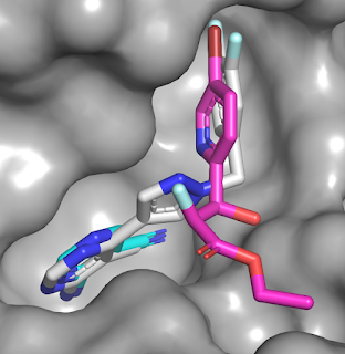
Initial SAR around fragment 1 revealed that growing toward the binding site of compound 2 would be possible, as illustrated by compound 9. Appending a pyridyl ring onto this molecule led to compound 16, with low micromolar affinity. The pyridyl moiety stacks onto an arginine side chain, and improving this interaction by replacing the pyridyl with a phenyl appended with electron-withdrawing fluorine atoms led to compound 27, with submicromolar activity. Overlaying the crystal structures of compounds 1 (cyan), 2 (magenta), and 27 (gray) reveals that the merged molecule does indeed bind in a similar manner to the component fragments.

Unfortunately, despite good biochemical activity against PurC, none of the compounds were particularly effective at inhibiting growth of either M. abscessus or M. tuberculosis. Such disconnects between biochemical and cell potency are unfortunately all too common, particularly for antimicrobial targets, as we wrote about here. The researchers suggest possible reasons including efflux and physicochemical properties. The paper ends by noting that work is continuing, and we look forward to hearing more.

07 February 2022

Automated scaffold hopping for fragments

A post last month covered high-throughput virtual screening, but most practitioners of FBLD still start with some sort of (bio)physical screen. These initial hits can’t be expected to be optimal, since the average fragment library contains a few thousand compounds at most. Indeed, as Xavier Barril and collaborators at Universitat de Barcelona and Oxford University write, “fragment hits should be seen as beacons indicating privileged areas of chemical space to be further explored.” They describe one way to expedite exploration in a recent J. Med. Chem. paper.
 
As we noted here, most good fragments make at least one essential interaction (such as a hydrogen bond) to the protein. The approach starts with a structure of a fragment bound to the target of interest, with that essential interaction identified.
 
Next, a virtual library is searched for similar molecules, with the definition of “similar” being rather loose (>50% Tanimoto similarity). Ideally the library is large enough to produce lots of hits; the researchers used ZINC15, which contains >15 million ostensibly commercial compounds. Also, only molecules within two non-hydrogen atoms of the starting fragment are considered. In other words, a fragment with ten “heavy” atoms would yield molecules with 8-12 non-hydrogen atoms. This search is similar though perhaps more permissive than Astex’s Fragment Network (which we wrote about here).
 
All the molecules are then superposed on the initial fragment structure and only those that maintain the key interaction and binding mode are kept. Aboout 500 molecules are then selected to represent the best and most-diverse hits. These are subjected to dynamic undocking (DUck), which weeds out fragments that have weaker interactions. If desired, each of the remaining hits can be subjected to further cycles.
 
To demonstrate the approach, the researchers turned to bromodomains, a popular target class for FBLD. They started with 1XA, a fragment Teddy highlighted back in 2013 that led to a clinical compound against BRD4. The isoxazole moiety makes a hydrogen bond with the side chain nitrogen of an asparagine that normally binds to acetylated lysine residues. After one cycle, 58 molecules were selected, but unfortunately only five were actually available commercially. Compound 3 had similar affinity and ligand efficiency as 1XA, and this scaffold had not been reported as a bromodomain ligand. A crystal structure of compound 3 bound to the first bromodomain of BRD4 confirmed the predicted binding mode.

Three additional successive iterations were conducted to look for more ligands, but experimental confirmation was challenging as overall only 17 of more than 100 ligands selected for purchase were commercially available. (Compound 23 was chosen for custom synthesis as it was related to a family of high-scoring molecules.) Encouragingly, eight molecules were active in a differential scanning fluorimetry (DSF) assay, a technique that works well for BRD4. Crystal structures of two of these were obtained: compound 9 contains an isoxazole moiety like 1XA (and indeed resembles this fragment) but compound 23 is quite distinct.


Overall this looks like a valuable method for scaffold hopping. Not only might the described approach lead to novel molecules, it could provide new growth vectors that may not be accessible from the original fragment. Before jumping immediately into chemistry with your fragment hits, it may be worth trying something like this.