Showing posts with label cryo-EM. Show all posts

23 February 2026

Twelfth Novalix Biophysics in Drug Discovery Conference

Last week the Twelfth Novalix Biophysics in Drug Discovery Conference was held for the first time in La Jolla, California. It’s been several years since I wrote about one of these, and I was happy to see that they’ve maintained their reputation for excellent science and convivial conversation. There’s no way to cover the two-dozen talks, but here are a few highlights.
 
One of the things I most enjoy about these meetings is learning about new and emerging methods, and these were well represented. Chris Brosey (AbbVie) discussed time-resolved high-throughput small-angle X-ray scattering (TR-HT-SAXS). As I discussed a couple years ago, the approach can be used to measure the kinetics of protein dimerization in response to fragment-sized ligands.
 
SAXS-based approaches typically require access to a synchrotron, but Takashi Sato (Rigaku) described a related approach, electron density tomography (EDT), using an in-house instrument. Using machine learning, EDT can provide more detailed structural information than standard SAXS, and Takashi provided examples for samples ranging in size from viruses to single-chain variable fragments (scFvs) smaller than 30 kD.
 
Another approach to examine protein complexes is microfluidic diffusional sizing (MDS), described by James Wilkinson of Fluidic Sciences. By assessing the amount of diffusion in disposable chip-based chambers, MDS can determine how the hydrodynamic radius changes in response to ligands. Each chip holds 24 samples, and data can be collected in less than an hour. The minimum observable size change is 5-10% so measuring small molecules directly is unlikely. Still, the technique is useful for observing induced proximity events such as those caused by molecular glues, and it is sufficiently robust that it can be run in pure serum.
 
Among solution-based methods, none have achieved such recent prominence as cryo-EM. Weiru Wang, my colleague at Frontier Medicines, described how this technique was used iteratively to design and characterize bivalent degrader molecules that covalently exploit the E3 ligase DCAF2. (We recently published this work in Structure.)
 
Cryo-EM has revolutionized the types of biological molecules that can be structurally characterized. According to Denis Zeyer (Novalix), the technique accounted for 40% of PDB entries last year. However, despite the “resolution revolution,” most structures are not as detailed as those from X-ray crystallography; Denis noted that only 20% of the new structures were solved to a resolution better than 3 Å, which may have negative implications for machine-learning methods trained on these lower resolution structures.
 
If cryo-EM is the new kid in town, NMR is the grizzled veteran. But proving that it is possible to find new applications for old methods, Matthew Eddy (University of Florida) described a clever 19F-labeling approach for GPCRs in nanodiscs to quantify the distribution of various states in response to anionic lipids and ligands. This has allowed him to distinguish between antagonists and inverse agonists, which can be difficult using cell-based assays.
 
Turning from solution-based to surface-based methods, SPR has moved into the number two slot for fragment-finding, as we noted in our recent poll. Just as there are new tricks for NMR, the same applies to SPR. Matthew Peterfreund (Bruker Biosensors) described switchSENSE, a fluorescence proximity assay built on an SPR chip that is useful for measuring the binding and kinetics of bifunctional ligands such as PROTACs to two or more proteins. He also introduced the Triceratops SPR#64 instrument, which as its name implies supports 64 sensor spots.
 
Kris Borzilleri (Pfizer) discussed SPR-microscopy (SPRm), which combines an optical microscope with an SPR instrument. This can be used to measure the affinities of ligands binding to receptors in cells grown on SPR chips, and Kris described applications to membrane proteins such as GPCRs and solute carriers. The technique is still quite slow though, at only 10-15 compounds in duplicate per week.
 
Another surface-based approach to screening cells was described by Volker Gatterdam of Lino Biotech. Focal molography relies on changes in diffraction from nanoengineered diffraction gratings, called molograms. Targets, which can include living cells, are immobilized to the molograms, and analyte is flowed over. The instrument contains 64 spots, and assays can be run in complex samples such as tissue lysates.
 
Covalent drug discovery also made an appearance, with talks by Landon Whitby (Lundbeck) and Ben Cravatt (Scripps). Landon provided an overview of chemoproteomics techniques to screen ligands in cells, such as those we wrote about in 2016. Ben continued the theme, including several success stories, and also discussed challenges for finding cryptic ligandable pockets. Despite impressive progress with machine learning, Ben noted that these methods often find only common solutions, while empirical chemoproteomics methods can find rare types of pockets.
 
Of course, as we’ve repeatedly emphasized, biophysics methods are best used in combination, as noted by Daniel Harki (University of Minnesota) and Ann Boriack-Sjodin (Takeda). Daniel presented a screen of 1056 fragments against the cancer target APOBEC3 using NMR and SPR. This yielded just a single validated hit, which interestingly turned out to be the same fragment found against KRAS in a paper we discussed in 2022. And Ann described how biophysics led to multiple clinical compounds against a variety of targets at Epizyme and Accent Therapeutics.
 
I’ll stop here, but please feel free to add your thoughts. And while the date for the next Novalix conference has not yet been scheduled, the location has, with a return to beautiful Strasbourg. Vive la biophysique!

05 December 2022

Fragments win in a virtual screen against the 5-HT2A receptor

Virtual screening is continuing to make impressive strides. The latest example, in Nature, comes from William Wetsel (Duke), John Irwin (UCSF), Georgios Skiniotis (Stanford), Brian Shoichet (UCSF), Bryan Roth (UNC Chapel Hill), Jonathan Ellman (Yale), and a large group of collaborators. The paper has received considerable attention (for example In the Pipeline), but in my opinion the connection to FBLD has been understated.
 
The researchers were interested in finding new agonists for the 5-HT2A receptor (5-HT2AR). This GPCR is the target for LSD and psilocybin, both of which have been shown to reduce depression and anxiety. Is it possible to find molecules with similar therapeutic activity but without the accompanying psychedelic properties?
 
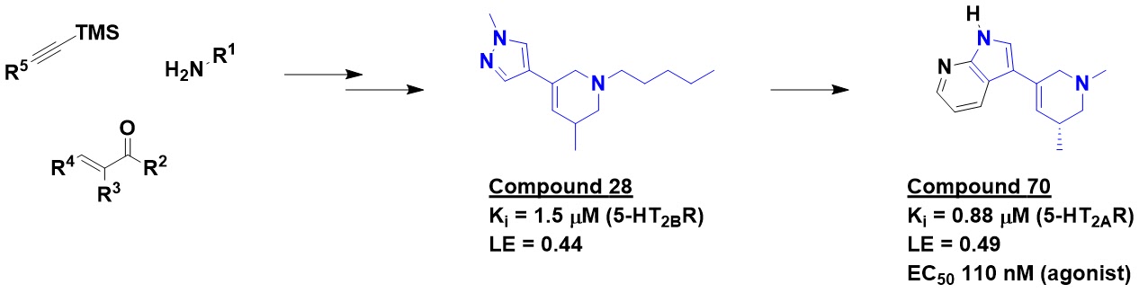
LSD contains a tetrahydropyridine (THP) moiety, which is relatively rare in screening libraries. The researchers developed convergent routes to THPs in which they could independently and efficiently vary multiple substituents. Using this chemistry, they constructed a virtual library of 4.3 billion compounds, all with molecular weights ≤ 400 Da and cLogP ≤ 3.5.
 
At the time the research began, there were no structures of 5-HT2AR, so the researchers built a homology model based on the closely related 5-HT2BR, which differs by only four amino acid residues in the orthosteric pocket where LSD binds. This model was then screened against a subset of the THP library, those ≤ 350 Da. Despite screening some 7.45 trillion complexes (sampling an average of 92 conformations and 23,000 orientations per molecule), the process took only nine hours on a 1000-core CPU cluster. The result was 300,000 hits in nearly 15,000 families. To ensure novelty, only compounds quite different from known ligands were further considered, and 17 “richly functionalized” THPs were synthesized and tested in radioligand assays. Four were active, including racemic compound 28. Searching the 4.3 billion compound library for analogs ultimately led to compound 70 and a related, slightly more potent molecule lacking the methyl substituent on the amine. A cryo-EM structure subsequently validated the predicted binding mode.
 

The paper spends considerable time characterizing these two compounds. Both are agonists and somewhat selective for 5-HT2AR over 5-HT2BR and 5-HT2CR. They are highly selective over 318 other GPCRs and 45 off-targets. GPCRs can signal through arrestin and/or G-protein, and while LSD works (mainly) through the arrestin pathway, the new molecules work (mainly) through the G-protein route. Importantly, the compounds showed anti-depressive and anti-anxiety effects in mouse models. Although you can’t ask mice if they are tripping, the molecules did not cause “head-twitch responses” and other behavioral effects seen with LSD, suggesting that they may not have hallucinogenic properties.
 
This is a lovely piece of work, and a few observations relevant to FBLD stand out. First, the best molecules are actually rule-of-three compliant, despite the fact that larger molecules were included in the virtual screen. Indeed, the top two molecules are actually smaller than the initial hits. This suggests that choosing more richly functionalized molecules may not have been the most efficient approach. We’ve written previously about V-SYNTHES, which entails stepwise selection and growing of fragments; it would be interesting to retroactively test whether this type of approach would have more quickly gotten to compound 70.
 
Finally, this approach can easily be extended to other scaffolds for which syntheses are readily available. Six years ago we wrote about the synthetic accessibility of dihydroisoquinolines, and last year Practical Fragments published our fifth “fragment library roundup.” The marriage of clever chemistry with virtual screening seems to have a bright future.

11 April 2022

Nucleophilic fragments vs SARM1: in situ inhibitor assembly

Recently Practical Fragments wrote about nucleophilic fragments that could react with proteins or cofactors. Previously we’ve also written about in situ chemistry, in which a protein catalyzes the formation of an inhibitor. An interesting marriage of these concepts has just been published (open access) in Mol. Cell by Robert Hughes (Disarm Therapeutics), Thomas Ve (Griffith University) and a group of international collaborators.
 
The researchers were interested in the protein SARM1, which is implicated in the axon degeneration associated with several neurodegenerative disorders. Last year the researchers published a Cell Rep. paper (also open access) in which a biochemical screen of roughly 200,000 molecules led to the discovery of isoquinoline as a 10 µM inhibitor of SARM1. Optimization led to 5-iodoisoqinoline, dubbed DSRM-3716, a 75 nM fragment-sized inhibitor. The paper goes on to demonstrate that the molecule not only prevents axonal degeneration but can even promote recovery of injured axons. The new paper explores the mechanism of action.
 
SARM1 is an NADase: it cleaves the critical cofactor nicotinamide adenine dinucleotide (NAD+). While using NMR to study the mechanism of inhibition, the researchers found that DSRM-3716 reacts with NAD+ to form the new compound shown. In this sense, DSRM-3716 acts as a prodrug, somewhat analogous to sulfanilamide antibiotics which act as PABA mimics to block folate biosynthesis.
 

What’s behind the inhibition of SARM1? A series of crystallographic and cryo-EM studies of SARM1 reveal that the protein can self-associate into multimers which are either inactive or active depending on the relative orientations of the individual proteins. NAD+ normally binds at the interface between two SARM1 proteins. The compound made from NAD+ and DSRM-3716 binds here as well, blocking further activity. The crystal structures also revealed a clear halogen bond (see here) with the iodine in DSRM-3716, explaining the increased activity over isoquinoline itself.
 
Unlike the nucleophilic fragments we wrote about last month, isoquinoline probably won’t raise too many eyebrows among medicinal chemists, as the moiety is found in a handful of approved drugs. The researchers also demonstrated that DSRM-3716 itself is selective for SARM1 in a panel of other enzymes that use NAD+.
 
This is a lovely case of high-throughput screening in which the hit turns out to be a fragment. Indeed, the highly charged compound that actually inhibits SARM1 would not be cell-permeable, but that's just fine since it is formed inside cells. It is worth noting that nearly 1000 approved drugs could be classified as fragments in terms of molecular weight. In the case of CNS drugs, small is beautiful, and it will be fun to watch how far DSRM-3716 derivatives will be able to advance.

24 May 2021

Sixteenth Annual Fragment-Based Drug Discovery Meeting

Last week Cambridge Healthtech Institute held its Sixteenth Drug Discovery Chemistry (DDC) meeting virtually for the second year in a row. The eight tracks, four in parallel, spanned three days. There were occasional kinks, and the 6:30 morning start time was painful for those of us on the Western edge of the US, but for the most part things went smoothly. Attendance was up 25% over last year, to 750 participants. Attendees from outside the US comprised 40%, similar to last year and up from 2019, while industry representation remained steady at 70%. As always I’ll just touch on a few broad themes – please feel free to add your impressions.
 
Methods
These meetings are always great venues to learn about new techniques, and one that stood out is “mass photometry” (MP). This label-free method allows visualization of single proteins in solution. The technology, which builds on interference reflection microscopy and interferometric scattering microscopy, rapidly provides information on the molecular weight of proteins to an accuracy of about 2 kDa. Stefan Geschwindner (AstraZeneca) used the method to follow a change in the monomer-dimer equilibrium of the protein PAD4 in response to ligand-binding. He noted that the instrument, sold by Refyn, provides measurements in about 10 seconds for proteins from 35 kDa to 4 MDa in size.
 
On the opposite end of reductionism, Bryan Roth (University of North Carolina, Chapel Hill) discussed customized high-throughput cell-based screens for 320 of the 358 GPCRs in the human genome. We’ve previously described how these assays have been used to screen molecules prioritized from massive computational screens. If you’d like to profile your own molecules, you can submit compounds here for free with no IP-entanglements.
 
Cryo-EM was the subject of our April 1 post last year, but as we discussed here the technique is no joke. Pamela Williams discussed how Astex has formed a consortium with several other companies to advance the technology. She noted that of the 270 membrane protein structures released last year with resolutions better than 3.5 Å, 167 came from cryo-EM while only 103 came from crystallography. Unlike in crystallography, resolution can vary over the protein; local resolution around the region of interest may be better or worse than global resolution. And there is still much to be done to truly industrialize the approach, particularly in sample preparation (specifically making the grids).
 
Speaking of crystallography, James Fraser (University of California, San Francisco) discussed a high-throughput crystallographic screen against the SARS-CoV-2 macrodomain protein, which we covered earlier this year. A fragment-linking program called “Fragmentstein” was used to generate molecules with low micromolar affinity.
 
At the last DDC meeting Frank von Delft (Diamond Light Source) described a high-throughput crystallographic screen of the SARS-CoV-2 MPro protein, and this year he provided an update on how the open-source COVID Moonshot effort has advanced hits to non-covalent 30 nanomolar binders with 100 nanomolar antiviral activity in cells. Current efforts are focused on improving metabolic stability and oral bioavailability, but Frank lamented the “astonishing inertia” on the part of funding agencies to support the effort. Although the spectacular success of vaccines should contain the outbreak, there are still occasional breakthrough infections, and not everyone can be vaccinated or mount a successful immune response. Small molecule drugs will be useful, and it would be nice if more people were working on them.
 
Success Stories
I’ll never turn down a crystal structure, but it is important to remember that fragments can be advanced even in the absence of structural information. As a case in point, Paolo di Fruscia (AstraZeneca) described the discovery of allosteric binders of MEK1; we wrote about these earlier this year.
 
On the other hand, Justin Dietrich described AbbVie’s discovery of TNFα inhibitors – an effort he said would not have been possible without structural information. As we discussed here, the team struggled with the lipophilic nature of some of their initial molecules; Justin noted that “if a protein wants grease you have to find a way to get it grease.” The physicochemical properties can then be improved during optimization.
 
Rod Hubbard (Vernalis) described the discovery of LpxC inhibitors. He emphasized the importance of using biophysical methods to carefully characterize and optimize the target protein, and noted that NMR can identify fragments that crystallography might miss. Also, as we noted when we wrote about this campaign last December, fragments can provide multiple starting points, and it can be useful to optimize fragments before embarking on a fragment-to-lead effort.
 
Jus Singh (Ankaa) provided a brief history of targeted covalent drugs. Some folks are still nervous about the potential for covalent molecules to cause idiosyncratic toxicity; indeed, this was one of the reasons Frank von Delft gave for pursuing non-covalent MPro inhibitors. However, Jus noted that five out of seven approved covalent kinase inhibitors are not associated with liver toxicity, while some non-covalent kinases inhibitors are. Jus also noted that all three FDA-approved BTK inhibitors are covalent, despite longstanding efforts to develop non-covalent inhibitors.
 
Finally, Micah Steffek (Genentech) described the discovery of LC3 binders, the first step in making an autophagy-based version of PROTACs. An NMR screen yielded hundreds of hits, but despite obtaining multiple crystal structures the team struggled to obtain molecules with better than mM potency. However, combining fragment information with results from a DEL-screen, akin to the story we described here, led to submicromolar inhibitors. Intriguingly, these covalently modified a lysine residue.
 
Odd and ends
Micah’s talk led to some discussion in a speaker panel as to the meaning of hit rates. Do you consider the primary hit rate or the confirmed hit rate? And what level of confirmation is required? As Rod Hubbard noted, different techniques may give different answers for good reasons, and you don’t want to throw away potential hits on the basis of the worst performing or least sensitive technique.
 
Mads Clausen (Danish Technical University) described his shapely library of fluorinated fragments (see here). Hit rates seem to be similar to flatter compounds, though with only four targets screened it is perhaps too early to draw conclusions.
 
But perhaps there is a limit to shapeliness: Justin Dietrich could not recall seeing any fully unsaturated Fsp3 = 1) fragments coming up as hits, despite being present in the AbbVie libraries. Andreas Lingel said he had occasionally seen these at Novartis, but they were never pursued. Similarly, no one could recall advancing any acyclic fragments. A quick glance at the 131 fragment-to-lead success stories captured in five annual reviews revealed that all of the fragments had at least one ring and one sp2-hybridized carbon.
 
And with that I’ll close. The Seventeenth installment of DDC is scheduled for April 18-21 of next year in San Diego, and the more biology-focused Discovery on Target is scheduled to take place both online and in-person in Boston from September 27-30. Hope to see you there!

24 August 2020

SAR by MicroED?

Of the dozen-plus methods to find and characterize fragments, only two have historically been able to provide detailed binding information: protein-detected NMR and X-ray crystallography. Earlier this year we described how researchers at Astex are using cryo-EM for FBLD. In a new open-access Communications Biology paper, Hongyi Xu and collaborators at Stockholm University, Lund University, and SciLifeLab describe another variation of cryo-EM.

Microcrystal electron diffraction (MicroED) is something of a cross between standard crystallography and cryo-EM. Like more “conventional” cryo-EM, an electron microscope is used to collect data on flash-frozen samples. But rather than painstakingly reconstructing thousands of images of individual protein molecules, MicroED uses diffraction of electrons from crystals that are far too small for standard X-ray crystallography. Even though electrons rather than photons are being scattered, diffraction is diffraction, so well-established X-ray crystallography methods can be used for processing MicroED data.

The researchers focused on human carbonic anhydrase II (HCA II), a popular model protein that has also been used to showcase X-ray crystallographic, native MS, and SPR methods. The microcrystals were less than 500 nm thick, smaller than most bacteria and at least 100-times smaller than typically used for X-ray crystallography. MicroED data were collected on microcrystals of native HCA II as well as microcrystals that had been soaked for 20 minutes with the known ligand acetazolamide.  At just 13 heavy atoms, this approved drug is still comfortably a fragment.

Data were collected to 2.5 Å resolution, which is modest especially compared with the 1.1 Å resolution of a published crystal structure. Nonetheless, the ligand density was clearly visible, and the refined model was very similar to the published crystal structure as well as another published structure of the complex determined by neutron diffraction. The researchers note that the observed features are similar to those that could be expected of a crystal structure solved at the same resolution.

The researchers note several potential advantages of MicroEM over X-ray crystallography. It can sometimes be difficult to obtain sufficiently large crystals for crystallography, particularly when protein is limited. Smaller crystals may allow faster diffusion of ligands into the crystals. And at higher resolution, individual hydrogen atoms are more easily resolved using electron diffraction than X-ray diffraction.

Will these advantages be enough to make MicroED a truly practical method for FBLD? It is not clear from the paper how long the data collection and processing took, HCA II is a friendly protein to work with, and acetazolamide is a high affinity ligand. That said, MicroEM was first described only in 2013, and this paper demonstrates that ligand binding modes can be determined. It will be fun to watch this technique develop.

01 April 2020

Fragment screening in cells with cryo-EM

Of all the biophysical advances so far this century, cryogenic electron microscopy (cryo-EM) has probably made the most impressive strides. Frequently dismissed as “blobology” just a few years ago, the technique now regularly produces three-dimensional structural models that rival those from X-ray crystallography. Indeed, it is rare to pick up an issue of Science or Nature that doesn’t contain a cryo-EM structure. Earlier this year, researchers from Astex described the structures of fragment hits against two proteins determined using cryo-EM. Now, the boffins from DREADCO (who previously brought us universal crystallography) have begun fragment screening in cells using cryo-EM.

Fragment screening in cells is not new: we previously highlighted work using either covalent or non-covalent fragments. However, figuring out which proteins the fragments bind can be challenging, which is one of the reasons structural information is so useful.

The researchers from DREADCO incubate their fragment library against cells – human or otherwise – for varying lengths of time. They then flash-freeze the cells in liquid ethane, collect, and process the data, using standard cryo-EM workflows. Of course, given the complexity of cells, the computational processing power needed is enormous – but nothing their SkyFragNet platform can’t handle.

One of the advantages of cryo-EM is that larger structures are more easily solved, so the researchers are focusing on organelles such as mitochondria, as well as ribosomes. Already they’ve found dozens of hits that resolve to high resolution, and they are in active fragment-to-lead optimization. Surely it is only a matter of time before our list of fragment-derived drugs includes one discovered with the aid of cryo-EM.

13 January 2020

FBLD by Cryo-EM

X-ray crystallography is the most popular fragment-finding method according to our latest poll. This is in part due to the previously unmatched level of detail a crystal structure can provide, and in part due to the increasing speed and automation for data collection and processing. But many proteins can never be coaxed into crystals, and while it is possible to advance fragments in the absence of structure, it is rarely easy.

In the past few years, cryogenic electron microscopy (cryo-EM) has come to rival crystallography in terms of resolution (see here for details). The first mention of the technique on Practical Fragments was in 2013, when Teddy wrote that he “could not figure out how you would use [it] in screening/FBHG. However, the point of emerging technology is to emerge…” And emerge it has. At FBLD 2018 researchers from Astex presented the first cryo-EM structure of a bound fragment, and in a new paper in Drug Disc. Today Harren Jhoti and collaborators at Astex and Isohelio provide details for two proteins.

The first target, β-galactosidase (Bgal), is a model protein that has previously been characterized by cryo-EM. The researchers solved the bound structures of three small molecules, two of them fragment-sized, to resolutions of 2.2-2.3 Å. The quality of the maps is such that they could easily be mistaken for crystallographic data: the density is clear and includes ordered water molecules. Induced conformational changes are evident, stereochemistry is unambiguous for all the ligands, and one piperidine ring even shows a hole in the middle. Even more impressive, the structures were solved using automated software.

But three structures do not a screen make. For this, the researchers chose the oncology target pyruvate kinase 2 (PKM2). They designed a small library of 68 highly soluble fragments and screened these at 5 mM. The structures of two complexes are shown, and while the resolution is lower than for Bgal, clear contacts with the protein are evident. Next, the researchers screened cocktails of four fragments, each at 25 mM. Fragments in mixtures were chosen to have diverse shapes, and two structures are shown demonstrating that the technique can distinguish the binders.

Throughput is still an issue: each round of data collection and processing – whether individual compound or cocktail – took about a day. However, the researchers expect that improvements in software and hardware should enable 400 fragments to be screened in less than a month. Indeed, given that much of the focus of cryo-EM has been on generating novel structures, one can imagine various shortcuts for screening, such as collecting smaller numbers of images to look for any evidence of bound ligands. I wonder too if something like PanDDA could be developed for cryo-EM.

Both proteins described have been previously characterized by crystallography, but these are still early days, so it is only a matter of time before we see fragment structures in proteins that haven’t been crystallized. That will open thousands of proteins heretofore inaccessible for structure-based design. The researchers conclude by predicting that “cryo-EM will have a transformative impact on the pharmaceutical industry in the near future.” I would not bet against this.

15 April 2019

Fourteenth Annual Fragment-based Drug Discovery Meeting

CHI’s Drug Discovery Chemistry (DDC) meeting took place last week in San Diego. I think this was the largest yet, with >825 attendees, a third from outside the US, and nearly 70% from industry. The initial DDC meeting in 2006 had just four tracks, of which FBDD is the only one that remains. This one had nine tracks and four one-day symposia, so it was obviously impossible to see everything. Like last year, I’ll just stick to broad themes.

Success Stories
As always, clinical compounds received deserved attention. Among two I’ve covered recently, Paul Sprengeler described eFFECTOR’s MNK1/2 inhibitor eFT508, while Wolfgang Jahnke discussed Novartis’s allosteric BCR-ABL1 inhibitor ABL001. As previously mentioned, ABL001 is a case study in persistence: the project started in stealth mode and was put on hold a couple times until seemingly intractable problems could be overcome.

Another story of persistence, albeit with a less happy outcome, was presented by Erik Hembre, who discussed Lilly’s BACE1 program. Teddy wrote about their first fragment-derived molecule to enter the clinic, LY2811376, back in 2011. Unfortunately this molecule showed retinal toxicity in three-month animal studies, so the researchers further optimized their molecule to LY2886721, which made it to phase 2 studies before dropping out due to elevated liver enzymes. Reasoning that a more potent molecule would require a lower dose and thus lower the risk of toxicity, the researchers used structure-based drug design to get to picomolar LY3202626, which also made it to phase 2 before being scuttled due to the apparent invalidation of BACE1 as an Alzheimer’s disease target.

Talks on BCL2 and MCL1 inhibitors from Vernalis, AstraZeneca, and Servier all involved fragments in some capacity, but unfortunately they were in the protein-protein interaction track which was held concurrently with the FBDD session I was chairing. Suffice it to say you can expect to hear more about the phase 1 compounds AZD5991 and S654315.

A few earlier-stage success stories included Till Maurer’s discussion of the Genentech USP7 program (see here), Santosh Neelamkavil on Merck’s Factor XIa inhibitors, and Rod Hubbard on Vernalis DYRK1A, PAK1, and LRRK2 inhibitors. We have previously written about how displacing “high-energy” water molecules can be useful, and this tactic was used by Sven Hoelder at the Institute of Cancer Research for their BCL6 inhibitors. Last week we highlighted halogen bonds, which proved important for transforming molecules that simply bind to MEK1 to molecules that bind and inhibit the protein, as described by AstraZeneca’s Paolo Di Fruscia.

Methods
The MEK1 story Paolo told began with a very weak (0.45 mM) fragment that the team was able to advance to 300 nM in the absence of structure, though they did eventually obtain a crystal structure that supported further optimization. On the topic of crystallography, Marc O’Reilly discussed the Astex MiniFrag approach, which we recently wrote about here. Only a couple of these fragments contain a bromine atom, but Marc did mention that, of the 10,051 X-ray complexes solved at Astex, a number show halogen bonds, including some to the hinge region in kinases.

At FBLD 2018 Astex’s Chris Murray showed the first cryo-EM structure of a fragment bound to a protein, and Marc confirmed that they have now obtained structures of fragments bound to two targets, with fragments as small as 120 Da and resolution as good as 2.3 Å. They are increasing automation, with turnaround times of less than 24 hours in some cases. Santosh also mentioned that Merck is applying cryo-EM to fragments.

Frank McCormick (UCSF) highlighted multiple fragment-finding methods used to discover inhibitors against RAS family proteins, which are responsible for more than a million cancer deaths each year. In addition to stalwarts such as crystallography and NMR, these include less common methods such as Tethering and the second harmonic generation (SHG) approach for detecting conformational changes used by Biodesy. RAS was reported as a cancer driver almost forty years ago, but only now are the first direct inhibitors entering the clinic – a testimony to both the challenging nature of the target and how far we’ve come.

SHG and Tethering were also highlighted elsewhere: Charles Wartchow described how SHG identified 392 hits from a collection of 2563 fragments against an E3 ligase bound to a target protein at Novartis, while Michelle Arkin described her use of Tethering at UCSF to find molecules that could stabilize a complex of 14-3-3 bound to a specific client protein (see here).

An effective sponsored talk was presented by Björn Walse of SARomics Biostructures and Red Glead Discovery, who described weak affinity chromatography (WAC). Once they saw the schedule for DDC, they looked for a target that would be presented shortly before their presentation, and chose the protein USP7 as a test case. Beginning in January, they screened a library of 1200 fragments to obtain 34 hits, of which 7 confirmed in a thermal shift assay. This led to an SAR-by-catalog experiment, and 11 of the 31 fragments tested showed activity, as did a Genentech positive control compound.

All methods can generate false positives and false negatives (see for example here and here), some of which were described in an excellent talk by Engi Hassaan of Philipps University. Engi discussed how improving the sensitivity of an STD assay by decreasing salt concentration identified more fragments that had previously been found by crystallographic screening. She also presented a case study of how introducing a tryptophan residue into a small protein to facilitate purification led to problems down the road when the tryptophan side chain blocked a key pocket in the crystal lattice. Gregg Siegal (ZoBio) also highlighted a case where a fragment bound to the dimer interface in a crystal structure, whereas in solution the fragment bound to the active site, as observed by NMR.

Finally, among computational methods, Pawel Sledz (University of Zurich) gave a nice overview of the SEED and AutoCouple methods, while Paul Hawkins (OpenEye) described rapid searching of more than 10 billion chemical structures using ROCS (rapid overlay of chemical features). SkyFragNet is looking closer with each passing year.

There is much more to say, so please feel free to comment. Several good events are still coming up this year, and mark your calendar for 2020, when DDC returns to San Diego April 13-17!

15 October 2018

FBLD 2018

Ten years ago, Vicki Nienaber (Zenobia) enlisted a small group of fellow enthusiasts to help her organize an independent fragment-based lead discovery conference in San Diego. That event was so successful that it was repeated in York in 2009, Philadelphia in 2010, San Francisco in 2012, Basel in 2014, and Cambridge (USA) in 2016. Last week, to celebrate its first decade, Derek Cole (Takeda), Rod Hubbard (University of York) and Chris Smith (COI) brought FBLD 2018 back to San Diego, along with some 200 fragment fans. With around 30 talks, more than 40 posters, and nearly 20 exhibitors, I won’t attempt to present a comprehensive overview, but just focus on broad themes.

Success Stories
I estimate that, in 2008, 14 fragment-based programs had entered the clinic, none of which had advanced beyond phase 2. That list has now grown to more than 40, so naturally success stories were a focus.

Andy Bell (Exscientia) discussed NMT inhibitors for malaria and the common cold (see here); the AI-driven approach took < 500 molecules to get to molecules with animal efficacy. Steve Woodhead (Takeda) revealed potent inhibitors of TBK1, a kinase involved in the innate immune response. It took just three months to go from a fragment hit to an animal-active lead, though unfortunately that molecule also showed apparent on-target toxicity. And Rosa María Rodríguez Sarmiento (Roche) described the discovery of COMT inhibitors (see here).

Mary Harner (BMS) described the discovery of sub-micromolar KAT II inhibitors in just a few months, enabled by parallel chemistry and the synthesis of 833 compounds. Several series turned out to be aggregators, and BMS has instituted a routine β-lactamase screen (an enzyme particularly sensitive to aggregators) to catch these early.

Keith McDaniel (AbbVie) described the discovery of the BET-family bromodomain inhibitor ABBV-075. This program also made rapid progress: just six months from the initial fragment hit, although the team did spend another year trying to find better molecules. This effort eventually paid off, as the same fragment has now led to a BD2-selective molecule, ABBV-744, that has recently entered the clinic.

And Paul Sprengeler (eFFECTOR) described the discovery of eFT508. This too was a rapid success: just 1 year and 170 compounds, enabled by 30 co-crystal structures, and in the end a dozen molecules competing for candidacy.

Notice that many of these projects moved quickly. Feel free to send this summary to anyone who worries that fragment programs move too slowly to be practical.

Technologies
Technologies have always had a starring role in FBLD conferences, and this one was no exception. Ben Cravatt (Scripps) discussed his fragment-based target discovery methods (see here and here). As I speculated recently, he is now using these approaches to discover new protein degraders. And his "fully functionalized fragments" are being adopted by others, as described in a poster by Emma Grant and collaborators at GlaxoSmithKline and University of Strathclyde.

Surface plasmon resonance (SPR) was used routinely by many of the speakers, but there is plenty of room for innovation. John Quinn (Genentech) described how to extend kinetic measurements to the very fast and the very slow. John also noted that gathering kinetic data earlier to deprioritize series with slow on-rates may be wise. And for those who wonder about the limits of detection for SPR, John measured the affinity of imidazole for NTA: just 13.6 mM!

Miles Congreve (Sosei Heptares) described multiple methods applied to GPCR targets along with a number of success stories. He also noted that, in the PAR2 program we mentioned recently, fragments were able to identify a buried pocket that could not be found using DNA-encoded libraries of several billion members, presumably because the pocket would not be accessible to a DNA-bound ligand. Interestingly, this pocket could be detected computationally using FTMap, as shown in a poster presented by Amanda Wakefield (Boston University).

Pedro Serrano (Takeda) described a variety of biophysical methods applied to GPCRs, the most stunning of which is an SPR microscope capable of performing kinetic binding assays on whole cells. He has tested this Biosensing Instrument on four different GPCRs, and although there are technical challenges, the data seem usable.

But the light shone most brightly on crystallography, illuminated by Stephen Burley (Protein Data Bank) among others. In order to justify continued public funding and free access (yes, there were suggestions to put the PDB behind a paywall), the PDB was asked to demonstrate its usefulness to society. Their analysis found that of the 210 new molecular entities (NMEs) approved by the FDA from 2010 through 2016, 184 had PDB entries for the target and/or the NME – for a total of 5914 structures, 95% of which were crystallographic. Most of these structures had been deposited at least 10 years before the drug was approved, so in many cases they probably played an important role.

John Barker described how Evotec has jumped into high-throughput screening by crystallography in a collaboration with the Diamond Light Source, which is now capable of doing 700 soaks per day. They have run 10 screens over the past 18 months with a small library of 320 fragments, with hit rates typically around 8%.

We have written about how high concentrations can improve success in crystal soaking experiments, and both Chris Murray and Dominic Tisi of Astex described how they’ve taken this to an extreme: 1 M soaks, with the fragment dissolved directly in the soaking solutions. Obviously this requires highly soluble fragments, so they’ve built a library of 81 “MiniFrags” having on average just 6.4 non-hydrogen atoms. They have tested these against five targets that diffract to high resolution and have found impressively high hit rates of 20-60%, compared to the 2-20% in the original 100 mM soaks for the same targets. Some of the sites are exploited by previously reported inhibitors or substrates, while others are new. And while the “universal fragment” 4-bromopyrazole did well, 1,2,3-triazole did even better – binding to all five targets in a total of 22 sites.

Crystallographers should not become complacent. Gabe Lander (Scripps) gave an update on cryo-EM, which we’ve written about here. The number of cryo-EM structures deposited in the PDB eclipsed those from NMR in 2016, and resolution continues to improve, with the current (as of late September) record at 1.56 Å. Still, the technique is not nearly as fast as crystallography: best case is 8 hours from data collection to refinement, although Gabe did think that 10 structures per day would be possible within the next few years. And Chris Murray noted that, if present trends continue, “we’ll all be doing cryo-EM in five years’ time.” Backing this up, he showed what I suspect may be the first clear density map of a fragment bound to a test protein.

This was the last major fragment event of the year, but next year’s calendar is already shaping up nicely. And mark your calendar for September 2020, when FBLD 2020 will move to the original Cambridge (UK).

18 June 2018

Fifth NovAliX Biophysics in Drug Discovery Conference

Last week NovAliX held its biophysics meeting outside of Strasbourg for the first time. Naturally they chose Boston, one of the most European of US cities and a major hub of drug discovery. The event brought together 118 participants from 15 countries, roughly 80% from industry. Although the food and drink could not compare to France, the science and discussion were every bit as satisfying. With 30 talks and 22 posters I won’t attempt to be comprehensive, but as with last year just try to capture a few themes. 

One particularly noteworthy session was devoted to single particle cryo-electron microscopy (cryo-EM), which was recently reviewed in Nat. Rev. Drug Discov. by conference chairman Jean-Paul Renaud and a multinational team of experts. The approach involves flash-freezing a thin film of sample and using transmission electron microscopy to capture two-dimensional “projection” images of your target. If the protein is randomly oriented you can computationally combine thousands of individual images into a three dimensional structure. Although the technique has been around for decades, until recently the resolution was too low to be useful for structure-based drug design. Recent advances in hardware and computation have led to what’s come to be known as the “resolution revolution,” explained Gabe Lander (Scripps).

One advance is the 300 keV Titan Krios – a massive (and massively expensive) instrument that is so widely coveted that Gabe showed pictures of happy scientists hugging newly delivered crates. Indeed, of the ~1000 structures solved to < 4 Å resolution, the vast majority of them were solved on one of more than 130 Krios instruments throughout the world. But Gabe showed that high resolution structures can be obtained with more common 200 keV instruments, including a 2.6 Å resolution structure of aldolase (150 kD), a 2.9 Å structure of hemoglobin (64 kD), and a 2.9 Å resolution structure of alcohol dehydrogenase (81 kD) with bound NAD+ cofactor. Although only a handful of sub-2 Å structures have been reported, he thought these would become routine in the next few years.

Bridget Carragher (New York Structural Biology Center) described challenges and how to overcome them. Currently it takes at best eight hours to go from data to structure, but she thought getting this to under one hour would be achievable. Moreover, cryo-EM can be used to characterize different conformational or oligomeric states present in a single sample, as Giovanna Scapin (Merck) demonstrated with insulin binding to its receptor. Indeed, even simple visualization – without fancy computational processing – can provide useful information about protein aggregation, as demonstrated by Wen-ti Liu (NovAliX).

Although primary fragment screening still looks a long way off for cryo-EM, it should start to provide useful structural information for fragments bound to targets less amenable to conventional biophysical techniques, such as membrane proteins – the topic of another session.

Miles Congreve (Heptares) discussed how their stabilized “StaR” GPCRs can provide high-resolution crystal structures suitable for FBDD (see for example here). This has allowed them to discover less lipophilic, more ligand-efficient drug candidates against a variety of targets.

According to Anass Jawhari, it isn’t even necessary to make mutant GPCRs: Calixar has developed proprietary detergents that can stabilize full length adenosine A2A receptor for a week – more than enough time to perform STD NMR screens of 100 fragments and identify 19 hits, some of which turned out to be functional antagonists. Matthew Eddy (University of Southern California) used two-dimensional NMR on this same protein to reveal dramatic differences in conformational dynamics when bound to agonists vs antagonists.

Indeed, conformational changes and dynamics were a running theme throughout the conference. Keynote speaker and Nobel-laureate Martin Karplus (Harvard) quoted fellow Nobelist Richard Feynman: “everything that living things do can be understood in terms of the jiggling and wiggling of atoms.” (As an aside, Martin’s MCSS method pioneered computational FBDD approaches, predating SAR by NMR.) Göran Dahl (AstraZeneca) described how large scale conformation changes well outside of the active site of PI3Kgamma were responsible for freakishly high selectivity of a class of inhibitors.

But how do you detect conformational changes? We’ve previously mentioned Biodesy’s SHG approach, and Parag Sahasrabudhe (Pfizer) described how this proved useful for classifying ligands for IL-17A. Gerrit Sitters (Lumicks) described a completely different “dynamic single-molecule” (DSM) approach, which involves trapping a single fluorescently labeled protein between DNA strands tethered to two microspheres. Changes in protein conformation caused by ligand binding change the distance between microspheres, and these can be detected to within 1 Å.

Kinetics is intimately linked to dynamics, but the factors responsible for slow binding and dissociation are still poorly understood. Chaohong Sun (AbbVie) examined an archive of 8000 data points and found that on-rates and off-rates each varied by more than five orders of magnitude. There was no correlation with ClogP of the ligands, though larger ligands were more likely to have slower kinetics. There were also significant target effects; on-rates were consistently slow for one target.

As we’ve previously discussed, off-rate screening (ORS) can be used to identify hits in crude reaction mixtures, and Menachem Gunzburg (Monash University) described how this technique is being used in hit-to-lead efforts. Lowering the temperature to 4 °C and adding 5% glycerol further slows dissociation, allowing weaker hits to be discovered.

At the extreme, irreversible inhibitors have an off-rate of 0, and Gregory Craven (Imperial College London) described quantitative irreversible tethering of electrophilic fragments to cysteine residues in proteins using a fluorimetric plate-based assay. As we’ve noted, one challenge with irreversible tethering is deconvoluting intrinsic reactivity from proximity-directed reactivity, which Gregory addresses using a reference thiol such as glutathione.

There is much more to say but in the interest of time I’ll stop here. If you missed the conference you have two chances next year: June 4-7 when it returns to Strasbourg, and November 20-22 when it will be held in Kyoto. And there are still excellent events coming up this year – hope to see you at one!