28 May 2024

Free computational fragment growing with ChemoDOTS

Back in 2018 we highlighted diversity-oriented target-focused synthesis, or DOTS, a combined computational and experimental method for growing fragments. The computational piece of this has now been turned into a free web server, called ChemoDOTS, and is described in Nucleic Acids Research by Xavier Morelli, Philippe Roche, and colleagues at Aix-Marseille University.
 
To get started, the user draws or uploads the structure of a fragment hit they wish to expand. ChemoDOTS identifies potentially reactive functionalities, such as amine groups. For each functionality, the program also provides compatible reactions, derived from a set of 58 commonly used in industry. The user then chooses one or more reactions of interest, at which point the program generates a list of molecules that could be created by linking the fragment to various building blocks using the selected chemistries. The building blocks themselves consist of 501,542 commercially available molecules from MolPort and 988,112 molecules from Enamine having between 4 and 24 non-hydrogen atoms.
 
The program generates molecules quite rapidly, between 1000-1500 per second. All of these can be downloaded at this point, but ChemoDOTS also allows further processing. Histograms showing molecular weight, cLogP, total polar surface area, the number of hydrogen bond donors and acceptors, and Fsp3 for the library are displayed, and the user can adjust sliders to select molecules having, for example, cLogP between 1 and 3 and 0-2 hydrogen bond donors. Finally, ChemoDOTS generates three dimensional conformers in a ready-to-dock format for each compound.
 
As a retrospective example, the researchers return to the BRD4 case study we wrote about here. Starting from the amine-containing fragment and the sulfonamidation reaction, ChemoDOTS generated 5546 molecules in just 5 seconds, including all 17 of those previously identified.
 
This is a nice approach, and I believe the researchers are correct when they say that to the best of their knowledge “ChemoDOTS is the only freely accessible functional and maintained web server to combine the design of medchem-compatible virtual libraries with an integrated graphical postprocessing analysis.” They plan to continue improving it, for example by adding new commercial building blocks from other sources.
 
If I could make one suggestion, it would be to include new types of chemistries beyond the 58, which came from a paper published in 2011. In particular, C-H bond activation methodologies have made impressive strides in recent years. Adding these is all the more important given that, according to a recent analysis, about 80% of successful fragment-growing campaigns involved growth from a carbon atom. But even in its current form, ChemoDOTS looks to be a useful approach for growing focused chemical libraries around fragment hits. Let us know how it works for you!
20 May 2024

Screening MiniFrags by NMR

Small is becoming big. Five years ago we highlighted MiniFrags, consisting of just 5-7 non-hydrogen atoms; FragLites and MicroFrags soon followed. Screening these tiniest of fragments at high concentrations can thoroughly explore hot spots on a protein and identify favorable molecular interactions. But because they are so extraordinarily small, experimental methods for screening them have been mostly limited to crystallography. In a new J. Med. Chem. paper, Annagiulia Favaro and Mattia Sturlese (University of Padova) turn to the most venerable of fragment-finding methods, NMR.
 
The researchers started with the 81 reported MiniFrags and removed those with aqueous solubility less than 250 mM or without protons observable by NMR (such as phosphate). The remaining 69 fragments were dissolved directly in phosphate buffer, mostly at 1 M concentration, though lower solubility fragments were dissolved at 250 mM. Importantly, the pH of each sample was carefully adjusted to 7.1 to ensure that any signals correspond to MiniFrag binding and not to changes in experimental conditions.
 
As a test case, the researchers chose the antiapoptotic target BFL1. This protein is related to BCL2, the target of venetoclax, which was discovered using SAR by NMR. BFL1 has a hydrophobic cleft with five subpockets and has been studied by NMR. Like other BCL2 family members it is a difficult target, as we noted earlier this year.
 
The actual screen was done using chemical shift perturbation (CSP) detected by two-dimensional 1H-15N HMQC. Fragments were screened at 100 mM, a 5000-fold excess above the protein concentration. Hits were confirmed at 20 mM (more on that below). As with the library preparation, pH was carefully controlled.
 
At such high ligand concentrations, any impurities could become a problem: a 2% contaminant would be present at 2 mM. To weed these out, the researchers performed WaterLOGSY experiments. These only produce a signal at ligand to protein ratios much lower than 1000 to 1, so any hits could only come from impurities.
 
Even at high concentrations, CSPs caused by weak fragments are small, so the researchers developed an analysis method to identify those that shift more than at least one standard deviation from the average. CSPs can shift in any direction on a two-dimensional map, but any one protein-ligand interaction should shift signals in the same direction. Here is where the 20 mM confirmation experiment comes into play: a “cosine similarity” assesses whether two CSPs are in the same direction and thus likely to be real.
 
Screening BFL1 led to 53 hits, a hit rate of 78%, similar to crystallographic screens of MiniFrags against other targets. Forty percent of MiniFrags bound to multiple sites on the protein; only 11 (16%) bound to a single site. The five subpockets were each liganded by 6-17 MiniFrags. In subsequent experiments, the researchers were also able to measure binding of two different fragments to different pockets simultaneously, akin to SAR by NMR.
 
This is an interesting approach, but while fragments with >5 mM dissociation constants have been advanced to drugs, the utility of a 100 mM binder remains to be seen. That said, the technique could be a boon for understanding protein-ligand interactions, and I look forward to seeing it applied more broadly. In particular, screening the same set of MiniFrags on the same protein by NMR, crystallography, and computational methods could be quite informative.

13 May 2024

Fragments in cells, writ large

Earlier this year we highlighted work in which a dozen fragments were screened against cells to look for noncovalent binders across the proteome. A new paper in Science by Georg Winter and collaborators at the Austrian Academy of Sciences, Pfizer, and several other organizations ups the game by more than an order of magnitude, and uses machine learning to make predictions about fragments’ cellular destinations and binding partners. (See also Derek Lowe’s post here.)
 
The researchers started with 407 diverse fully functionalized fragments (FFFs), which as we previously discussed consist of a variable fragment coupled to a photoreactive group and an alkyne moiety that can be used to pull down any bound proteins using click chemistry. These were selected from a larger set of ~6000 FFFs available from Enamine. The FFFs were incubated at 50 µM with intact HEK293T cells, followed by ultraviolet crosslinking.
 
Next, cells were lysed and treated with a biotin-azide probe that reacts with the alkyne on the FFFs. Covalently modified proteins were captured on streptavidin resin and proteolytically digested. Tandem mass tag (TMT) proteomics, which we wrote about here, was used to identify captured proteins. Unlike earlier methods, the researchers did not pinpoint the specific fragment binding sites on proteins.
 
In total the researchers found 2667 proteins bound to one or more fragments, of which ~86% had no reported ligands. Both proteins and ligands varied considerably in promiscuity: some proteins bound to more than half of the FFFs, and some fragments bound to hundreds of proteins, while others bound only a few, or none. To look for specific interactions, the researchers focused on proteins bound by fewer than 10 different ligands.
 
Three protein-ligand interactions were analyzed in some detail: the kinase CDK2 (and other CDK family members), the adapter protein DDB1, and the solute carrier protein SLC29A1. In each case the researchers confirmed the results from their chemoproteomic screens. Follow-up studies with related molecules led to more potent derivatives, with a CDK2 inhibitor showing low micromolar activity in a biochemical assay and an SLC29A1 inhibitor showing micromolar activity in a cell-based assay.
 
The researchers also found patterns in their larger data set. Armed with 47,658 protein-ligand interactions, the researchers were able to use machine learning to start to predict which molecular features were associated with binding. They ranked fragments as promiscuous or nonpromiscuous and built a promiscuity model. Molecules with higher lipophilicity and a greater fraction of aromatic carbon atoms tended to be more promiscuous, but the model could correctly categorize compounds as promiscuous even if they had lower ClogP values, or nonpromiscuous even if they had higher ClogP values.
 
Beyond promiscuity, the researchers used machine learning to predict other behavior, such as subcellular localization. A relatively easy case was to predict which molecules would accumulate in lysosomes; these tended to be hydrophobic basic amines. More impressively, the researchers could predict fragments likely to bind to transmembrane transporters, RNA binding proteins, and even intrinsically disordered proteins. And this is just the start: they hope one day to predict “target proteins from an input chemical structure alone.”
 
Perhaps most exciting, all of the data and models are available for free at Ligand Discovery. You can explore the proteins bound across all 407 fragments, input one or more proteins and find ligands, predict whether any given FFF is likely to be promiscuous or not, and even “build a machine learning model on the fly to predict potential interactions.” 
 
Check it out and let us know your experience.

06 May 2024

Covalent fragments vs WRN

Last week Practical Fragments highlighted a covalent clinical compound from Vividion and Roche against the oncology target WRN. Another series of inhibitors against this protein are described in a recent Cancer Discov. paper by Gabriele Picco, Mathew Garnett, and collaborators at the Wellcome Sanger Institute, GSK, IDEAYA, and several academic institutes.
 
As we described in more detail last week, WRN is a synthetic lethal target for microsatellite instability (MSI) cancers. In contrast to the Vividion paper, which started by screening covalent fragments against cell lysates, here the researchers incubated purified WRN protein against each member of their covalent library (at 20 µM for 24 hours at 21 ºC) and analyzed the reactions by intact protein mass spectrometry. The fragment library was based around the methyl acrylate warhead, which, as we discussed a decade ago, has a narrower range of reactivities than more common acrylamides.
 
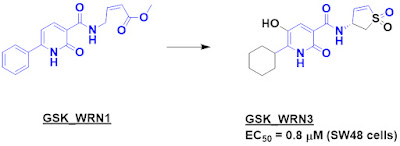
GSK_WRN1 was one of the prominent hits, with 81% modification. Tryptic digestion revealed that it modified C727, the same cysteine found by the Vividion researchers. Medicinal chemistry led to GSK_WRN3, with sub-micromolar activity in MSI SW48 cells. (Unfortunately no other details on the chemistry are provided; the paper states that these will be written up separately.)
 
GSK_WRN3 or a closely related compound were tested in a battery of assays and found to be inactive against three other helicases, which is not surprising given that C727 is unique to WRN. Chemoproteomic studies in cells also revealed the compound to be quite selective towards WRN vs other proteins. The compounds selectively inhibited MSI cancer cell lines and patient-derived organoids while sparing microsatellite stable (MSS) cell lines and organoids. One of the compounds showed activity in a mouse xenograft model.
 
In a useful public service, the researchers tested two previously reported WRN inhibitors, MIRA-1 and NSC617145, in the same set of several dozen cell lines and found that they were not only ineffective, they lacked selectivity for MSI cells over MSS cells. Although Dr. Saysno might object, I nominate these molecules to be added to the “Unsuitables” bestiary at the Chemical Probes Portal.
 
I do wish more details about the molecules were provided, especially the kinact/Ki values. It is interesting that GSK_WRN3 bears remarkable structural similarities to VVD-109063. IDEAYA recently announced that their collaboration with GSK has resulted in a development candidate targeting WRN, and it will be fun to see the full story emerge.