24 February 2014

Fragments vs bacterial DNA ligase: triumph of structure-based design

Fragment approaches have been used successfully against several anti-bacterial targets (see for example here, here, and here). In a recent issue of ACS Med. Chem. Lett., a team of researchers from Astex and GlaxoSmithKline report another potential weapon in the ongoing war against bugs.

The researchers were interested in bacterial DNA ligase (LigA), which is essential for DNA replication, is highly conserved among numerous types of bacteria, and is quite different from its human counterpart. They started by screening ~1500 fragments against S. aureus LigA using a combination of X-ray crystallography (soaking), ligand-observed NMR (WaterLOGSY), and thermal shift assays. Hits that made it through this gauntlet were then evaluated by isothermal titration calorimetry (ITC) and prioritized in part by ligand efficiency. One of the best molecules was compound 3.

The chlorine atom of compound 3 bound in a hydrophobic pocket of the enzyme. Wary of the potential reactivity of this motif, the researchers replaced it with a trifluoromethyl group; they also removed a nitrogen from the pyrazine ring to provide a vector for fragment growing. The resulting compound 10 had slightly improved potency.


Examining the structure of the initial fragment also revealed a water-mediated hydrogen bond, and by enlarging the triazole to a 6-azaindole 6-azaindazole (compound 12) the researchers were able to make this hydrogen bond directly while also more effectively filling the pocket, providing a satisfying 70-fold boost in affinity. However, close inspection of the crystal structure and computational modeling suggested that this molecule was binding in an energetically unfavorable conformation. Simply adding a nitrogen to the pyridine ring alleviated this problem, providing another 15-fold boost in potency (compound 13). This molecule also showed antibacterial activity against a number of Gram-positive pathogens.

This is a brief but elegant paper that demonstrates the power of crystallography and modeling to drive a fragment-derived medicinal chemistry effort. It will be fun to watch this story progress.

19 February 2014

Poll results: how do you store your fragment libraries?

The results are in, and it looks like there is wide diversity in how folks store their working fragment libraries:


Of the 79 votes, the largest number, roughly 42%, keep their compounds at -20 °C. The next largest category was room temperature (29%), with -80 °C (16%) and +4 °C (13%) rounding out the list.

There were also some good comments to the original post giving more details as to solvent, use of inert gas, etc. These parameters are more difficult to capture in a multiple-choice poll, so please keep those examples coming.

17 February 2014

Druggable is as Druggable Does; Or a Million Ways to use NMR

As we all know, the closure of sites is a bad thing for those of us in Pharma.  One very small silver lining is that this frees up a lot of very nice work to be published.  The former BI site in Laval has been closed for a year and we are still seeing great papers coming out.  In this one in JMed ChemLaPlante and co-workers tell us about their fragment efforts against HCV helicase

HCV has recently had drugs approved for its treatment, but as with any virus, different modes of treatment are important.  The ATP-dependent helicase activity is found in the C-terminal 2/3 of the NS3 protein. Helicase activity is straight forward to measure and there has been some success in terms of non-viral specific inhibitors.  The inhibitors found to date have been found to act through undesireable mechanisms, but with a wealth of structural information there is no reason why helicase is inherently undruggable.  With this information in hand, they decided to target site 3+4 (green sticks are DNA from the structure), near the most conserved residue W501.  The ATP-binding site is 1+2 for reference. 
 Their first approach was to screen the 1,000,000+ corporate compound collection.  As you would expect for a paper blogged about here, they failed to find anything interesting (all the inhibitors worked by undesireable modes).  So, on to the FBDD campaign, to save the day once more.  The used a "shotgun" approach with their fragment screen:

One source of compounds came from an earlier HTS where they rejected fragment-like molecules for lack of potency, additional HCS screening of in house fragment collection, commercial fragments were screened in an SPR assay, virtual screening, and NMR.  They had a stringent workflow aimed at producing quality compounds for X-ray.  [The in-house fragment collection was 1000 compounds.]  This, along with NMR, validated ligands that bound to site 3+4.  They note one particularly noteworthy problem: high false positive rates due to the high ligand concentrations needed for the assays.  This lead to aggregation, solubility, and promiscuity.  This lead them to implement specific assays designed to eliminate these compounds (two NMR papers published in 2013, ref 18). 

They then clustered the best hits into 9 chemotypes:

 They used an "Analog by Catalog" approach and soaked or co-crytallized the best compounds into crystals.  S6, S7, and S9 were not found to bind to helicase in the crystallization trials and were deprioritized.  S5 was found at Site 3+4, but also others.  S1-4, and S8 were found to bind solely to site 3+4 (12 examples shown overlain). The key feature of this is the compounds are centralized in a wide groove over W501.  The topology of the binding site (wide groove and small lipophilic pocket) meant that optimizing for potency could be challenging.
From this, they decided S2-S4 were the most promising.  In the end, the focused on the S2 indole series as the most promising.  The S2 stereotype 1
was found from an STD-NMR screen of 3 fragment per sample (300 uM fragment and 3.5 uM helicase).  They then, much to my heart's delight, they reached into the NMR cabinet for line broadening and competition experiments confirming it binds in site 3+4.  X-ray confirmed the binding mode, but potency was not improved with chemistry.  So back into the NMR cabinet they went: a methyl resonance assay, 
 15N TROSY showing peaks shifting upon addition of a derivative of 1, and 19F NMR!  OMG, how awesome is this?  

In terms of the chemistry, removing the Br does not change the potency, but did change the orientation of the compound in the binding site.  Further elaboration led to this compound 19 (3 uM and 0.23 LE):
It contains a nitro group, think what you may.  In order to confirm the binding affinity of the compound without immobilizing protein, they used the methyl resonances to do the titrations.  The two separate peaks they followed gave values of 32 and 28 uM (+/- 8).  Given the broadness of these peaks, I think this is a pretty decent assay, although it is an order of magnitude different than the biochemical Kd.  However, subsequent structural studies revealed that there is significant structural dynamic differences between pH 6.5 and 7.5.  ITC gave the same number (33 uM and enthalpy driven); however, the ITC had to be run at high compound concentration and a different pH.  They then went off the deep end and decided to use CD (I can't link to a previous post of using CD because we have never had a post where someone used it).  With a horrible assay (don't even get me started on near-UV CD as a readout of tertiary structure), they got reasonably close to the Kds determined by ITC and methyl-NMR.  

This is a very nice example of not being afraid of a target and using all available tools to advance hits against it.  It also shows the WIDE range of NMR experiments that can be used and that are easy and practical.  In terms of full disclosure, Steven LaPlante is a FOT (Friend of Teddy) and I have been working with him. 

12 February 2014

Fragments vs NAMPT, maximum ligand efficiencies, and off-target activities

The enzyme nicotinamide phosphoribosyltransferase (NAMPT) is essential for the synthesis of the important cofactor NAD and thus an intriguing target for blocking cancer cell metabolism. In two recent papers, researchers from Genentech, Forma Therapeutics, Pharmaron Beijing, and Crown Biosciences describe how they used fragment-based approaches to discover new inhibitors of this enzyme.

In the first (J. Med. Chem.) paper, Peter Dragovich (Genentech) and collaborators start with a screen of 5000 fragments using surface plasmon resonance (SPR) at the relatively low concentration of 100 µM. This yielded 283 hits which were retested at 150 µM and also competed with a known high-affinity inhibitor; noncompetitive fragments, which presumably bind outside the active site, were discarded. This winnowed interesting hits to 118 fragments, each of which was characterized in full dose-response curves. Only 6 were extremely weak (KD> 2 mM) or nonspecific, while 35 were quite potent (KD< 100 µM).

As an interesting aside, the substrate for NAMPT is nicotinamide, and this was characterized by SPR as having a remarkably high ligand efficiency (LE) approaching the “soft limit” Teddy recently discussed. The researchers suggest:

The LE exhibited by nicotinamide for NAMPT is the highest we have observed for a fragment lead and, given that NAMPT is highly optimized to efficiently bind this substrate, may approach an upper limit of this parameter for such molecules.

Keep in mind that Genentech has done lots of screens, so this is a significant statement. Indeed, I can think of only a few fragments (here and here) with comparable LE values.

But back to NAMPT. More than 30 co-crystal structures of fragments bound to the enzyme were solved, and several of these fragments were advanced. In doing so a variety of information was used, including data from molecules previously discovered in-house and elsewhere. Lots of nice SAR are presented, and if you’re into structure-based drug design I’d strongly encourage a close reading of the paper. Just to give you a flavor, compounds 12 and 13 (blue), despite their structural similarity, bound in very different orientations. A bit of engineering led to compound 15, and crystallography revealed that only a single enantiomer of a racemic mixture binds to the enzyme. Borrowing information from other NAMPT inhibitors led to the potent single enantiomer compound 17; the other enantiomer is 250-fold less active. Further modification yielded an orally active molecule with activity in a mouse xenograft model.


In the Bioorg. Med. Chem. Lett. paper, members of the same team describe two other series of molecules derived from fragments – and provide some important warnings about interpreting data.

One series (not shown here) was optimized to nanomolar potency in biochemical assays and antiproliferative cell assays. However, the team did a series of careful follow-up studies to show that these molecules are probably acting through off-target mechanisms. For example, the molecules do not reduce NAD levels as they should, and addition of the product of NAMPT did not rescue the cells, as it would were NAMPT the primary target.

For the other series, compound 7 (red above) was characterized crystallographically bound to NAMPT. Initial attempts to improve affinity were unsuccessful, but the co-crystal structure of another fragment suggested that replacing the pyrazole moiety with a simple phenyl group would be tolerated, leading to compound 25. Subsequent fragment growing ultimately led to Compound 51, with low nanomolar potency in both biochemical and cell-based assays. Importantly, this molecule did reduce NAD levels in cells, and the antiproliferative effects could be rescued by adding the product of NAMPT. Taken together, these data show that compound 51 is a nanomolar inhibitor of NAMPT both biochemically and in cells.

The importance of such rigorous characterization is driven home by a footnote, in which the researchers reveal that compound 51 was previously alleged to be an inhibitor of glucose transporter 1 (GLUT1). This was published in a high-profile journal, and several chemical suppliers now sell this compound (called STF-31). Although the current paper does not explicitly say so, it is possible the results in the earlier paper could be attributed to NAMPT inhibition rather than GLUT1 inhibition.

In the hope that views on STF-31 will evolve, I’ll close this Darwin Day post with a quote from The Descent of Man:

False facts are highly injurious to the progress of science, for they often long endure; but false views, if supported by some evidence, do little harm, as every one takes a salutary pleasure in proving their falseness; and when this is done, one path towards error is closed and the road to truth is often at the same time opened.

10 February 2014

Pushing the Limit

Dan's recent post discusses the limits of fragments binding to PPIs.  This paper from a while back came to my attention recently and I think it is important to bring up for discussion.  In it, they discuss the physical limits of binding.  They start with this:
Protein−ligand binding is a delicate balance between the loss of entropy resulting from complexation and the enthalpy gained by forming favorable contacts with the protein.
Entropy changes comes from linking two fragments, loss of internal flexibility, and reorganizing water in the binding site.  Current thinking (2 years ago) provides that in terms of favorable energy, van der Waals forces are the primary driver of affinity, while H-bonding and electrostatic interactions drive specificity.  Then they revisit this seminal paper from Kuntz et al but with the intent of exploring ALL biophysical properties rather than drug like ones.  For this study, Ligand Efficiency is DeltaG divided by the HAC. 

If van der Waals forces are the primary driver of affinity, there should be a correlation between affinity and size/contact area. 
There is not.  In terms of efficiencies, the median efficiency is -0.34 kcal/mol*atom.  Putting that in terms of buried surface area (BSA), they determined that the median efficiency is -23 cal/mol*A^2 (Angstrom squared).  To compare, if you look at only solvent accessible area, this value goes down to -7 cal/mol*A^2.  However, despite their inherently larger binding areas, macromolecules do not bind with greater inherent affinity than small molecules.  They argue that this is due to better "burying" of the small molecules.  As expected the most ligand efficient compounds are small, highly charged compunds buried in highly charged sites.  The limit is -1.75kcal/mol*atom, but a soft limit of -0.83 kcal/mol*atom is proposed.  

For maximal binding efficiency, they found that 90% of these interactions involve a charge-charge interaction or a metal ion.  In fact, the most efficient have several charge-charge interactions.  It is known the most efficient ligands are small, but not all small ligands are highly efficient.  So, what makes this difference?  As would be expected (at least I expected it), the longer the distance between charged groups the less efficient the interaction. 
For every 1A drop in average contact distance, the maximal efficiency goes down -0.41 kcal/mol*atom.  Wait!  What about desolvation you ask?  Isn't the entropic cost of desolvation in charged molecules very high?  In many of the highest efficiency complexes, there is water in the binding sites, so not all of the water is displaced, the authors state.  They also state that the charges in the binding pocket may not be fully solvated because the pockets around the charge are so small.  Yeah, I am not happy with that explanation either. 

So what makes maximally affinity?  Kuntz et al. said after 15 heavy atoms your affinity plateaus.  In fact, Kuntz showed that it is exceedingly rare to find an affinity >-15 kcal/mol, arguing that this is due to biological effects, namely clearance.  This paper argues that that cutoff is "seredipitously random or manmade".  Other people have argued that as ligands increase in size the maximal efficiency would drop because the number of interactions that need to be optimized increases and the only way to do this is through structural compromises and thus reduced affinity.  The authors of this paper begrudgingly admit this hyopthesis fits their data.

So, what implications does this mean for fragments? Does Kuntz's data mean that fragment libraries should be no bigger than 15 heavy atoms? Should we consider adding charged moieties or even metals?  They argue that this is a source of vast potential improvement for drug design. 

03 February 2014

How weak is too weak for PPIs?

Ben Perry brought up an interesting question in a comment to a recent post about fragments that bind at a protein-protein interface: “At what level of binding potency does one accept that there may not be any functional consequence?” I suspect the answer will vary in part based on the difficulty and importance of the target, and many protein-protein interactions (PPIs) rank high on both counts. In a recent (and open-access!) paper in ACS Med. Chem. Lett., Alessio Ciulli and collaborators at the University of Dundee, the University of Cambridge, and the University of Coimbra (Portugal) ask how far NMR can be pushed to find weak fragments.

The researchers started with a low micromolar inhibitor of the interaction between the von Hippel-Lindau protein and the alpha subunit of hypoxia-inducible factor 1 (pVHL:HIF-1α), an interaction important in cellular oxygen sensing. The team had previously deconstructed this molecule into component fragments, but they were unable to detect binding of the smallest fragments.

In the new study, the researchers again deconstructed the inhibitor into differently sized fragments and used three ligand-detected NMR techniques (STD, CPMG, and WaterLOGSY) to try to identify binders. As before, under standard conditions of 1 mM ligand and 10 µM protein, none of the smallest fragments were detected. However, by maintaining ligand concentration and increasing the protein concentration to 40 µM (to increase the fraction of bound ligand) or increasing concentrations of both protein (to 30 µM) and ligand (to 3 mM), the researchers were able to detect binding of fragments that adhere to the rule of three.

Of course, at these high concentrations, the potential for artifacts also increases, but the researchers were able to verify binding by isothermal titration calorimetry (ITC) and competition with a high-affinity peptide. They were also able to use STD data to show which regions of fragments bind to the protein, suggesting that the fragments bind similarly on their own as they do in the parent molecule. (Note that this is in contrast to a deconstruction study on a different PPI.) Even more impressively for a large (42 kDa) protein, the researchers were able to use 2-dimensional NMR (1H-15N HSQC) to confirm the binding sites.

Last year we highlighted a study that deconstructed an inhibitor of the p53/MDM2 interaction. In that case, the researchers were only able to find super-sized fragments, and they argued that for PPIs the rule of three should be relaxed. The current paper is a nice illustration that very small, weak fragments can in fact be detected for PPIs, though you may need to push your biophysical techniques to the limit.

But back to the original question of how weak is too weak. With Kd values from 2.7-4.9 mM, these are truly feeble fragments. Nonetheless, they could in theory have been viable starting points had they been found prospectively. That assumes, though, that these fragments would have been recognized as useful and properly prioritized. The ligand efficiencies (LE) of all the fragments, while not great, are not beyond the pale for PPIs. Previous research had suggested that much of the overall binding affinity in compound 1 comes from the hydroxyproline fragment (compound 6, which was originally derived from the natural substrate). Not discussed in the paper, but perhaps more significantly, the LLE (LipE) and LLEAT values are best for compound 6, which despite having the lowest affinity is the only compound that could be crystallographically characterized bound to the protein. In the Great Debate over metrics, this suggests that LLE and LLEAT may be more useful than simple LE for comparing very weak fragments.

29 January 2014

Kill Them Bugs!

Bugs are bad.  I hate bugs.  Bugs of all kinds.  In our part of the world we have a particularly noxious, invasive bug called the stink bug.  Ewwww.  And they are everywhere.  And in the winter they are particularly prevalent because they get in your attic, soffets, etc. and then creep into your house.  I would love to be part of a global effort to eradicate these horrible creatures.  I may lose my green bona fides advocating the genocide of an entire species, but so be it.  It is also not so practical, so really not germane to this blog.

However, targeting bacteria is practical, and important.  Antibiotic resistance is on the rise globally and only two antibiotics with novel modes of action have been approved in this century.  Dire consequences meet pressing need.  Many antibiotics with improved efficacy are due to higher to higher potency or resistance to degradation.  But, this avenue has a limited lifespan and novel targets are needed.  Into this breach steps Astra Zeneca, with this paper.  The topoisomerases DNA gyrase and Topisomerase IV Top IV) have already been shown clinically to be validated targets.  The A subunits contains the DNA cleavage domain while the B subunits contain the ATP binding and hydrolysis domain.  DNA gyrase inhibitors also typically inhibit TopIV.  Fluoroquinolones (the DNA complex) and aminocoumarins (the ATP site) target these enzymes. Aminocoumarins have not received much attention to due PK and safety issues. There are a wide variety of ATP-targeting compounds.

Cpds 1 and 2 have been shown by X-Ray to bind in the ATP site and extend outside that site to generate additional interactions with R144.  The team's design goal was a new scaffold that would merge these two compounds attributes.  They chose 2-pyridylureas which had not previously been explored.  Modeling showed that 5-substitution reaches towards R144 with a carboxylate and 4-substitution allows for exploration into more open space.  6-substitution abuts a hydrophobic region and should not be messed with.

Cpds 3-13 were synthesized (or were commercially available) to test these hypotheses with Cpd 6 clearly the best.  Then they explored the 4 and 5 substitutions 9see the actual paper for Tables 1-3).  The chemistry and isozyme exploration they performed was very detailed.  The two best compounds ended up being 31 and 35

Then the paper gets into the details (it's 24 pages long and the results/discussionare pp 5-13).  I highly recommend reading that part on your own.  I am really impressed by the work.  As they discuss, the Xtal structures support many of the design hypotheses.  This cannot be understated.  Fragment-based drug design (and in this case it really is DESIGN) was effective and robust.  In the end, their compounds were able to realize potent inhibition of 4 topoisomerases across three bacterial species. Importantly, bacterial growth was realized through inhibition of both the gyrase and Top IV which is the key criterion for continued optimization.  Efficacy in a mouse model was demonstrated with 35.   

22 January 2014

Fragments vs procaspase-6: preventing activation

The caspases are involved in a plethora of cellular processes and have been targeted by many groups using a variety of methods. They are cysteine proteases that cleave substrates immediately after an aspartic acid residue – and they pose several challenges for drug hunters. Because of their active site cysteine, they are particularly susceptible to non-specific inhibitors such as PAINs. Another difficulty is that their predilection for negatively charged substrates (such as aspartate) complicates efforts to identify cell-permeable inhibitors. To address both these issues, Jeremy Murray and colleagues at Genentech collaborated with Adam Renslo and colleagues at UCSF to avoid the active site altogether. They’ve recently published this work in ChemMedChem.

Caspases are initially translated as inactive zymogens that form dimers. These are cleaved to form a dimer of dimers, and this complex works as the active protease. The researchers wanted to see if they could find molecules that bind to the zymogen dimer interface and so block activation.

They started by performing a surface plasmon resonance (SPR) screen using 2300 fragments against both the active and inactive (procaspase) forms of the protein. Initial screening was done at a relatively low concentration of fragment (50 µM), with full dose-response assays being run on hits from either assay. This resulted in 84 hits against procaspase-6, with dissociation constants from 3 µM to 2300 µM, most of which were selective for the inactive zymogen.

Crystallography revealed that several of the fragments were in fact binding to a pocket at the dimer interface. In particular, fragments 1 and 6 bound (separately) at partially overlapping sites, suggesting the possibility of merging. This led to compound 8, with an improvement in affinity, albeit at a cost to ligand efficiency. However, modeling suggested that the bound conformation of this molecule would be strained, a situation which could be rectified by moving the position of the nitrogen atoms in the pyrimidine ring. Gratifyingly, this led to nearly a 10-fold boost in potency (compound 11), and further tweaking improved the dissociation constant to sub-micromolar (compound 12).


At the same time, the researchers noticed that the dimer interface is symmetrical, and that compound 6 binds near the center of the symmetry axis. This led them to design symmetrical compound 14, which displayed a modest improvement in potency but at a cost to ligand efficiency. Again modeling came to the rescue, this time suggesting a larger central element to yield sub-micromolar binders such as compound 16.

Whether or not targeting procaspase-6 will be useful therapeutically, this paper is a nice example of fragment merging against an unconventional binding pocket. It is also an excellent example of cooperation on multiple levels: first between biologists, biophysicists, chemists, and modelers, and second between academia and industry.

15 January 2014

Poll: how do you store your fragment libraries?

Following up on the recent post on pool size, we thought we’d poll readers on the very practical question of how they store their libraries. I’ve heard some vigorous debates, so it will be interesting to see the results. Please vote on the right side of the page. Also, please note that this question refers to working libraries as opposed to master stocks.

13 January 2014

There can be too much of a Good Thing

One nice thing about being a consultant is that I get paid to think about things for people (sometimes).  One of the things I have been thinking about lately (on the clock) is the optimal size of fragment pools.  I got to started wondering if there can be too many fragments in a pool:
You know how you mother always said, don’t eat too much it will make you sick?  I never believed her until my own child was allowed to eat as much easter candy as possible and it actually made him sick.  [It was part of a great experiment to see how much "Mother Wisdom" was true, like Snopes.]  I have been working lately in library (re)-optimization and one thing that keeps coming up is how many fragments should go in a pool.  As pointed out here and discussed here, there are ways to optimize pools for NMR (and I assume the same approach can be done for MS).  So, we have always assumed that the more fragments in a pool the better off you are, and of course the more efficient.  
But is that true?  Is there data to back this up?  Probably not, I don’t [know if] anyone wants to run the control.  So, let’s do the gedanken. If you have 50 compounds in a pool (nice round number and easy to do math, so its my kind of number) you would expect for a “ligandable” target to have a 3-5% hit rate.  That means out of that pool you would expect 1.5-2.5 fragments to hit.  So, that means that you have 2 fragments that hit, these two would then compete and your readout signal would be reduced by 50%.  So, if you are already having trouble with signal you are going to have more.  Also, can you be sure that  negatives are real, or did they “miss” because of lowered signal due to the competition.  And what if one of the hits is very strong?  Also, how do you rank order the hits?  Do you scale the signal by the number of hits in the pool?
  I then reached out to the smart people I know who tend to be thinking about the same things I do, but in far greater depth.  I spoke to Chris at FBLD and he was putting together a large 19F library, aiming to get up 40 or more 19F fragments in a pool.  Well, Chris Lepre at Vertex was already thinking about this exact problem. He shared his thoughts with me and agreed to let me share them here (edited slightly for clarity).  

To accurately calculate the likelihood of multiple hits in a pool, I [Ed:Chris] used the binomial distribution.  For your hypothetical pools of 50 and a 3% hit rate, 44% of the samples will have multiple hits (25.6% with 2 hits, 12.6% with 3, 4.6% with 4); at a 5% hit rate this increases to 71% (26.1% = 2 hits, 22% = 3, 13.6% = 4, 6.6% = 5, 2.6% = 6).  So, the problem of competition is very real.  It's not practical to deconvolute all mixtures containing hits to find the false negatives:  the total number of experiments needed to screen and deconvolute is a minimum when the mixture contains approximately 1/(hit rate)^0.5 (i.e., for a 5% hit rate, mixtures of 5 are optimal). [Ed:Emphasis mine!] 

Then there's the problem of chemical reactions between components in the mixture.  Even after carefully separating acids from bases and nucleophiles from electrophiles in mixtures of 10, Mike Hann (JCICS 1999) found that 9% of them showed evidence of reactions after storage in DMSO. This implies a reaction probability of 5.2%, which, if extended to the 50 pool example, would lead one to expect reactions in 70% of those mixtures.  If this seems extreme, keep in mind that the number of possible pairwise interactions = npairs = n(n-1)/2 (n-1/2)*n [Ed: fixed equation], where n = the number of compounds in the pool.  So, a mixture of 10 has 45 possible interactions,  while a mixture of 50 has 1200.  Even with mixtures of only five, I've seen a fair number of reacted and precipitated samples.  Kind of makes you wonder what's really going on when people screen mixtures of 100 (4950 pairs!) by HSQC.  [Ed: I have also seen this as I am sure other people have.  I think people tend to forget about the activity of water.  For those who hated that part of PChem, here is a review.  Some fragment pools could be 10% DMSO in the final pool, and are probably much higher in intermediate steps.]

Finally, there's the problem of chemical shift dispersion.  Even though F19 shifts span a very large range and there are typically only one or two resonances per compound, the regions of the spectrum corresponding to aromatic C-F and CF3 become quite crowded.  And since the F19 shifts are relatively sensitive to small differences in % DMSO, buffer conditions, etc. it's necessary to separate them by more than would be necessary for 1H NMR.  Add to that that need to avoid combining potentially reactive compounds (a la Hann) and the problem of designing non-overlapping mixtures becomes quite difficult.  [Ed: They found that Monte Carlo methods failed them.]

I've looked at pools as large as 50, but at this point it looks like I'll be using less than 20 per pool.  I'm willing to sacrifice efficiency in exchange for avoiding problems with competition and cross-reactions.  The way I see it, fragment libraries are so small that each false negative means potentially missing an entire lead series, and sorting out the crap formed by a cross-reaction is a huge time sink (in principle, the latter could yield new, potentially useful compounds, but in practice it never seems to work out that way).  The throughput of the F19 NMR method is so high and the number of good F-fragments so low that the screen will run quickly anyway.  Screening capacity is not a problem, so there's not really much benefit in being able to complete the screen within 24 hrs vs. a few days.
The most common pool size (from one of our polls) was 10 fragments/pool.  This would mean that the expected hit rate was 1% or less.  This is a particularly low expected hit rate, or people are probably putting too many fragments in a pool.  So, is there an optimal pool size?  I would think that there is: between 10-20 fragments.  You are looking for a pool size that maximizes efficiency but you don't want to have so many that you also raise the possibility of competition. 

09 January 2014

Poll results: affiliation, fragment-finding methods, and library size

Here’s a summary of the latest poll.

Readership demographics have not changed significantly since 2010, aside from a slight shift towards industry (~58% today vs 51% in 2010).


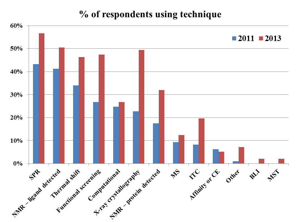
The next question asked about screening methods, and here things get more interesting.


The first thing to notice is that, with one minor exception, most fragment-finding techniques are being used more, with SPR and ligand-detected NMR now being used by more than half of all respondents. As a consequence, the average number of techniques being used jumped from 2.4 in 2011 to 3.6 in 2013.

The overall order of popularity doesn’t seem to have changed much, the major exception being X-ray crystallography, which is way up. However, this may be an artifact; a comment on the 2011 poll suggested that some voters interpreted the question to be about primary screening methods, as opposed to all methods.

(Technical disclosure: feel free to skip this paragraph unless you’re a data geek. Due to issues with polling in Blogger, this year’s poll was run in Polldaddy, the free version of which gave total votes for this question but not the number of individual respondents. However, since the two other questions in the poll allowed only single answers, the number of responses was equal to the number of respondents: 95 for the demographic question and 97 for the library question below. I thus assumed 97 respondents for this question, which is coincidentally identical to the number in 2011. Note also that the categories BLI and MST were new for 2013.)

Finally, the question on fragment library size shows that most folks are using libraries of 1000-2000 fragments, with only ~10% of respondents using very small (≤500) or very large (≥5000) collections of fragments.

This result is strikingly similar to the median of 1300 fragments that Jamie Simpson, Martin Scanlon, and colleagues found in an analysis of 22 published libraries. Teddy’s notes from FBLD 2012 put the median slightly higher: around 2500 for 17 libraries. Perhaps people with larger libraries tend to broadcast their size? Of course, in the end, it’s not the size of your library that matters; it’s what’s in it, and what you do with it.

Thanks again for participating, and if you have ideas for new polls, please let us know.

06 January 2014

That's One Way How to Do It

There is no right way to do science, that's what makes science awesome.  However, when you are entering a new field, or trying something new the first thing you do is find a current review.  I remember in grad school whenever it was my turn to present literature at group meeting, I would search the topic in TIBS (Trends In Biochemical Sciences).  That was always the best starting point.  However, when you want to really do something, as in practical applications you looked for a Methods in Enzymology paper or Current Protocols.  This always give you a way to do something, with in depth technical hints and tricks of the trade.  On this blog, we discuss a lot of different techniques and oftentimes it is out of the users area of expertise.  We try to make it understandable and I think we are largely successful.  

In this paper, yours truly, Darren Begley, and colleagues from Emerald put forth one way to run and analyze Saturation Transfer Difference NMR for fragment campaigns.  STD is the subject of MANY posts here.  One of the things I want to point out is that opinions are like belly buttons, everyone has one.  So, in this Protocol we put forth a way to perform STD it is not the only way to do, but I think it is a rather robust method.  Not everything will translate to every company.  For example, most companies don't have extremely small, highly soluble fragments like the Fragments of Life and thus 500mM stocks will not be achievable.  I believe 100 mM is a better generic concentration.  However, I would love to hear in the comments what other people think.  Additionally, there are computational approaches that make the manual creation of pools unnecessary.  In terms of analysis, there are a million different approaches.  Most companies that make NMR software have some sort of automation.  I really like the implementation from Mnova.  However, it is important to keep in mind that your results are really only as good as your understanding of the experiment. 

What this all really comes down to is that NMR, and STD in particular, is not a black box.  You still need an expert user running the experiments and analyzing the data.  My goal with this paper though is to enable better understanding of the STD experiment for the lay user.  Hopefully, this leads to greater use of the experiment and concomitant increased success in screening.  I would really like to hear comments about what people do differently and why. 

02 January 2014

Fragment events in 2014 and 2015

Lots of great events coming up this year and next, so start making your travel plans soon!

2014

February 18-19: Select Biosciences Discovery Chemistry Congress will be held in Barcelona, Spain, and includes a number of fragment-based presentations as well as a short course taught by Ben Davis on February 17.

April 24-25: CHI’s Ninth Annual Fragment-Based Drug Discovery will be held in San Diego. You can read impressions of last year's meeting here and here, the 2012 meeting here, the 2011 meeting here, and 2010 here. Also, Teddy and I will be teaching a short course on the topic over dinner on April 24.

May 21-22: Cambridge Healthtech Institute’s Fourteenth Annual Structure-Based Drug Design will be held in Boston, with several talks on FBLD. 

June 1-4: Newly added! Developments in Protein Interaction Analysis (DiPIA 2014) will be held in La Jolla, CA. This event is organized by GE Healthcare Life Sciences so it should be a great opportunity to learn about recent developments in fragment screening by SPR, ITC, and DSC.

July 19-22: Zing conferences is holding its first-ever Fragment Based Drug Discovery and Structure Based Drug Design in Punta Cana, Dominican Republic. In addition to the amazing location there are some great speakers, so definitely check this one out.

September 21-24: Finally, FBLD 2014 will be held in Basel, Switzerland. This marks the fifth in an illustrious series of conferences organized by scientists for scientists, the last of which was in San Francisco in 2012.  I believe this will also be the first major dedicated fragment conference in continental Europe. You can read impressions of FBLD 2010 and FBLD 2009.

2015

June - TBA: NovAliX will hold its second conference on Biophysics in Drug Discovery in Strasbourg, France. Though not exclusively devoted to FBLD, there is lots of overlap; see here, here, and here for discussions of last year's event.

December 15-20: Finally, the first ever Pacifichem Symposium devoted to fragments will be held in Honolulu, Hawaii. The Pacifichem conferences are held every 5 years and are designed to bring together scientists from Pacific Rim countries including Australia, Canada, China, Japan, Korea, New Zealand, and the US. There is lots of activity in these countries, and since travel to mainland US and Europe is onerous this should be a great opportunity to meet many new folks - in Hawaii no less!

Know of anything else? Add it to the comments or let us know!


30 December 2013

Review of 2013 reviews

The year is coming to an end, and as we did last year, Practical Fragments is looking back at notable events as well as reviews that we haven’t previously highlighted.

The fragment calendar started in March in Oxfordshire, at the RSC Fragments 2013 conference, closely followed in April by CHI’s FBDD meeting in San Diego (here and here). Closing out the year for conferences that Teddy or I attended was the Novalix conference on Biophysics in Drug Discovery in Strasbourg (here, here, and here).

There weren’t any new books published (though the special issue of Aus. J. Chem. practically counts as one), but there were several notable reviews.

Stephen Fesik and colleagues at Vanderbilt University published “Fragment-based drug discovery using NMR spectroscopy” in J. Biomol. NMR. This is an excellent overview that covers library design, NMR screening methodologies, and compound optimization. The researchers make an interesting case for including multiple similar compounds and allowing for larger, more lipophilic fragments, while always being careful to avoid “bad actors”. They also do a good job of summarizing the various NMR techniques, including their strengths and limitations, in language accessible to a non-spectroscopist. Finally, the section on fragment linking discusses the theoretical gains in affinity, the practical challenges to achieving these, and strategies to overcome them.

Turning to the other high-resolution structural technique, Rocco Caliandro and colleagues at the CNR-Istituto di Cristallografia in Italy published “Protein crystallography and fragment-based drug design” in Future Med. Chem. This provides a fairly technical description of X-ray crystallography and its role in FBDD, along with a table summarizing around 30 examples, five of which are discussed in some detail.

Of course, it’s always best to use multiple techniques for finding fragments, so it’s well worth perusing “A three-stage biophysical screening cascade for fragment-based drug discovery,” published in Nature Protocols by Chris Abell and colleagues at the University of Cambridge. This expands on a gauntlet of biophysical assays (involving differential scanning fluorimetry (DSF), NMR, crystallography, and isothermal titration calorimetry (ITC)) that we discussed earlier this year. Nature Protocols are highly detailed, with lots of troubleshooting tips, so this is a great resource if you’re exploring any of these techniques.

Finally, Christopher Wilson and Michelle Arkin at the University of California San Francisco published “Probing structural adaptivity at PPI interfaces with small molecules” in Drug Discovery Today: Technologies. Protein-protein interactions are frequent targets for FBLD: see for example here, here, here, here, and here – and that’s just for 2013! The current review gives a nice overview of the technology called Tethering, focusing on the cytokine IL2 and an allosteric site on the kinase PDK1.

And with that, Practical Fragments thanks you for reading and says goodbye to 2013. May your 2014 be happy and fulfilling!

23 December 2013

Fragments in Australia

Last year we highlighted the first FBDD conference held in Australia. That meeting has now led to a dozen papers in the December issue of Aus. J. Chem. Many of the papers use the same fragment libraries, so this is a good opportunity to survey a variety of outcomes from different techniques and targets.

The collection of papers (essentially a symposium in print) starts with a clear, concise overview of fragment-based lead discovery by Ray Norton of Monash University. Ray also outlines the rest of the articles in the issue.

A well-designed fragment library is key to getting good hits, and the next two papers address this issue. Jamie Simpson, Martin Scanlon, and colleagues at Monash University discuss the design and construction of a library built for NMR screening. Compounds were selected using slightly relaxed rule-of-three criteria, and special care was taken to ensure that at least 10 analogs of each were commercially available to facilitate follow-up studies. Remarkably, of 1592 compounds purchased, only 1192 passed quality control and were soluble at 1 mM in phosphate buffer. The properties of the final library are compared with nearly two dozen other libraries reported in the literature; this is the most extensive summary I’ve seen on published fragment libraries. The paper also analyzes the results of 14 screens on various targets using saturation transfer difference (STD) NMR. As the researchers note, this technique is prone to false positives, and indeed the average hit rate of 22.5% is high, with only about 50% confirming in secondary assays. There is also a nice analysis of what features are common to hits, along with a list of the 24 compounds that hit in more than 90% of screens.

The other paper on library design, by Tom Peat, Jack Ryan and others (including Pete Kenny), discusses library design at CSIRO. The researchers started with 500 fragments commercially available from Maybridge and supplemented these with roughly the same number of fragments from a collection of small heterocycles that had been synthesized internally; additional “three-dimensional” fragments are also being constructed. At CSIRO the primary screening method appears to be surface plasmon resonance (SPR), in particular the ProteOn instrument that allows simultaneous analysis of six fragments against six targets. Eight of about ten targets have yielded confirmed hits. The researchers show examples of specific (good), nonspecific (probably bad) and ill-behaved (ugly) fragments.

Next up is an excellent discussion of PAINS by Jonathan Baell (at Monash) and collaborators. Although Practical Fragments has covered this topic repeatedly (here, here, here, here, here, and here) it is a sad fact that more examples appear in the literature every day, so there is always something new to write about.

Fragment-finding methods make up the next several papers, starting with a nice overview of native mass spectrometry by Sally-Ann Poulsen at Griffith University. This paper covers theory, practical issues, and recent examples. Roisin McMahon and Jennifer Martin at University of Queensland, along with Martin Scanlon, describe thermal shift assays. In addition to highlighting a number of published examples, the paper also delves into some of the technical challenges and issues with false positives and false negatives, concluding with a nuanced discussion of how to deal with conflicting data.

The subject of conflicting data is central to the work of Olan Dolezal and Tom Peat, both of CSIRO, and their collaborators. They screened the protein trypsin against 500 Maybridge fragments using SPR. Unfortunately they couldn’t go higher than 100 micromolar without running into problems of solubility and aggregation, but even at this relatively low concentration they found 18 hits. X-ray crystallography validated 9 of them, and isothermal titration calorimetry (ITC) also validated 9, with 7 confirmed by all three techniques. (Incidentally, there are lots of great experimental details here.) Four of the SPR hits could not be confirmed by either ITC or X-ray, and 3 turned out to be false positives when repurchased and tested; in one case this appeared to be due to cross-contamination with a more potent compound. In general, the more potent compounds tended to be the ones that reproduced best, and solubility seemed to be a limiting factor for ITC. Despite the imperfect agreement of biophysical techniques, these were still superior to computational approaches on the same target with the same library. As they conclude:

It is gratifying to know (at least for these authors) that experimental data are still of enormous value in the area of fragment-based ligand design and that the modelling community still has a way to go before the experimentalists are put out to pasture.

But experimentalists should not get too cocky: the next paper, by Jamie Simpson and collaborators at Monash University, describes some of the things that can go wrong. An STD NMR screen of the antimicrobial target ketopantoate reductase (KPR) using the same Maybridge library of 500 compounds revealed 196 hits! The 47 with the strongest STD signals were then tested in a 1H/15N-HSQC NMR assay, leading to 14 hits, of which 4 gave measurable IC50 values in an enzymatic assay. Unfortunately, follow-up SAR was disappointing, and subsequent experiments revealed that aggregation was to blame: when the biochemical experiments were rerun in the presence of 0.01% Tween-20, only a single fragment gave a measurable IC50 value. The researchers redid their STD-NMR screen in the presence of detergent, resulting in 71 hits, all of which were tested in the biochemical screen. This led to the identification of a new (and fairly potent) hit that had previously been missed. This nicely illustrates the fact that false positives are not just a problem in terms of wasted resources, they can also overwhelm the signal from true positives. The moral? Always use detergent in your assay!

The question of whether structure is needed to prosecute fragments has come up before, and the next paper, by Stephen Headey, Steve Bottomley, and collaborators at Monash University, addresses this question directly. The target protein, a mutant form of α1-antitrypsin called Z-AAT, unfolds and polymerizes in vivo, causing a genetic disease. The researchers used an STD NMR fragment screen of 1137 fragments to identify several hundred hits, and focused on those that bound to the mutant form of the protein rather than the wild-type. They then used a technique called Carr-Purcell-Meiboom-Gill (CPMG) NMR (which relies on line broadening when fragments bind to a protein) to confirm 80 hits, the best of which had a dissociation constant of 330 micromolar. If you’ve stuck through this post thus far you’ll recall that the Monash library was designed for “SAR by catalog”, and 100 analogs of this fragment were purchased and tested, leading to several new hits, one with a dissociation constant of 49 micromolar. Although there is still a long way to go, metastable proteins are tough targets, so this is a nice start.

The next paper, by Ray Norton at Monash University and collaborators, describes a fragment screening cascade against the antimalarial target apical membrane antigen 1 (AMA1). An initial STD NMR assay of 1140 fragments produced 208 hits, but competition experiments with a peptide ligand whittled this number down to 57 that confirmed in both STD and CPMG NMR assays. Of these, 46 confirmed in an SPR assay, and although most are fairly weak, some SAR is starting to emerge as new analogs are synthesized.

Another antimicrobial target, 6-hydroxymethyl-7,8-dihydropterin pyrophosphokinase (HPPK), is the subject of a paper by James Swarbrick at Monash and collaborators. An initial STD NMR screen gave an unnervingly high hit rate (notice any themes emerging?), so 2D 15N-HMQC experiments were performed on 750 Maybridge fragments, yielding 16 hits. Competition experiments using CPMG NMR and close analyses of the chemical shifts suggested that these fragments bind in the substrate binding site, and SPR confirmed binding for some of the fragments.

Finally, Martin Drysdale of the Beatson Institute highlights some of the success stories of FBDD, including clinical compounds, and ends with a call for shapelier fragments.

All in all this is a great collection of papers, particularly for those relatively new to the field. It will be fun to revisit some of these projects in a few years to see how they’ve progressed.

19 December 2013

Undruggable? Pshaw, Fragments can do it.

Bromodomains, as well as other epigenetic targets, are hot right now.  This paper adds to the growing library of fragment success against bromodomains.  As in any nascent field, some of the targets do not have a known biological role.  BAZ2B (bromodomain adjacent to zinc finger domain protein 2B) is one of these.  BAZ2B is interesting compared to the 41 other bromodomains where there is structural information.  Its KAc binding pocket is smaller than other bromodomains (92-105 Angstrom vs 130-220 Angstrom for the other bromodomains and lacks features of BET bromodomains, like the ZA channel and the hydrophobic groove adjacent to the WPF motif.  
BAZ2B (Left), BRD2-BD1 (Right)
Due to these structural differences, the strategies that have been applied successfully to other bromodomains will not transfer to BAZ2B; thus, it considered one of the least druggable bromdomains.  Hence, fragments to the rescue!  

They screened 1300 commercially available (and thus Voldemort Rule compliant) with an alpha screen with hits being defined as 50% activity at 1mM.  10 compounds were identified and confirmed using STD, Waterlogsy, and CPMG NMR experiments (0.8% hit rate).  
(The same library was screened against BRD2-BD1 and CREBBP and had hits rates of 1.8% and 6.1% respectively.)  The ten fragments were then soaked into BAZ2B crystals yielding structures for 1,3,6 and KAc. 
a) KAc, b) 1, c) 3, d) 6
Fragment 6 had poor solubility, so direct ITC was not possible, instead a competition ITC study was used and yielded a 65 uM Kd.  They attempted to optimize this fragment from the 1 position of THgammaC.  All of these modifications resulted in worse affinity.  They then attempted to replace the Chlorine with aryl substituents (based on modeling results).  These compounds showed some improvement in solubility, but no significant improvements in affinity.  They then tried to change the electronic properties of the aromatic substituent.  EWG showed the expected reduction in affinity, but EDG did not show an increase in affinity.  

The most ligand efficient fragments (7, 8, and 10) all contained thioamides, so they synthesized thioamide and thiourea analogs of fragment 6.  Both of these molecules did not bind to the protein. Finally, they attempted to merge 3 and 6 putting the KAc mimetic on the scaffold of 6 . 

This urea containing compound (40) showed improved solubility, and 8-fold reduction in Kd, and a corresponding increase in ligand efficiency. 
This paper is a nice story of using fragments, structural biology, and modeling to generate useful compounds as tools.  I think it also points to "druggability" being a useless term when it comes to fragments.  Archimedes may have wanted a lever long enough, but for me, I just want fragments diverse enough.