Showing posts with label SPR. Show all posts

23 February 2026

Twelfth Novalix Biophysics in Drug Discovery Conference

Last week the Twelfth Novalix Biophysics in Drug Discovery Conference was held for the first time in La Jolla, California. It’s been several years since I wrote about one of these, and I was happy to see that they’ve maintained their reputation for excellent science and convivial conversation. There’s no way to cover the two-dozen talks, but here are a few highlights.
 
One of the things I most enjoy about these meetings is learning about new and emerging methods, and these were well represented. Chris Brosey (AbbVie) discussed time-resolved high-throughput small-angle X-ray scattering (TR-HT-SAXS). As I discussed a couple years ago, the approach can be used to measure the kinetics of protein dimerization in response to fragment-sized ligands.
 
SAXS-based approaches typically require access to a synchrotron, but Takashi Sato (Rigaku) described a related approach, electron density tomography (EDT), using an in-house instrument. Using machine learning, EDT can provide more detailed structural information than standard SAXS, and Takashi provided examples for samples ranging in size from viruses to single-chain variable fragments (scFvs) smaller than 30 kD.
 
Another approach to examine protein complexes is microfluidic diffusional sizing (MDS), described by James Wilkinson of Fluidic Sciences. By assessing the amount of diffusion in disposable chip-based chambers, MDS can determine how the hydrodynamic radius changes in response to ligands. Each chip holds 24 samples, and data can be collected in less than an hour. The minimum observable size change is 5-10% so measuring small molecules directly is unlikely. Still, the technique is useful for observing induced proximity events such as those caused by molecular glues, and it is sufficiently robust that it can be run in pure serum.
 
Among solution-based methods, none have achieved such recent prominence as cryo-EM. Weiru Wang, my colleague at Frontier Medicines, described how this technique was used iteratively to design and characterize bivalent degrader molecules that covalently exploit the E3 ligase DCAF2. (We recently published this work in Structure.)
 
Cryo-EM has revolutionized the types of biological molecules that can be structurally characterized. According to Denis Zeyer (Novalix), the technique accounted for 40% of PDB entries last year. However, despite the “resolution revolution,” most structures are not as detailed as those from X-ray crystallography; Denis noted that only 20% of the new structures were solved to a resolution better than 3 Å, which may have negative implications for machine-learning methods trained on these lower resolution structures.
 
If cryo-EM is the new kid in town, NMR is the grizzled veteran. But proving that it is possible to find new applications for old methods, Matthew Eddy (University of Florida) described a clever 19F-labeling approach for GPCRs in nanodiscs to quantify the distribution of various states in response to anionic lipids and ligands. This has allowed him to distinguish between antagonists and inverse agonists, which can be difficult using cell-based assays.
 
Turning from solution-based to surface-based methods, SPR has moved into the number two slot for fragment-finding, as we noted in our recent poll. Just as there are new tricks for NMR, the same applies to SPR. Matthew Peterfreund (Bruker Biosensors) described switchSENSE, a fluorescence proximity assay built on an SPR chip that is useful for measuring the binding and kinetics of bifunctional ligands such as PROTACs to two or more proteins. He also introduced the Triceratops SPR#64 instrument, which as its name implies supports 64 sensor spots.
 
Kris Borzilleri (Pfizer) discussed SPR-microscopy (SPRm), which combines an optical microscope with an SPR instrument. This can be used to measure the affinities of ligands binding to receptors in cells grown on SPR chips, and Kris described applications to membrane proteins such as GPCRs and solute carriers. The technique is still quite slow though, at only 10-15 compounds in duplicate per week.
 
Another surface-based approach to screening cells was described by Volker Gatterdam of Lino Biotech. Focal molography relies on changes in diffraction from nanoengineered diffraction gratings, called molograms. Targets, which can include living cells, are immobilized to the molograms, and analyte is flowed over. The instrument contains 64 spots, and assays can be run in complex samples such as tissue lysates.
 
Covalent drug discovery also made an appearance, with talks by Landon Whitby (Lundbeck) and Ben Cravatt (Scripps). Landon provided an overview of chemoproteomics techniques to screen ligands in cells, such as those we wrote about in 2016. Ben continued the theme, including several success stories, and also discussed challenges for finding cryptic ligandable pockets. Despite impressive progress with machine learning, Ben noted that these methods often find only common solutions, while empirical chemoproteomics methods can find rare types of pockets.
 
Of course, as we’ve repeatedly emphasized, biophysics methods are best used in combination, as noted by Daniel Harki (University of Minnesota) and Ann Boriack-Sjodin (Takeda). Daniel presented a screen of 1056 fragments against the cancer target APOBEC3 using NMR and SPR. This yielded just a single validated hit, which interestingly turned out to be the same fragment found against KRAS in a paper we discussed in 2022. And Ann described how biophysics led to multiple clinical compounds against a variety of targets at Epizyme and Accent Therapeutics.
 
I’ll stop here, but please feel free to add your thoughts. And while the date for the next Novalix conference has not yet been scheduled, the location has, with a return to beautiful Strasbourg. Vive la biophysique!

27 October 2025

Fragments vs FEN1: A chemical probe

Synthetic lethality is a relatively new approach to treating cancer by targeting proteins whose inhibition is lethal to cancer cells that have specific mutations. Disruption of flap endonuclease 1 (FEN1), an enzyme important for DNA replication and repair, becomes synthetic lethal combined with BRCA1 and BRCA2 loss of function mutations. However, although a few inhibitors have previously been reported, these had poor cell activity and physicochemical properties. In a new J. Med. Chem. paper, Sam Mann and collaborators at Artios Pharma, Merck KGaA, and several other organizations describe a chemical probe.
 
FEN1 is a member of the RAD2 nuclease family, all of which contain two magnesium atoms in the active site. Thus, the researchers set out to build a metal-chelating library, a strategy we’ve written about previously. Noting that a bivalent metal chelator requires at least three hydrogen bond acceptors, the researchers included fragments that were not strictly rule-of-three compliant. More than 300 fragments were screened in a biochemical assay against FEN1 and three related proteins, EXO1, GEN1, and XPG. Hits were selected based on potency, ligand efficiency, and selectivity. Two related fragments came out on top, one of which was characterized crystallographically bound to FEN1, confirming engagement with the catalytic magnesium ions


Compound 6 was trimmed back to the chelating core compound 7 before attempts were made to grow the molecule in several directions, leading eventually to compound 21, the first molecule to show target engagement in cells. Modeling suggesting the presence of a high-energy water that might be displaced, which was attempted by expanding the core to include a morpholine moiety. Further modulation of properties ultimately led to MSC778, with modest oral bioavailability in mice, rats, and dogs. The paper describes some nice medicinal chemistry that goes beyond the scope of this post. For example, there was a correlation between cellular target engagement and off-rates as determined by surface plasmon resonance (SPR). One wonders if a covalent inhibitor, with essentially no off-rate, could be even more effective.
 
MSC778 is at least 65-fold selective for FEN2 over related RAD2 family members. It is also clean in a panel of off-target safety assays. The molecule is cytotoxic to a cancer cell line in which the BRCA2 gene had been knocked out but less so to the same cell line carrying wild-type BRCA2. Surprisingly though, no tumor growth inhibition was seen in a mouse xenograft model using the mutant BRCA2 cell line at the highest tolerable dose. However, tumor stasis was seen when the compound was dosed in combination with niraparib, a PARP inhibitor, consistent with earlier cell experiments suggesting that PARP inhibitors could synergize with FEN1 inhibitors.
 
The lack of single agent activity seen with MSC778 was undoubtedly disappointing, though the researchers note that it is unclear whether this is “due to insufficient target coverage, or an unexpected disconnect between the phenotypic consequences of FEN1 inhibition in vitro and in vivo.” Nonetheless, MSC778 looks to be a useful chemical probe for further understanding the biology of FEN1. This paper is also a nice application of building and screening a metal-chelating fragment library, which could be useful for targeting additional metalloproteins.

22 September 2025

Fragment merging without crystallography for CGRP receptor antagonists

Migraines are the third leading cause of disability worldwide. Although the pathology is complex, blocking the interaction of calcitonin gene-related peptide (CGRP) with its receptor, thereby decreasing vasodilation, has proven successful in the clinic. However, some of the early small molecule antagonists were discontinued due to hepatotoxicity. In a recent J. Med. Chem. paper, Naohide Morita, Isao Azumaya, and collaborators at Kissei Pharmaceutical and Toho University describe a new class of inhibitors.
 
CGRP binds at the interface of a heterodimeric receptor comprised of the calcitonin receptor-like receptor (CLR) and receptor activity-modifying protein 1 (RAMP1). To find hits, the researchers screened a library of 2500 fragments (which could be up to 350 Da) at 500 µM against the extracellular CLR/RAMP1 domains using SPR. This yielded 565 hits, which were clustered based on similarity, and 250 were chosen for dose-response studies, leading to 38 confirmed hits. Competition studies with a known CGRP antagonist whittled this number down to just four, with compound 1 being chosen for further study due to ease of analog synthesis.
 
Compound 1 was confirmed as a binder using isothermal titration calorimetry (ITC). Unfortunately, co-crystallography with CLR/RAMP1 was unsuccessful, so the researchers turned to docking using information from known small molecule inhibitors. This work suggested that compound 1 binds to the CGRP receptor but does not interact with RAMP1, a conclusion further supported by mutagenesis studies.
 
To find fragments that bind RAMP1, the researchers performed a second fragment screen, again using SPR. This time the fragments were chosen from those in the first set that had not been tested in dose-response studies, supplemented with several hundred more selected based on structures of known CGRP antagonists. Of 784 fragments screened, 114 were taken into dose-response studies, leading to 8 hits. Compound 2 was the most potent, and mutagenesis studies suggested it interacted with RAMP1.
 
Crystallography of compound 2 was also unsuccessful, but docking, supported by NMR studies, suggested a possible binding mode. Compounds 1 and 2 were merged to yield compound 3, which had a satisfying 2000-fold improvement in potency compared to compound 1. Compound 3 also showed cell activity.


Compound 3 contains three stereocenters, so the researchers sought to simplify the molecule. They also needed to improve potency and metabolic stability. Multiparameter optimization ultimately led to compound 15, with picomolar(!) affinity for the receptor, subnanomolar activity in cells, and good pharmacokinetic properties. A standard model for migraine is inhibition of facial blood flow in marmosets, and compound 15 was active. The compound was also clean in tests for hepatotoxicity.
 
Although no further development of compound 15 is reported, this is a nice case study in fragment merging. As the researchers note, it is also one of just a handful of examples that succeeded in the absence of crystallographic data (we wrote about another one here). Hopefully this will further embolden researchers to pursue fragment merging and linking without direct structural information.

12 May 2025

From fragment to macrocyclic Ras inhibitors

At the Drug Discovery Chemistry meeting last month chemist John Taylor described efforts against the oncology target RAS. This story was recently published in J. Med. Chem. by John, Charles Parry, and a team of some three dozen collaborators at CRUK Scotland Institute, Novartis, and Frederick National Laboratory for Cancer Research.
 
Practical Fragments has highlighted multiple Ras efforts, including the development and approval of sotorasib, which inhibits the G12C mutant of KRAS. Sotorasib binds in the so-called switch II region, next to the site where the nucleotides GDP and GTP bind. Before the discovery of this site, researchers had identified fragments that bind to a different site, switch I-II. 
 
Most of the ligands that bind to either site only inhibit the off-form of Ras proteins, in which the proteins are bound to GDP. One mechanism of resistance for cancer cells is to increase the amount of protein in the active, or GTP-bound state. Thus, the researchers focused on the oncogenic G12D mutant of KRAS bound to a GTP analog and screened it against 656 fragments using SPR. Ligand-detected NMR confirmed five of the hits, including compound 5.
 

Two dimensional 1H-15N HSQC NMR revealed that compound 5 binds in the switch I-II pocket; merging this with a literature fragment generated compound 6. SAR studies led to compound 11, which was characterized crystallographically bound to the protein. The structure suggested trying to make a salt bridge with an aspartic acid residue, leading to compound 13, with sub-micromolar affinity for the inactive form of the protein. A crystal structure of a related compound suggested the possibility of macrocylization, and this turned out to be successful, with compound 21 being the most potent. (All values shown here are determined by NMR or SPR on the G12D KRAS mutant bound to either GDP or the GTP analog GMPPMP.)
 
A number of different macrocycles were made and tested, and all of them were more potent against the inactive than the active form of KRAS. Crystal structures suggested that a glutamic acid side chain adopts a conformation in the the GTP-bound form of KRAS that impedes ligand interactions.
 
Interestingly though, building off the molecules in another direction led to the opening of a small subpocket that had not previously been reported in the literature. Exploiting this “interswitch” region led to compound 36, with a nearly 10-fold preference for the active form of KRAS.
 
Most of the macrocycles in both series were able to block nucleotide exchange in a biochemical assay, meaning they could prevent the exchange of GDP for GTP. A few of the compounds were tested in cell-based assays and could block binding between RAF and multiple Ras isoforms, including two mutants of KRAS as well as wild-type KRAS, HRAS, and NRAS.
 
Unfortunately, and not surprisingly given their high polar surface areas, the compounds had low permeability, high efflux, and high clearance in vitro. Mouse studies on one compound confirmed these liabilities in vivo.
 
Although the compounds could not be advanced, this is still a nice fragment to lead story. The fact that a new pocket could be identified despite so much previous effort on this target is a good reminder that no matter how much you know, there is always room for surprises.

04 December 2023

Screening tough proteins by SPR

Surface-plasmon resonance (SPR) is among the most popular methods for finding fragments. However, as we have noted, SPR can be very prone to operator error and misinterpretation. In a recent (open access) SLAS Discovery paper, U. Helena Danielson (Uppsala University) and a who’s-who team of biophysicists from across Europe provide experimental strategies for screening difficult proteins.
 
The researchers chose five different proteins, some of which were screened in two or three different forms for a total of nine protein constructs. Six of these were screened against their FL1056 library, a custom-built 1056-member library which include molecules from the FragNet program. The library includes a number of “three dimensional” molecules as assessed by principal moment of inertia (PMI). The other library, FL90, is a small set of commercially available fragments we highlighted here.
 
Before screening compounds against proteins, the researchers conducted a “clean screen.” This involved injecting fragments (at 500 µM each) over the sensor surface using the same buffer that would be used in the actual screen to pre-identify fragments that stick to the surface. This typically disqualified about 1% of fragments, though for one set of conditions the number was closer to 3%.
 
That work done, the researchers turned to the actual screens. After proteins were immobilized on the sensor chips, the fragments were typically screened at a single concentration of 250 µM each. The threshold for the initial hit cutoff was set low, often around 10% of the library, to minimize false negatives. Subsequent follow-up studies at varying concentrations were used for confirmation. This led to a significant winnowing, with the final number of confirmed hits between 0.5 and 7% of the library.
 
The proteins themselves were intentionally chosen to present various difficulties. Acetylcholine binding protein (AChBP, which we wrote about here) forms a large (125 kDa) pentameric complex with multiple binding sites. Lysine demethylase 1 (LSD1) is a multidomain, cofactor-dependent protein that requires a partner protein, CoREST, for activity. LSD1 was screened in the presence or absence of CoREST. Farnesyl pyrophosphate synthase (FPPS, which we wrote about here) is a target for cancer and osteoporosis, and the microbial forms are targets for trypanosomiasis drugs. Human as well as Trypanosoma cruzi and Trypanosoma brucei proteins were screened. Protein tyrosine phosphatase 1B (PTP1B, which we recently wrote about here) is a difficult enzyme with a couple allosteric sites. The C-terminal region is intrinsically disordered, and the protein was screened with or without this region. Finally, human tau is both intrinsically disordered and prone to aggregation. As we noted earlier this year it is of interest due to its potential role in Alzheimer’s disease.
 
Happily, hits were identified against all the proteins, some with ligand efficiency values above 0.5 kcal/mol per heavy atom. The chemical structures for selected hits are shown, and the researchers do appropriately caution that validating them using orthogonal (non-SPR) methods is essential before further studies.
 
I do wish the researchers had noted whether shapely hits were enriched or depleted among the confirmed hits. To my eye most seemed fairly flat, and some seemed dubiously PAINS-like, including an eyebrow-raising dinitro-catechol. Nonetheless, the paper is a nice summary of multiple SPR campaigns. If you’re about to embark on one yourself, it is worth carefully perusing.

22 August 2022

Fragments vs human Adensoine 2a Receptor using SPR

Last week we highlighted the use of surface plasmon resonance (SPR) to find ligands against RNA. Although RNA is not a typical protein target, it is at least normally free in solution. Targets such as GPCRs are more technically challenging because they are bound within membranes. Challenging, but not impossible, as illustrated by this post from 2012. A new ACS Med. Chem. Lett. paper by Reid Olsen, Iva Navratilova, and colleagues at Exscientia, University of Dundee, and AstraZeneca provides the latest example.
 
Navratilova and colleagues previously described using SPR to screen the β2 adrenergic receptor. In the new paper, the researchers studied the human adenosine 2a receptor (hA2AR), a “rheostat for energy homeostasis” that also plays a role in cancer immunotherapy. hA2AR is one member of a small family of adenosine receptors, and the researchers expressed all four of them, each with a polyhistidine tag that could be captured in the SPR instrument using a nickel-NTA sensor chip. Other labs (such as Heptares) have used mutant, stabilized forms of GPCRs, but here the researchers used native proteins and stabilized them by crosslinking them to the surface of the chip. They confirmed that these GPCRs bound known ligands with similar affinities to those reported in the literature.
 
Next the researchers screened a library of 656 fragments, each at 50 µM, against hA2AR. This led to 72 potential hits taken into dose-response experiments, of which 17 confirmed with affinities ranging from 1.1 to 410 µM. All the sensorgrams are shown, as are the structures of the fragment hits. These confirmed hits were also screened against A1, A2B, and A3; most of the fragments bound to all the receptors, though two were selective for hA2AR.
 
To assess where the fragments bind, the researchers added a known high-affinity ligand; ten of the fragments could be competed, while seven showed less or no competition, suggesting that they may bind to an allosteric site.
 
GPCRs biology is complicated, and just because a ligand binds does not mean it will have any effect on signaling. In cell experiments, none of the fragments behaved as agonists, but five fragments could act as antagonists of a known agonist. Another fragment seemed to increase the signal, suggesting it is an allosteric modulator. As the researchers conclude, “while SPR can screen fragment-like molecules that allow for extrapolation of extremely large and diverse chemical spaces, it cannot predict the biological activity of these binders."
 
Nonetheless, this paper provides a nice guide on how to use SPR, with its low protein requirements, to screen GPCRs. And the fragments disclosed could be interesting starting points for medicinal chemistry.

15 August 2022

Fragments vs RNA with SPR: A guide

Fragment-based lead discovery on RNA has a long history: the first mention on Practical Fragments was in 2009. Most often, various NMR methods have been used (see this example from last year), though isothermal titration calorimetry (ITC) is also effective. However, both of these techniques generally require considerable amounts of RNA. In a recent Biochemistry paper, J. Winston Arney and Kevin Weeks describe using SPR, which could increase the speed and ease of screening RNA.
 
Non-specific binding is a significant problem in characterizing RNA ligands. RNA is negatively charged, and many ligands are positively charged, leading to non-specific interactions. In a typical SPR experiment, the target is bound to a surface and the analyte is allowed to flow over the immobilized target; binding causes a change in refractive index that can be detected. However, if the analyte interacts non-specifically with the target, this will also be detected. For high affinity ligands the non-specific interactions may be minimal at low concentrations, but for low-affinity ligands such as fragments, it can be difficult to differentiate specific from non-specific binding.
 
SPR experiments generally use a reference cell, in which the analyte is allowed to flow over the surface in the absence of target; this signal is then subtracted from the target channel. Arney and Weeks decided to use a reference cell containing mutant RNA not expected to bind to the ligand.
 
The researchers developed their approach using two different riboswitches, each with known high-nanomolar ligands. Immobilizing the riboswitches to the chip and flowing ligand led to non-specific binding at concentrations of 100 µM or so. However, when the reference cell contained a mutant riboswitch designed not to bind to the ligands, this non-specific binding could easily be subtracted, leading to simple single-site binding models.
 
Of course, creating a mutant RNA assumes you already know where your ligand binds, which is not true if you are looking for ligands to a new target. To increase the generality of their approach, the researchers used a different riboswitch or a completely arbitrary RNA for the reference. These also worked, though not quite as well as the targeted mutants.
 
Finally, the researchers tested a dozen RNA-ligand pairs that had previously been rigorously characterized. Importantly, these varied considerably in affinity, from 8 nM to 2 mM. Most of them were also fragment-sized, with molecular weights as low as 119 Da. The correlation between SPR dissociation constants and those reported in the literature was excellent.
 
The technique does have limitations. First, the RNA-bound surfaces do seem somewhat unstable over a period of days. Also, larger RNAs present technical challenges, though the researchers do state that they have been able to examine molecules as large as 300 nucleotides. Overall this looks like a nice approach for measuring RNA-ligand affinities.

25 July 2022

Fragments vs TEAD: noncovalent this time

Last week we described a fragment-derived covalent probe that targets the four closely related TEAD transcription factors, which are part of the Hippo signaling pathway implicated in some cancers. A new paper in J. Med. Chem. by Timo Heinrich and collaborators at Merck KGa, iBET, and Cancer Research Horizons brings us another fragment-derived probe, this one noncovalent.
 
The researchers started by screening 1930 fragments, each at 2 mM, against TEAD1 and TEAD3 using SPR. Perhaps not surprisingly given the high concentration used, this led to a whopping 560 hits. These were then tested in dose-response format against TEAD1 with or without the coactivator YAP; 254 compounds showed differential affinity, among them compound 1. This molecule was crystallized bound to TEAD3, which revealed that it binds to the hydrophobic pocket normally occupied by a covalently-bound palmitoyl group required for activity. Despite being a fragment, compound 1 was active in a cell reporter assay, and the researchers state that further optimization was done using cellular assays rather than biophysical or biochemical experiments.
 

Analysis of the crystal structure suggested that enlarging the cyclopentyl moiety could fit more snugly into a hydrophobic pocket, while adding a small propyl moiety could extend into a separate pocket, leading to compound 6, with a 10-fold boost in activity. Replacing the propyl with an additional ring led to sub-micromolar compound 9. Finally, replacing the saturated ring with a substituted phenyl moiety led to MSC-4106, with low nanomolar activity in the cell reporter assay.
 
Thermal stabilization (specifically, nanoDSF) assays showed that MSC-4106 stabilized TEAD1 and TEAD3 but not TEAD2 or TEAD4. Palmitoylation assays confirmed this selectivity profile. The paper also includes a nice table comparing experimental selectivities of seven other non-covalent TEAD inhibitors, which vary from having activity only against TEAD1 to activity against all four homologs.
 
MSC-4106 was clean when tested at 10 µM against a panel of 58 receptors and 1 µM against nearly 400 kinases. It did not inhibit hERG or any of the common CYP450s. Finally, PK studies in mice, rats, and dogs showed that the compound is orally bioavailable with a long half-life. Given these favorable properties it was taken into xenograft studies, where it showed tumor growth inhibition at 5 mg/kg and tumor regression at 100 mg/kg. Analysis of tumor tissue showed downregulation of a TEAD-regulated gene, Cyr61.
 
Can we draw any lessons from comparing covalent MYF-03-176 (discussed last week) with non-covalent MSC-4106? Probably not, given that the former hits all TEAD homologs while the latter is selective for TEAD1 and TEAD3. Both molecules look to be excellent chemical probes for further dissecting Hippo signaling. I look forward to seeing how TEAD inhibitors ultimately fare in the clinic.

23 January 2022

Fragments (almost) in the clinic: MRTX1719

Synthetic lethality is a relatively new approach to cancer therapy. The idea is to inhibit a protein that is necessary for cancer cells but dispensable for normal cells, thereby minimizing toxicity. Last year we described one example, and in a just-published open access J. Med. Chem. paper Chris Smith and colleagues at Mirati describe another.
 
The biology gets a bit complicated, so please bear with me. Protein arginine methyl transferase 5 (PRMT5) is an epigenetic writer that adds two methyl groups to arginine residues in a wide variety of proteins. It is essential for cell survival. PRMT5 uses a cofactor, S-adenosyl-L-methionine (SAM), that is converted to methylthioadenosine (MTA) during the reaction. In certain cancers a gene called methylthioadenosine phosphorylase (MTAP) is deleted, causing an accumulation of MTA and – through product inhibition – a decrease in PRMT5 activity. The idea is to develop a drug that binds to and further stabilizes the (inactive) PRMT5•MTA complex, which is abundant in cancer cells, while not interfering with the active form of the protein, which predominates in normal cells. Told you it was complicated! [Note added: as befits the complicated biology I got a couple things wrong, corrected in the comment on 26 Jan.]
 
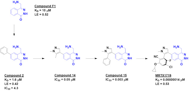
The researchers started with an SPR screen of 1000 commercially available fragments, each at 100 µM. PRMT5 was immobilized on the chip, with MTA added to the buffer to form the PRMT5•MTA complex. This screen yielded 17 hits, and based on this encouraging result a further set of nearly 1900 fragments was screened at 500 µM. The higher concentration yielded significantly more hits, and when these were tested in dose response experiments 100 were found with dissociation constants better than 1 mM. The best 24 of these were then screened against PRMT5 loaded with either MTA or the cofactor SAM. Compound F1 proved to be 5-fold selective for the MTA-bound protein over the SAM-bound protein.
 
Crystallography revealed that this molecule binds in the substrate-binding site in the vicinity of MTA and suggested that it would clash with SAM binding, thus providing an explanation for its selectivity. The crystal structure also revealed a nearby pocket that could be targeted through fragment growing, and this was accomplished with compound 2, which also showed activity in a biochemical assay. Further structure-based design led eventually to compound 14, which was 26-fold selective for the MTA-bound protein.
 

Crystallography revealed another lipophilic pocket, and adding a phenyl group provided a nice increase in potency in the form of compound 15. This molecule also showed low micromolar cell activity. Further structure-based drug design ultimately led to MRTX1719; the medicinal chemistry is elegant but beyond the scope of this post. Chemists will recognize that the final molecule is an atropisomer. This type of stereoisomer is uncommon in drugs in part because they can be difficult to separate; the researchers note assessing 70 different conditions before abandoning one series in favor of a more tractable one.
 
The dissociation constant of MRTX1719 was measured by SPR as 0.14 pM and 9.4 pM for the PRMT5•MTA and PRMT5•SAM complexes, respectively. We don’t encounter femtomolar binders very often; the dissociation half-life for the MTA-bound protein is 14 days! The 67-fold difference in binding was in good agreement with 70-80-fold differences in cells without or with MTAP.
 
MRTX1719 was quite selective in a panel of 42 methyltransferases. Pharmacokinetics and oral bioavailability were good in mice, dogs, and cynomolgus monkeys. The molecule was well tolerated in a mouse tumor model and caused tumor growth inhibition. Based on these results, an IND for the molecule has been submitted to the FDA.
 
This is a lovely fragment-to-candidate story, and Practical Fragments wishes everyone involved good fortune in the clinic!

19 April 2021

Fragments vs KEAP1: deconstruction and merging

One of the more challenging protein-protein interactions targeted by drug hunters is the interface between the transcription factor NRF2 and its repressor KEAP1. This is part of the cellular defense against reactive oxygen species; increasing NRF2 activity may be useful for treating a variety of diseases. Unfortunately, the binding site on KEAP1 that interacts with NRF2 is large and has a predilection for carboxylic acids. Thus, many of the molecules reported as inhibitors tend not to be druglike. Anders Bach (University of Copenhagen) and a multinational team of collaborators sought to do better, and have just published some of their journey in J. Med. Chem.
 
The researchers had previously tested 19 reported small-molecule KEAP1 inhibitors, of which only nine confirmed. (This is a salutary reminder to take any individual publication with a large grain of salt.) The nine fell into six chemical series (two shown below), and the researchers decided to fragment some of these molecules into 77 fragments. The fragments were then tested in four assays: fluorescence polarization (FP), a thermal shift assay (TSA), saturation transfer difference (STD) NMR, and surface plasmon resonance (SPR).
 
Primary hit rates were generally high, from 25%-64%, but long-time readers will not be surprised that the overlap was not great: no fragments hit in all four assays, and only eight hit in three. As the researchers point out, this could reflect differences in sensitivity, conditions (from 3-8% DMSO and from 0.5 to 8 mM fragment), and different types of false positives and false negatives. Interestingly, and in contrast to previous work, overlap was good between STD NMR and SPR.
 
Crystal structures of seven hits were solved bound to the protein, and compounds 4c and 1m (from different precursor molecules) were merged to provide compound 8, with low micromolar affinity. Compound 8 was the subject of considerable medicinal chemistry, with five different vectors chosen for growing. Despite being structurally enabled, the researchers struggled; changes that improved affinity in one context did not do so in another. After considerable effort, the researchers obtained compound 77o, with mid-nanomolar activity.
 

Compound 77o is stable in human plasma and mouse liver microsomes. Unfortunately, and unsurprisingly given the two carboxylic acids, it has poor permeability. Indeed, a fragment-derived KEAP1 inhibitor we described previously has only a single carboxylic acid, as does precursor compound 7. As the researchers themselves acknowledge, “the physicochemical properties of our compounds are not favorable for membrane permeability.”
 
Nonetheless, this paper is a lovely example of fragment-based deconstruction reconstruction (FBDR) and is well worth studying for the thorough descriptions of fragment screening in orthogonal assays and structure-based design. Another lesson may be that despite considerable effort, the final molecule is far from a chemical probe, let alone a drug. Perhaps some targets truly are undruggable. Or maybe – as for other seemingly undruggable targets – a change in strategy is needed.

25 January 2021

Fragments vs TNFα advanced with biophysics, linking, and growing

The cytokine tumor necrosis factor α (TNFα) is a key mediator of inflammation and has long been a target for rheumatoid arthritis, Crohn’s disease, psoriasis, and a host of other inflammatory diseases. Several biologic drugs, such as adalimumab, are approved but these monoclonal antibodies and fusion proteins can be immunogenic or induce neutralizing antibodies. Small molecules could avoid these pitfalls and also reach organs, such as the brain, less accessible to biologic agents. Impressive efforts towards this goal have just been reported in J. Med. Chem. by Justin Dietrich, Chaohong Sun, and colleagues at AbbVie. (Andrew Petros presented this work at the CHI DoT meeting last September.)
 
The researchers began by screening 18,000 fragments using two-dimensional (13C-HSQC) NMR against TNFα in which the methyl groups of isoleucine, valine, leucine, and methionine were isotopically labeled. Only 11 fragments caused significant perturbations, an 0.06% hit rate reflecting the difficulty of finding hits against this target. All the fragments were characterized by SPR, and compound 1 turned out to have reasonable affinity and ligand efficiency. Synthesis of a few dozen analogs led to compound 2, with improved activity.
 

TNFα forms a homotrimer, and a crystal structure of compound 2 bound to TNFα revealed that two copies of the fragment bind within a large hydrophobic cavity at the interface of the three protein monomers. Not present in the apo-form of the protein, this central pocket is formed by the movement of tyrosine side chains, causing desymmetrization of the protein trimer. The researchers linked the two nearby fragments to produce compound 3 with improved affinity but decreased ligand efficiency. Further optimization led to compound 4, which was active in cells. But perhaps not surprisingly given its size and lipophilicity, this molecule had high clearance and poor oral bioavailability in mice. 
 
 
A second fragment, compound 6, had lower affinity than fragment 1, and parallel chemistry efforts generated only flat SAR. A crystal structure revealed that compound 6 bound in a similar manner as compound 1, with two copies in the central cavity. Surprisingly, a crystal structure of compound 8, which differs from compound 6 only by a single methyl group and actually has slightly lower affinity, revealed a singly copy bound in the central void. Scaffold hopping led to compound 9, which was ultimately optimized through structure-based design and careful attention to drug-like properties to compound 12. This molecule is orally bioavailable and showed activity in a mouse arthritis model.
 
This is a lovely paper that illustrates several important lessons. First, as the researchers note, “we have learned from multiple programs, including this one, aimed at developing small-molecule inhibitors of protein-protein interactions, that biophysical methods, when used to drive a fragment-based approach, offer the greatest chance of success.” NMR was essential for finding the initial fragments, SPR provided necessary thermodynamic and kinetic information, and crystallography led to the breakthrough discovery of the binding mode of compound 8.
 
Second, although the initial series generated by fragment linking ultimately did not advance, it proved critical for developing chemical tools, validating assays, and providing structural insights.
 
And finally, this paper is a paean to persistence for difficult targets. As the researchers note, scientists have been seeking small molecule inhibitors of TNFα for decades, and although compounds were reported as early as 2005, most of these have had poor physicochemical properties. Seemingly undruggable targets can sometimes be unlocked. But it usually takes time.

16 March 2020

Fragments vs a Pseudomonas aeruginosa virulence factor

The world is understandably focused on SARS-CoV-2; see for example last week’s post. But there are many other threats out there, including infectious Pseudomonas aeruginosa, which is particularly problematic for immunocompromised people. A recent (open access!) ChemMedChem paper by Martin Empting and collaborators at the Helmholtz Centre for Infection Research and elsewhere describes a clever approach to tackle this pathogen.

An age-old problem for antibiotics is that they provoke resistance: nothing like death to kick evolution into high gear. One way to sidestep this is to develop drugs that target virulence rather than essential microbial pathways. The protein PqsR is part of the Pseudomonas Quinolone Signal Quorum Sensing system, and is important for pathogenicity.

A previously published screen of 720 fragments by SPR yielded about 40 hits, including compound 3. Not only does this compound have impressive ligand efficiency, it also has high enthalpic efficiency; the binding is largely enthalpy-driven. Although the utility of thermodynamics for lead optimization is questionable, the researchers were cognizant of the hydrophobic nature of the ligand binding site for PqsR, and sought molecules that would make polar interactions from the start rather than having to engineer them; a similar strategy proved successful for Astex.


Crystallography with compound 3 was unsuccessful, but SAR by catalog led to compound 7, which has higher affinity for PqsR as assessed by isothermal titration calorimetry (ITC) and also shows activity in a reporter gene assay. Fragment growing led to compound 11, which the researchers were able to characterize crystallographically. The two aromatic rings are at a sharp angle to one another, and attempts at rigidifying the linker proved unsuccessful. But further growing led to compound 20, with submicromolar activity in the reporter assay. This molecule also reduced release of a toxic virulence factor from a clinical isolate of P. aeruginosa.

Interestingly, despite the increased activity of compound 20 over compound 11 in the reporter assay, it seems to have lower affinity for PqsR by ITC. The researchers suggest that the full protein in cells likely behaves differently than the truncated version studied in the biophysical assays.

The researchers also emphasize that flexible linkers were more successful than rigid linkers in improving potency – a phenomenon we’ve previously highlighted here and here. Intuitively a more flexible linker is likely to be more forgiving, as a fraction of an ångström can make the difference between binding or not.

There is still much to do: in particular, activity will need to be improved further, and no pharmacokinetic or other animal data are provided. Moreover, a clinical trial with an anti-virulence strategy would be difficult to design. Still, this is an interesting approach, and I hope the authors or others will follow up on it.

27 January 2020

Three dimensional fragments revisited

A long-running debate in the fragment world centers on the utility of “three dimensional” fragments. Proponents argue that these (often aliphatic) fragments may be more novel, have better physicochemical properties, and have more vectors for elaboration than “flatter” (mostly aromatic) molecules. Skeptics retort that hit rates are likely to be lower for these more complex molecules, and good luck making analogs. Two papers published late last year add more data to the debate.

The first paper, published in J. Med. Chem. by William Pomerantz and collaborators at the University of Minnesota and Eli Lilly, describes the results of a fragment screen against the bromodomain BRD4(D1), a popular member of the BET family. The 467 fragment library was enriched for shapely fragments as assessed by plane of best fit (PBF), which is the “average distance of a non-hydrogen atom from a plane drawn through the compound such as to minimize the average.” For example, "flat" benzene has a PBF of 0 while the cofactor NADPH has a PBF of 1.53.

The library was screened using ligand-observed (CPMG) NMR, and 34 hits were confirmed using protein-observed fluorine (PrOF) NMR. All of these were competitive with the known ligand (+)-JQ1, consistent with binding at the acetylated lysine recognition site. The average PBF of the hits was 0.44, essentially the same as the library itself (0.46). This is higher than the average PBF (0.36) of all fragments crystallized with BRD4 in the protein data bank.

Structures of all the hits are provided, and some of them are indeed quite unusual. The researchers characterized a substituted thiazepane crystallographically and were able to optimize this to a 32 µM binder with good ligand efficiency. This fragment was also selective against a handful of other bromodomains.

The researchers had previously screened BRD4(D1) under identical conditions with a more traditional, “flatter” library with an average PBF of 0.26. Interestingly, in that case the hits were less shapely than the library as a whole, with an average PBF of 0.17. The confirmed hit rate was also higher: 20% vs 7%. That said, the fragments in the traditional library tended to be smaller (averaging 180 Da vs 241 Da), so the molecular complexity of this library was likely to be lower, which could account for the higher hit rate.

The second paper, published in Bioorg. Med. Chem. Lett. by Ulrich Grädler and collaborators at Merck KGaA, EMD Serono, Edelris, and Proteros, focuses on cyclophilin D (CypD), which has been implicated in cardiovascular disease and multiple sclerosis. Unlike BRD4, this is a tough target: an HTS screen of 650,000 compounds in a biochemical assay yielded just 178 hits, none of which confirmed. Undeterred, the researchers screened 2688 fragments by SPR at 2 mM, resulting in 58 confirmed hits, all quite weak (millimolar). Crystallography was attempted on most of them, yielding six structures, including such shapely specimens as compounds 3 and 7.


Compound 3 binds in the lipophilic S2 pocket of CypD, overlapping with the aniline moiety of previously reported compound 2. Fragment merging led to compound 14, with nearly 40-fold improved affinity over compound 2. A similar strategy merging compound 3 with fragment 8 led to low micromolar compound 27, two orders of magnitude more potent than the starting fragments. Perhaps most impressively, fragment linking compound 3 with compound 7, a shapely fragment which binds in the S1’ pocket, led to submicromolar compound 39, with affinity more than 10,000-fold higher than either fragment.

So in the end, fanciers of shapely fragments and detractors alike can feel vindicated by these papers. Hit rates might be lower for three dimensional fragments, but the resulting hits are likely to be less precedented. In the case of CypD, a shapely fragment led to three different series for a target that had resisted HTS. Of course, there is still some way to go: no cell, permeability, or stability data are provided for any of the molecules, and medicinal chemists may blanch at the seven stereocenters in compound 39. But these are interesting starting points, and it will be fun to see where they end up.

18 November 2019

Fragment-based Drug Design Down Under 2019

The last major fragment meeting of 2019 took place at the Monash Institute of Pharmaceutical Sciences, Monash University, in marvelous Melbourne last week. This was the third Australian meeting devoted to fragments; you can read about the first, in 2012, here. With some 125 participants from four continents, two dozen talks, and nearly as many posters I’ll just try to capture major themes.

Biophysics played a starring role – if you haven’t already voted (right side of page) on which fragment-finding techniques you use please do so. Sarah Piper (Monash) discussed cryo-electron microscopy and showed some lovely high-resolution structures of proteins with bound ligands, though not yet with fragments. Sally-Ann Poulsen (Griffith University) described using native-state ESI mass spectrometry to discover new carbonic anhydrase binding fragments (see here). She uses a 96-well “nanoESI” chip to generate 5 µm droplets as opposed to the ~100 µm droplets typically fed into the instrument. Smaller droplets contain fewer molecules of salt and buffer, and thus generate cleaner spectra.

NMR screening is the go-to method for screening at Monash University, as highlighted by Martin Scanlon and multiple other speakers. Indeed, Monash has built their own version of Astex’s MiniFrag library – their MicroFrags include 92 compounds with 5-8 non-hydrogen atoms. Rebecca Whitehouse has screened these at 300 mM (yes, millimolar) by 15N-1H HSQC against the E. coli protein DsbA (EcDsbA) and found numerous hits, including at an internal cryptic site previously identified by Wesam Alwan (Monash). Encouragingly, the results were consistent with a crystallographic screen of the same library done at 1 M.

SPR was highlighted by Nilshad Salim (ForteBio) and in a separate Biacore user day, and is an essential tool for off-rate screening (ORS). ORS facilitates screening of crude, unpurified reaction mixtures, since the off-rate of a compound bound to a protein is not dependent on compound concentration (see here). Compound purification is a major time-sink, and avoiding it is a key component of REFiL, or Rapid Elaboration of Fragments into Leads.

As Bradley Doak (Monash) discussed, REFiL entails the parallel synthesis of compound libraries around a selected fragment in 96-well plates using diverse reagents and high-yielding chemistries such as amide bond formation, alkylation, and reductive amination. Reaction mixtures are evaporated, resuspended in DMSO, and screened using ORS; this has led to affinity improvements of ten-fold or better compared with the original fragment for four projects tested thus far.

Beatrice Chiew (Monash) presented a case study against the oncology target 53BP1. Screening 1198 fragments led, after catalog-mining and rescreening, to 25 hits, all quite weak. Applying REFiL improved affinities by up to 15-fold, with the best molecules around 10 µM. Beatrice noted that because SPR provides “on-chip purification,” active compounds could be identified even when the reaction yields were less than 10%. She did note that examining the raw data (sensorgrams in SPR-speak) is important to recognize and avoid false positives.

Similarly, Luke Adams (Monash) applied REFiL to the bromodomain BRD3-ET. After two cycles, he was able to improve a 230 µM fragment to a 1.5 µM binder. Importantly, the off-rates were similar for the purified molecules and the crude reaction mixtures.

And Mathew Bentley (Monash) is exploring the potential of REFiL using crystallography, or REFiLX. This led to a 60 µM binder against the notoriously difficult EcDsbA. That affinity is more impressive given that the previous structure-based design and synthesis of more than 100 compounds – aided by 25 crystal structures – had failed to break 250 µM.

Vernalis pioneered off-rate screening, and Alba Macias described the company’s latest developments in this area. In the case of tankyrase, a 700 µM fragment was used to generate 80 compounds, which took one chemist a couple days. This yielded a 350 nM binder, the structure of which bound to the enzyme was solved using the crude reaction mixture for soaking.

Following up on this success, Vernalis is exploring the limits of crude reaction mixtures for high-throughput crystallography. Although promising, Alba noted caveats for the two proteins tested. Unlike off-rates, crystallographic success is dependent on compound concentration, so low-yielding reactions can lead to false negatives. And as anyone who has spent time working with fragments can attest, a beautiful co-crystal structure is no guarantee of high affinity, so false positives (ie, no improvement in affinity over the starting fragment) can be a problem too.

Alba also gave a brief summary of the discovery of S64315/MIK665, a fragment-derived MCL-1 inhibitor discovered by Vernalis, Servier, and Novartis that is currently in phase 1.

MCL-1 is a member of the BCL-2 of family proteins, and BCL-2 itself is targeted by the second fragment-derived drug to be approved. Guillaume Lessene (Walter & Eliza Hall Institute) spoke about both of these proteins, as well as BCL-xL. Long-time readers may remember this selective BCL-xL inhibitor, discovered using second-site NMR screening. Blocking this protein leads to platelet cell death, but AbbVie researchers are ingeniously side-stepping this liability by conjugating a related small molecule to an antibody to reduce systemic exposure. The resulting ABV-155 may be the first antibody drug conjugate derived from fragments, and was said to be in phase 1.

There was quite a bit more, though in the interest of time (and readers’ patience!) I’ll stop here. But I must note before closing that this meeting launched the Australian Research Council-funded Centre for Fragment-Based Design. This is in some ways an Antipodean version of FragNet, though with a longer (five-year) funding period and the opportunity to include a few postdocs as well as graduate students. If you’re interested, please contact them.

22 October 2018

Fragments vs Ras – part 3

Six years ago we highlighted papers from Genentech and Steve Fesik’s group reporting fragments that bind to Ras-family proteins, which are among the best validated but most difficult anti-cancer targets. The fragments bind some distance from the GTP-binding site, but can block Ras signaling by interfering with important protein-protein interactions. However, the most potent molecules reported bound at this site with just ~200 µM affinity, and we concluded by musing that “it still remains to be determined whether this is a ligandable site on the protein.” As reported recently in Nature Communications by Terence Rabbitts and collaborators at the University of Oxford, St. James University, Domainex, and the University of Aberystwyth, the answer appears to be yes.

The researchers screened HRas against 656 fragments, each at 200 µM, using SPR, resulting in 26 initial hits. These were tested again by SPR against active-form protein (bound to the GTP mimetic GTPγS) or inactive protein (bound to GDP). A single compound, Abd-1, was selective for the activated form of the protein, and did not bind when the protein was complexed to an antibody the researchers had previously generated that binds at the same PPI site.


Abd-1 had low affinity and was not particularly soluble, so the researchers looked for analogs with better properties, resulting in Abd-2, which binds to both HRas and KRas. Further growing in the direction taken by the Fesik group did not lead to significant improvements, but a breakthrough occurred when the researchers grew off a different region of the fragment, towards what looked to be the wall of the small pocket. As Trevor Perrior mentioned at the DOT meeting last month, this led to the opening up of a new channel and a substantial boost in affinity for Abd-5. Further growing allowed the researchers to trim off the right-hand portion entirely, leading to Abd-7, with mid-nanomolar activity and good ligand efficiency. Crystallography revealed that, despite the conformational changes, the core of Abd-7 still binds in the same location as Abd-2.

Not only did Abd-7 bind tightly to KRas, it also inhibited the pathway in cell-based assays (albeit at 100-fold higher concentrations), presumably by blocking interactions with Ras-effector proteins. The compound also showed low micromolar activity against cancer lines with different Ras mutations in cell viability assays. The researchers note that “the observed discrepancy between affinity (in vitro Kd) and efficacy (IC50 in cells) is a known challenge that can be addressed through chemistry.” Other possible challenges include metabolic stability and oral bioavailability, neither of which is discussed. Nonetheless, the paper reveals that this site in Ras family proteins is ligandable. It is also a useful reminder that proteins can be remarkably plastic, and sometimes the best route forward really is by slamming into what appears to be a solid wall.

15 October 2018

FBLD 2018

Ten years ago, Vicki Nienaber (Zenobia) enlisted a small group of fellow enthusiasts to help her organize an independent fragment-based lead discovery conference in San Diego. That event was so successful that it was repeated in York in 2009, Philadelphia in 2010, San Francisco in 2012, Basel in 2014, and Cambridge (USA) in 2016. Last week, to celebrate its first decade, Derek Cole (Takeda), Rod Hubbard (University of York) and Chris Smith (COI) brought FBLD 2018 back to San Diego, along with some 200 fragment fans. With around 30 talks, more than 40 posters, and nearly 20 exhibitors, I won’t attempt to present a comprehensive overview, but just focus on broad themes.

Success Stories
I estimate that, in 2008, 14 fragment-based programs had entered the clinic, none of which had advanced beyond phase 2. That list has now grown to more than 40, so naturally success stories were a focus.

Andy Bell (Exscientia) discussed NMT inhibitors for malaria and the common cold (see here); the AI-driven approach took < 500 molecules to get to molecules with animal efficacy. Steve Woodhead (Takeda) revealed potent inhibitors of TBK1, a kinase involved in the innate immune response. It took just three months to go from a fragment hit to an animal-active lead, though unfortunately that molecule also showed apparent on-target toxicity. And Rosa María Rodríguez Sarmiento (Roche) described the discovery of COMT inhibitors (see here).

Mary Harner (BMS) described the discovery of sub-micromolar KAT II inhibitors in just a few months, enabled by parallel chemistry and the synthesis of 833 compounds. Several series turned out to be aggregators, and BMS has instituted a routine β-lactamase screen (an enzyme particularly sensitive to aggregators) to catch these early.

Keith McDaniel (AbbVie) described the discovery of the BET-family bromodomain inhibitor ABBV-075. This program also made rapid progress: just six months from the initial fragment hit, although the team did spend another year trying to find better molecules. This effort eventually paid off, as the same fragment has now led to a BD2-selective molecule, ABBV-744, that has recently entered the clinic.

And Paul Sprengeler (eFFECTOR) described the discovery of eFT508. This too was a rapid success: just 1 year and 170 compounds, enabled by 30 co-crystal structures, and in the end a dozen molecules competing for candidacy.

Notice that many of these projects moved quickly. Feel free to send this summary to anyone who worries that fragment programs move too slowly to be practical.

Technologies
Technologies have always had a starring role in FBLD conferences, and this one was no exception. Ben Cravatt (Scripps) discussed his fragment-based target discovery methods (see here and here). As I speculated recently, he is now using these approaches to discover new protein degraders. And his "fully functionalized fragments" are being adopted by others, as described in a poster by Emma Grant and collaborators at GlaxoSmithKline and University of Strathclyde.

Surface plasmon resonance (SPR) was used routinely by many of the speakers, but there is plenty of room for innovation. John Quinn (Genentech) described how to extend kinetic measurements to the very fast and the very slow. John also noted that gathering kinetic data earlier to deprioritize series with slow on-rates may be wise. And for those who wonder about the limits of detection for SPR, John measured the affinity of imidazole for NTA: just 13.6 mM!

Miles Congreve (Sosei Heptares) described multiple methods applied to GPCR targets along with a number of success stories. He also noted that, in the PAR2 program we mentioned recently, fragments were able to identify a buried pocket that could not be found using DNA-encoded libraries of several billion members, presumably because the pocket would not be accessible to a DNA-bound ligand. Interestingly, this pocket could be detected computationally using FTMap, as shown in a poster presented by Amanda Wakefield (Boston University).

Pedro Serrano (Takeda) described a variety of biophysical methods applied to GPCRs, the most stunning of which is an SPR microscope capable of performing kinetic binding assays on whole cells. He has tested this Biosensing Instrument on four different GPCRs, and although there are technical challenges, the data seem usable.

But the light shone most brightly on crystallography, illuminated by Stephen Burley (Protein Data Bank) among others. In order to justify continued public funding and free access (yes, there were suggestions to put the PDB behind a paywall), the PDB was asked to demonstrate its usefulness to society. Their analysis found that of the 210 new molecular entities (NMEs) approved by the FDA from 2010 through 2016, 184 had PDB entries for the target and/or the NME – for a total of 5914 structures, 95% of which were crystallographic. Most of these structures had been deposited at least 10 years before the drug was approved, so in many cases they probably played an important role.

John Barker described how Evotec has jumped into high-throughput screening by crystallography in a collaboration with the Diamond Light Source, which is now capable of doing 700 soaks per day. They have run 10 screens over the past 18 months with a small library of 320 fragments, with hit rates typically around 8%.

We have written about how high concentrations can improve success in crystal soaking experiments, and both Chris Murray and Dominic Tisi of Astex described how they’ve taken this to an extreme: 1 M soaks, with the fragment dissolved directly in the soaking solutions. Obviously this requires highly soluble fragments, so they’ve built a library of 81 “MiniFrags” having on average just 6.4 non-hydrogen atoms. They have tested these against five targets that diffract to high resolution and have found impressively high hit rates of 20-60%, compared to the 2-20% in the original 100 mM soaks for the same targets. Some of the sites are exploited by previously reported inhibitors or substrates, while others are new. And while the “universal fragment” 4-bromopyrazole did well, 1,2,3-triazole did even better – binding to all five targets in a total of 22 sites.

Crystallographers should not become complacent. Gabe Lander (Scripps) gave an update on cryo-EM, which we’ve written about here. The number of cryo-EM structures deposited in the PDB eclipsed those from NMR in 2016, and resolution continues to improve, with the current (as of late September) record at 1.56 Å. Still, the technique is not nearly as fast as crystallography: best case is 8 hours from data collection to refinement, although Gabe did think that 10 structures per day would be possible within the next few years. And Chris Murray noted that, if present trends continue, “we’ll all be doing cryo-EM in five years’ time.” Backing this up, he showed what I suspect may be the first clear density map of a fragment bound to a test protein.

This was the last major fragment event of the year, but next year’s calendar is already shaping up nicely. And mark your calendar for September 2020, when FBLD 2020 will move to the original Cambridge (UK).