26 August 2019

Biophysics beyond fragments: a case study with ATAD2

Three years ago we highlighted a paper from AstraZeneca arguing for close cooperation of biophysics with high-throughput screening (HTS) to effectively find genuine hits. A lovely case study just published in J. Med. Chem. shows just how beneficial this can be.

Paul Bamborough, Chun-wa Chung, and colleagues at GlaxoSmithKline and Cellzome were interested in the bromodomain ATAD2, which is implicated in cancer. (Chun-wa presented some of this story at the FragNet meeting last year.) Among epigenetic readers, bromodomains are usually quite ligandable, but ATAD2 is an exception, and when this work began there were no known ligands.

Recognizing that they might face challenges, the researchers started by carefully optimizing their protein construct to be stable and robust to assay conditions. This included screening 1408 diverse compounds, none of which were expected to bind. Disturbingly, a TR-FRET screen at 10 µM yielded a 4.1% hit rate, suggesting many false positives. Indeed, when an apparently 30 nM hit from this screen was tested by two-dimensional 15N-1H HSQC NMR, it showed no binding. The researchers thus made further refinements to the protein construct to improve stability and reduce the hit rate against this “robustness set.”

This exercise illustrates an important point: make sure your protein is the highest quality possible!

Having done this, the researchers screened 1.7 million compounds and obtained a relatively modest 0.6% hit rate. Of these 9441 molecules, 428 showed dose response curves and were tested using SPR and HSQC NMR. In the case of SPR, the researchers also tested a mutant form of the enzyme that was not expected to bind to the acetyl-lysine mimics being sought. Most of the hits did not reproduce in either the SPR or the NMR assays, and at the end of the process just 16 closely related molecules confirmed – a true hit rate of just 0.001%!

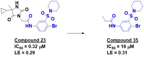
Compound 23 is the most potent molecule disclosed, but the researchers mention a manuscript in preparation that describes further optimization. The compound shows promising selectivity against other bromodomains; it certainly doesn’t look like a classic bromodomain binder. X-ray crystallography revealed that the binding mode is in fact different from other bromodomain ligands. Trimming down compound 23 produced compound 35, which shows reasonable activity and ligand efficiency.

This paper nicely demonstrates the power of biophysics to discern a still small signal in a sea of noise. As the researchers note, PAINS filters and computational approaches would not have worked due to the sheer diversity of the misbehaving compounds. (That said, if the library had been infested with PAINS, the false positive rate would have been even higher!)

The paper is also a good argument for FBLD. Compound 35 is probably too large to really qualify as a fragment, but perhaps related molecules could have led to this series. And GSK also discovered a different series of potent ATAD2 inhibitors from fragments, which Teddy wrote about.

19 August 2019

Fragments in the clinic: Navoximod

A good fragment can be a useful starting point for creative scientists, no matter where it comes from. Indeed, we recently described how fragment-derived molecules discovered at one institution were used to discover a clinical compound at another. A similar story from Mario Mautino and collaborators at NewLink Genetics and Genentech has recently appeared in J. Med. Chem.

The researchers were interested in the protein indoleamine 2,3-dioxygenase 1 (IDO1), whose immunosuppressive properties may allow cancer cells to survive and proliferate. They took their starting point from a 2006 publication of a crystal structure of compound 1 bound to IDO1. Structure-based design led to potent molecules such as compound 11, but (not unexpectedly) these phenol-containing molecules tended to have high clearance.


Abandoning the phenols, the researchers instead began rigidifying the series, leading to modest improvements in potency as exemplified by compound 37. Modeling suggested that fragment growing could be productive, and this was confirmed by compound 46.

IDO1 is a heme-containing enzyme, and in fact the imidazole moiety of compound 1 interacts with the heme iron. Other human heme-dependent enzymes include the CYPs, and since these are often involved in metabolizing drugs, it is important to avoid inadvertently inhibiting them. The researchers spent considerable effort further optimizing their molecules for potency, selectivity against CYPs, and metabolic stability. This is described in extensive detail – it makes for an excellent case study in lead optimization. (The separation of stereoisomers and absolute assignment is an impressive piece of work.) Ultimately they arrived at navoximod (NLG-919 or GDC-0919), which showed activity in mouse xenograft models and suitable properties for oral dosing in humans, and entered the clinic in 2014.

Unfortunately, although several IDO1 inhibitors have entered clinical development, those that have made it to late stage trials have proven disappointingly ineffective. Whether some combination with other drugs or a new biomarker will reinvigorate interest in this target, or whether, like BACE1, a good idea meets an unforgiving reality, remains to be seen. There is still no shortcut to avoid the massively expensive experiment of putting a drug into the clinic to test a therapeutic hypothesis.

12 August 2019

Achieving maximum diversity with minimum size

One theoretical advantage of fragment-based drug discovery is the ability to efficiently explore chemical space: there are vastly fewer possible fragment-sized molecules than lead-sized molecules. That said, even fragment space is daunting; the number of possible molecules with up to 17 non-hydrogen atoms is about three orders of magnitude larger than the largest computational screen. Maximizing diversity is thus a key goal in designing fragment libraries, but how do you actually do this? A new open-access paper in Molecules by Yun Shi and Mark von Itzstein at Griffith University provides a practical new approach.

As the researchers point out, diversity itself can be a slippery concept. Functional diversity (ie, what targets are bound) is important but hard-won knowledge. Physicochemical diversity is by definition limited for fragments. That leaves structural diversity, as defined by “molecular fingerprints.” These can be as simple as the presence or absence of a fluorine atom, or can require complicated calculations involving, say, the distance between a hydrogen bond donor and acceptor in the lowest energy conformation of a molecule. In their paper the researchers focus on “extended-connectivity” fingerprints, which take into consideration the physical connectivity between different types of atoms.

But how can you actually quantify structural diversity? One possibility is by comparing molecules to see how different they are, as used for example in Tanimoto similarity assessments. Each additional molecule would be chosen to be least similar to those in a library. Alternatively, one could consider “richness,” how much of chemical space is covered, by calculating how many unique structural features (such as specific bond connectivities) are represented. Each additional molecule would be chosen to provide as many new molecular fingerprints as possible. Shi and von Itzstein propose a third approach, “true diversity,” that considers the number of unique features as well as their proportional abundances. In other words, a library with a higher true diversity would have a “more even distribution of proportional abundances.” The researchers note that this approach has been used in ecology for decades.

To see how their approach performs, the researchers started with a set of 227,787 commercially available fragments, all of which were roughly rule-of-3-compliant and scrubbed of undesirable functionalities. They also considered a subset of 47,708 fluorine-containing fragments. For both sets, they then assessed structural diversity as a function of increasing fragment library size using Tanimoto similarity, richness, and true diversity, as well as random sampling.

Naturally, as the size of a fragment library rose, the diversity increased. As expected, applying Tanimoto similarity or richness led to greater diversity at a smaller library size than did random sampling. This was even more true for true diversity. Interestingly, true diversity reached a maximum at 8.8% or 15.7% (for the full and fluorinated libraries) and then began to decline. This conceptually makes sense because commercial compounds themselves are unlikely to be truly diverse.

More importantly, just 1% or 2.5% of fragments were sufficient to achieve the same true diversity as the full sets. This corresponds to 2052 fragments for the complete commercial set, the structures of which are provided in the supplementary material. As the researchers note, this is comparable to the size of many commonly used fragment libraries.

The method is computationally inexpensive (it runs on a desktop), and should be a useful tool for both building and curating fragment libraries, real and virtual. Of course, diversity is not everything, and it probably makes sense to include privileged pharmacophores even at the cost of lower diversity. But as Lord Kelvin said, “when you can measure what you are speaking about, and express it in numbers, you know something about it.” This paper provides a quantitative approach for measuring diversity.

05 August 2019

Fragments vs RAS family proteins: A chemical probe

RAS family proteins are considered a holy grail of oncology research. Way back in 2012 we discussed a couple papers disclosing low affinity fragments that bind in a small, shallow, polar pocket found in KRAS, NRAS, and HRAS. At the time we wondered “whether this is a ligandable site on the protein.” Last year we highlighted a paper proving that the site is, in fact, ligandable, as exemplified by the mid-nanomolar molecule Abd-7. A paper just published in Proc. Nat. Acad. Sci. USA by Darryl McConnell and collaborators from Boehringer Ingelheim and Vanderbilt University (including Steve Fesik, who published one of the 2012 reports) describes successful development of another ligand. (See here for a fun animated description set to music.)

Consistent with the “undruggable” reputation of RAS family proteins, a high-throughput screen of 1.7 million compounds failed to find anything useful. In contrast, a library of just 1800 fragments screened using STD NMR and MST identified 16 fragments that bind to an oncogenic mutant form of KRAS, as confirmed by 2-dimensional (HSQC) NMR. A separate HSQC NMR screen of 13,800 fragments identified several dozen more, though all the fragments from both screens have dissociation constants weaker than 1 mM. SAR by catalog led to amine-substituted indoles such as compound 11, which modeling suggested could form a salt bridge to an aspartic acid side chain.


The pocket in which all of these molecules bind, between the so-called switch I and switch II regions of KRAS, is much smaller than typical drug-binding sites, but modeling suggested that fragment growing could pick up an additional hydrogen bond, leading to compound 15. Crystallography confirmed the predicted binding mode of this molecule, and informed additional structure-based design, leading first to compound 18 and ultimately to BI-2852, with low or sub-micromolar affinity for wild-type and mutant KRAS, NRAS, and HRAS as assessed by ITC. The researchers also confirmed that the enantiomer is about 10-fold less potent, thereby providing a control compound. Commendably, the researchers have made BI-2852 and the enantiomer available (for free!) to the research community as a chemical probe.

A crystal structure of KRASG12D bound to BI-2852 (cyan) compared with Abd-7 (magenta) reveals how shallow the pocket is; both molecules are largely surface-exposed. The conformational flexibility of the protein is also interesting: Abd-7 would not be accommodated by the protein conformation bound by BI-2852.

The biology is also quite interesting – and complicated. RAS family proteins behave as molecular switches, cycling between the “on” (GTP-bound) state and the “off” (GDP-bound) state, with these transitions assisted by other proteins. On-state RAS drives cell-proliferation and survival. Molecules that bind at the switch I/II pocket block the transition from off to on, but they also block the transition from on to off. Thus, cellular effects are modest. Moreover, BI-2852 hits all RAS isoforms, which could lead to unacceptable toxicity in animals.

This is a lovely paper, but I do quibble that the promise of the title – “drugging an undruggable pocket on KRAS” – remains to be demonstrated. First, both the biochemical and cell-based potency need to be further improved. As the molecule is already large, gaining this needed potency could come at the cost of physicochemical properties. Indeed, the researchers do not discuss the pharmacokinetics of BI-2852. And finally, as the authors themselves note, they will probably need to improve selectivity to spare one or more wild-type RAS isoforms.

What this work does establish indisputably is that the switch I/II pocket is ligandable, though not without effort, as indicated by the 42 authors. Whether or not the site is actually druggable may require another seven years to determine.