Showing posts with label kinase. Show all posts

27 July 2026

Ligand affinities for isolated proteins vs in cells and cell lysates

Earlier this year we highlighted a study that compared the affinities of kinase inhibitors for isolated proteins vs those same proteins in cells and found differences – sometimes dramatic. A new open-access paper in J. Am. Chem. Soc. by Blake Peterson and colleagues at Ohio State University addresses the same question, and explores insights from cell lysates as well.
 
To measure affinity in cells, the earlier paper used NanoBRET, which relies on a fusion protein consisting of the kinase and a luminescent protein. It is possible that this artificial protein may behave differently from the native protein, so the Peterson group developed the Fluorescent Probe Cellular Binding Assay (FPCBA). This technique makes use of a fluorophore conjugated to a ligand for the protein of interest. The protein of interest is overexpressed, the binding of fluorescent probe causes cells to become fluorescent, and the level of fluorescence can be quantified using flow cytometry. Adding a (non-fluorescent) ligand displaces the fluorescent probe, which is rapidly effluxed, and the loss of fluorescence can be used to calculate the affinity of the ligand.
 
For the sake of brevity I’m skipping lots of important details, such as detailed characterization of efflux and use of a second fluorescent protein to correlate expression of the target, but these are described extensively in the paper. The researchers focus on a fluorescent probe based on dasatinib, an approved drug that binds to dozens of kinases.
 
The researchers compared the affinity of dasatinib and imatinib, another approved drug, to the kinase ABL1 using both FPCBA and NanoBRET. The results were reassuringly similar, though dasatinib bound 3 or 4-fold more tightly in the PBCBA assay than the NanoBRET assay while imatinib bound up to 2-fold less tightly, which might be due to changes in the regulatory mechanism of the ABL1 fusion protein required for NanoBRET.
 
Whole cell assays tend to be lower throughput, so cell lysates are often used instead. For kinases, a common assay makes use of “kinobeads.” Native kinases are captured from cell lysates on beads and quantified using mass spectrometry. An inhibitor blocks binding of the kinase to the bead, and this reduction in binding can be used to calculate the affinity. The researchers measured the affinity of dasatinib and imatinib against a score of kinases using both FPCBA and kinobeads.
 
Of 21 kinases, 14 showed similar affinities (within four-fold) for dasatinib by both FPCBA and kinobeads. Six of the remaining seven showed up to 13-fold tighter binding in FPCBA than kinobeads, which the researchers attribute to the kinases being in a more active state in cells than in lysates. (A similar phenomenon has been seen for the helicase WRN.)  In contrast, the kinase SRC showed 11-fold weaker activity in cells than lysates, which the researchers attribute to autoinhibitory regulation.
 
The situation becomes even more discordant when comparing affinities in cells to the affinities of isolated recombinant kinase domains using the KINOMEscan assay. Since a picture is worth several hundred words (the length of this blog post), I’ve graphed the results. While possible to see a correlation, it is also possible to see dragons in the shapes of clouds.
 
The affinity of dasatinib was generally higher (often by more than 10-fold) in the KINOMEscan than the FPCBA assay, which is not surprising given the lack of ATP in the biochemical assay. But there were exceptions, most notably EPHA4, which binds slightly more tightly to dasatinib in cells than in the KINOMEscan assay. Interestingly, the affinity in the cell lysate assay was lower; the researchers suggest that in cells the kinase is in a more active form than it is in lysates, perhaps due to membrane localization.
 
In the end, this paper is another useful reminder that assays with isolated proteins do not necessarily translate to cells. And of course, activity in cells does not necessarily translate to activity in vivo. Understanding why is part of what makes drug discovery both frustrating and exciting.

09 March 2026

Selectivity in cells may vary

Last year we celebrated the ten-year anniversary of the Chemical Probes Portal. One of the key requirements for a chemical probe is selectivity, which was set to >30-fold vs related targets when the Portal launched in 2015. For enzymes such as kinases, selectivity is often measured in cell-free assays. A new open-access J. Med. Chem. paper by Matthew Robers, Alison Axtman, and collaborators at Promega and University of North Carolina at Chapel Hill suggests that such data don’t necessarily translate to cellular assays.
 
Kinases are one of the most heavily mined classes of targets this century; five of the eight FBLD-derived approved drugs target kinases. With more than 500 in the human proteome, selectivity has long been a focus. One common method for assessing selectivity in cell-free assays is the Eurofins DiscoverX panel, which currently includes more than 450 kinases. Each kinase has a DNA tag and is paired with a promiscuous high affinity binder attached to a solid support. Test compounds are added, and qPCR is used to assess and quantify which kinases are displaced. The competition assay allows determination of dissociation constants.
 
To measure the binding of compounds to kinases in living cells, the researchers turned to the NanoLuc-bioluminescence resonance energy transfer (NanoBRET) assay. This is also a displacement assay that relies on a bivalent molecule containing a kinase ligand and a fluorophore. Kinases are tagged with NanoLuc, which causes luminescence of the fluorophore when it is in close proximity (ie, bound to the kinase). Ligands that bind to the kinase displace the bivalent molecule, decreasing luminescence.
 
The researchers started with four promiscuous kinase inhibitors, two of which (dasatinib and sorafenib) are approved drugs. They ran these against 240 or 300 kinases in the NanoBRET assay and compared the values with published DiscoverX dissociation constants. Most of the compounds were more potent in the DiscoverX assay than in the cell-based assay, and the researchers suggest several possible reasons. First, the DiscoverX assay is run in the absence of the cofactor ATP, which can compete with ligands that bind to the active site. Second, cell (im)permeability could decrease cellular potency. Finally, most of the DiscoverX kinases are truncated, whereas the NanoBRET kinases are full length.
 
For these and other reasons, it is common for compounds to be less active in cell assays than biochemical or biophysical assays. Surprisingly though, for a few kinases the compounds were actually more potent in the cellular assay than they were in the DiscoverX assay.
 
To extend these findings, the researchers tested additional kinase inhibitors and found that three kinases were particularly susceptible to inhibition in cells. One of these kinases, PIP4K2C, was engaged at mid nanomolar potency by cabozantinib, and the researchers suggest this could be useful for immuno-oncology. More worrisome, several approved drugs bind to the tumor suppressor STK11 in cells, raising the potential that these compounds could inhibit exactly the wrong pathway.
 
On the bright side, the researchers find that some molecules that look moderately selective in the DiscoverX assay are actually quite selective in cell assays, and they propose new chemical probes for the little-studied kinases BRSK1/2 as well the kinases DDR1/2.
 
Kinases are certainly not the only class of targets for which compounds’ performance differs outside vs inside cells; we wrote about covalent WRN inhibitors here. This paper is a good reminder that as useful as cell-free assays are, things can go weird once you go into cells – for better or for worse.

14 April 2025

A library of covalent fragments vs a library of kinases

Protein kinases have proven to be a fruitful class of targets, as evidenced by more than 80 FDA-approved drugs, five of which came from fragments. Because all protein kinases bind ATP, selectively inhibiting just one of the more than 500 family members can be challenging. This is a bit easier for the 215 protein kinases that contain a cysteine within the ATP-binding pocket capable of reacting with covalent ligands. In a recent (open access) Angew. Chem. Int. Ed. paper, Matthias Gehringer, Stefan Knapp, and collaborators at Johann Wolfgang Goethe-University and Eberhard Karls University Tübingen provide such starting points for dozens of kinases.
 
The researchers built a small library of 47 fragments consisting of six classic hinge-binding moieties such as pyrazole and azaindole coupled through nine aryl linkers at varying positions to an electrophilic acrylamide warhead. Although most of the compounds are rule-of-three compliant, the researchers note they “reside at the upper end of fragments space,” similar to what we discussed last week. Chemical reactivity towards the abundant cellular thiol glutathione was tested and found to be lower than the approved drug afatinib, meaning the fragments might be good starting points for optimization.
 
Each member of the fragment library was screened against 47 different protein kinases chosen to present cysteine residues at a variety of positions around the ATP binding site. Two types of screens were conducted: intact protein mass spectrometry to assess covalent binding and differential scanning fluorimetry (DSF) to assess protein stabilization. Screens were run at fairly high concentrations, 50 µM protein and 100 µM fragment.
 
The results, plotted as a two-dimensional figure with kinases on one axis and compounds on the other, provide a wealth of information. Some compounds hit multiple kinases while others hit few or none. Similarly, some kinases are hit by multiple compounds while others are recalcitrant.
 
A couple more general observations emerged. First, there was little if any correlation between the inherent reactivity of a given fragment (as assessed by reactivity with glutathione) and the number of kinases hit, suggesting that covalent modification was driven by specific interactions rather than nonspecific reactivity. Second, there was also no clear correlation between the ability of a fragment to stabilize a given kinase and the ability of the same fragment to covalently bind to that kinase. This latter observation isn’t surprising, since one could imagine a fragment binding noncovalently to a kinase and stabilizing it without forming a covalent bond.
 
Most proteins contain multiple cysteine residues, and the researchers confirmed that the fragments were covalently modifying the cysteines in the ATP-binding pocket using mutagenesis, trypsin digestion, or, for MAP2K6, RIOK2, MELK, and ULK1, crystallography. The crystal structures were particularly informative in showing hydrogen bond interactions between the covalently-bound fragments and the hinge region.
 
As we’ve noted, the best metric for characterizing irreversible covalent inhibitors is kinact/KI, and the researchers determined these for covalent inhibitors of PLK1, PLK3, RIOK2, CHEK2, and CSNK1G2. The values ranged from 2 to 8 M-1s-1, comparable to other early covalent fragments.
 
This is a lovely, systematic paper that is in some ways an irreversible complement to a study we wrote about in 2013 focused on reversible covalent kinase inhibitors. The fact that hit rates are relatively high likely reflects the fact that all the fragments contain privileged hinge-binding pharmacophores.
 
Perhaps most importantly, all the data are available in the supporting information. If you’re interested in pursuing any of these 47 kinases, you may find good starting points here.

22 January 2024

Covalent complexities for kinase inhibitors

Covalent drugs are becoming increasingly popular. But as more researchers search for them, they may encounter pitfalls. A new paper in J. Med. Chem. by  David Heppner and collaborators at the State University of New York Buffalo, AssayQuant Technologies, and Eberhard Karls Universität Tübingen provides a nice roadmap for avoiding them.
 
The researchers focus on covalent inhibitors of epidermal growth factor receptor (EGFR), a kinase that is frequently mutated in cancer. The first drugs against this target, such as erlotinib, were non-covalent, and these have been largely displaced by more effective covalent molecules such as afatinib. Unfortunately, these earlier drugs are not effective against a common mutant (T790M), spurring the development of third generation molecules such as osimertinib, which was approved by the FDA in 2015. Osimertinib has been extensively studied, with more than 2800 references in PubMed. Yet it is not as well understood as you might expect.
 
The team uses this system to demonstrate how characterizing irreversible inhibitors is not simple. For reversible enzyme inhibitors, researchers frequently discuss IC50 values or, when they are being more precise, inhibition constants (Ki). The latter are in theory absolute values that do not depend on concentrations of cofactors such as ATP. But for irreversible inhibitors, the IC50 values change depending on how long (and at what concentration) incubation occurs. The proper assessment of an irreversible inhibitor is kinact/KI, which takes into account both the irreversible inactivation step (kinact) as well as the inhibition constant (KI). Note that Ki is not the same as KI ; the former describes only the initial reversible association between protein and inhibitor, while KI incorporates the irreversible step. Told you it was complicated!
 
And it gets worse. The researchers examined three irreversible covalent inhibitors under various conditions. In one condition, the inhibitors were pre-dissolved in 10% DMSO before being added to the assay mixture to give a final DMSO concentration of 1%. In another condition, the inhibitors were dissolved in pure DMSO before being added to the assay. Despite the final concentration of DMSO being the same (1%), the second condition gave kinact/KI values up to 11-times greater (more potent).
 
If subtle experimental variations in one lab can change values by more than an order of magnitude, you might expect the literature to vary even more, and you’d be right. In the case of osimertinib, the reported values of kinact/KI vary by nearly 500-fold. Some of the experimental parameters the researchers consider are concentrations of reducing agents such as DTT, which can react with covalent inhibitors, and serum albumin, which also contains a free cysteine residue. Although these did not seem to be problematic for osimertinib itself, they could affect other molecules.
 
Another consideration for kinases in particular is the concentration of the cofactor ATP. The value of kinact/KI itself will vary depending on [ATP], and the researchers describe how to calculate a “true” kinact/KI which could be used to compare the potency of a given inhibitor against the wild-type vs mutant forms of the enzyme. But while this is more theoretically rigorous, it may be less biologically relevant, since physiological ATP concentrations are less variable than differences in the Michaelis constant (KM) for ATP for different kinases and mutants.
 
There is lots more to digest in this paper, including analyses of structure-kinetic relationships (SKR, akin to structure-activity relationships, or SAR) for different inhibitors and thorough experimental descriptions. The take-home message is that, due in part to different and often incomplete details, “potency measurements are generally difficult to compare among literature studies,” and “any potency assessments should include appropriate controls under the same conditions as the experimental inhibitors.”

20 June 2022

KinaFrag: a free, searchable database of kinase fragments

Four of the six approved fragment-derived drugs are kinase inhibitors, and three of these bind in the active site. Despite these successes, there are plenty of opportunities for new kinase-directed drugs, particularly those targeting cancer resistance mutations. In a recent Brief Bioinform. article, Guang-Fu Yang and colleagues at Central China Normal University describe a new tool to facilitate these discoveries.
 
The researchers started by trawling multiple databases such as kinase.com, DrugBank, ChEMBL, and the Protein Data Bank for kinase inhibitors. The results were combined and collated to yield a set of 7783 kinase-inhibitor fragment complexes, with more than 3000 unique fragments. Most of these bind in the “front cleft” of the active site, where the adenine of ATP normally binds, but several hundred also sit in the so-called back pocket or the intervening area.
 
What’s nice is that all this information is available on a free website called KinaFrag. You can download the structures yourself, but the site can also be browsed or searched. Fragments are annotated with links to various databases; here’s an example.
 
 
There are some bugs. While I was able to search by physicochemical parameters such as molecular weight and number of hydrogen bond donors, I could not get the substructure search to work. I’d be curious as to whether readers could do so.
 
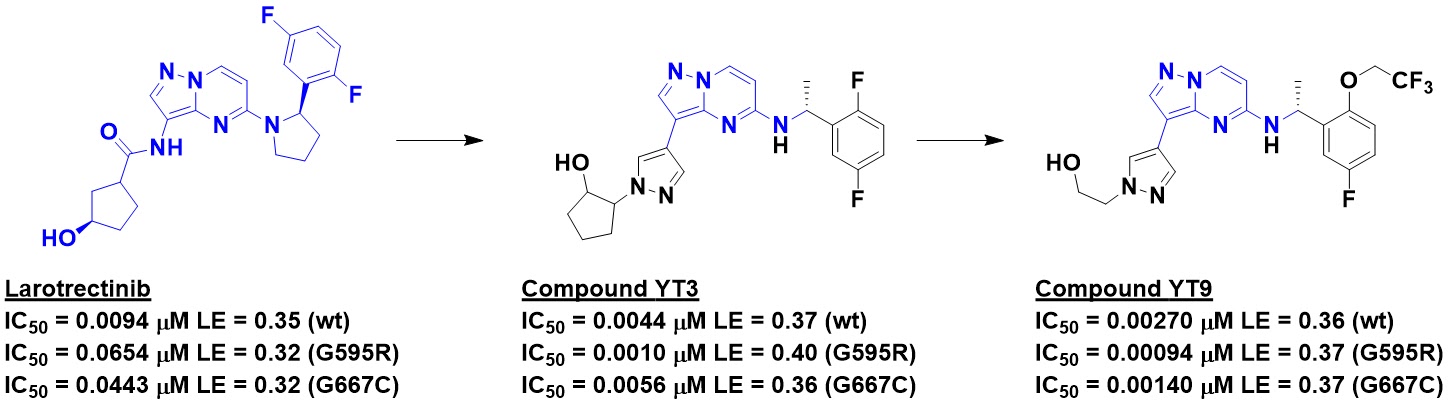
To demonstrate the utility of KinaFrag, the researchers describe a case study in which they started with the anticancer drug larotrectinib, which inhibits TRK family kinases. However, the molecule is less effective against several mutations observed in the clinic. Examining the bound structure revealed that the mutations introduce steric clashes. Retaining the hinge-binding fragment while performing virtual screening of fragments from KinaFrag led to molecules such as YT3, potent against both wild type TRKA and two resistance mutants, and further optimization resulted in YT9. 
 

Not only was YT9 active against the wild type and mutant forms of TRKA, it showed good oral bioavailability and pharmacokinetics in rats. Encouragingly, the molecule slowed tumor growth in both wild type and mutant TRKA mouse xenograft models.
 
One could debate whether this is an example of FBLD; the discovery of YT9 could also be considered a classic case of scaffold hopping. But semantics aside, this is a nice example of thinking in terms of fragmenting molecules. More broadly, KinaFrag looks like a useful tool for work on kinases – especially if the substructure search works.

30 December 2021

Review of 2021 reviews

As the year winds down SARS-CoV-2 continues its relentless drive through Greek letters and the planet. But there is hope: vaccines seem to be holding, for those who have access, and two oral drugs have been granted emergency use authorization by the US FDA, one of which (PF-07321332) is covalent and looks remarkably effective. As is our custom, Practical Fragments ends the year by highlighting conferences and reviews.
 
Conferences started the year online only (CHI’s Sixteenth Annual Fragment-based Drug Discovery), moved to hybrid (CHI’s Nineteenth Annual Discovery on Target) and sadly returned to virtual (Pacifichem 2021).
 
This year produced more than twenty FBLD-related reviews, and these are grouped thematically: NMR and crystallography, computational methods, targets, library design, and covalent fragments. The most general is the sixth installment in a series of annual reviews in J. Med. Chem. covering fragment-to-lead success stories from the previous year. Iwan de Esch (Vrije Universiteit Amsterdam) took the lead (pardon the pun) on the most recent review, which details 21 examples from 2020. In addition to the centerpiece table showing fragment, lead, and key parameters, this open-access paper also includes an analysis on the molecular complexity of fragment hits.
 
NMR and crystallography
Consistent with its central role in FBLD, several reviews covered NMR. Ben Davis (Vernalis), one of the leading practitioners, discusses fragment screening in Methods Mol. Biol. The chapter is written for a non-specialist, so you won’t see detailed pulse sequences. Instead, Ben provides a very accessible and practical guide covering everything from sample preparation through data analysis and validation.
 
A more detailed description of solution NMR in drug discovery by Li Shi and Naixia Zhang (Shanghai Institute of Materia and Medica) is published (open access) in Molecules. With 180 references, this review covers considerable ground, including various ligand-detected and protein-detected methods for screening as well as for hit-to-lead and mechanistic studies. The paper also includes a nice summary of in-cell (!) NMR.
 
The Pacifichem meeting had several talks on fluorine NMR, and speaker Will Pomerantz, together with Caroline Buchholz, has published a thorough, open-access review in RSC Chem. Biol. Will has been a leading developer of protein-observed 19F NMR, so naturally this topic is well-covered, but there is plenty on ligand-observed 19F NMR as well as a good background section and musings on the future of the field.
 
And if you’re looking for a detailed how-to guide for NMR-based fragment screening, Harald Schwalbe and colleagues describe the platform they’ve built at the Center for Biomolecular Magnetic Resonance (BMRZ) at Johann Wolfgang Goethe-University Frankfurt in J. Vis. Exp. This open-access paper also describes quality control experiments of the iNEXT library, which we’ve discussed here.
 
Switching gears to crystallography, J. Vis. Exp. carries a paper by Frank von Delft and collaborators describing the XChem platform at the Diamond Light Source. This high-throughput fragment screening platform has delivered a 95% success rate on more than 150 screens, with hit rates varying from 1-30%. In addition to technical details, this open-access article also provides tips on successfully getting your screening proposal through peer review.
 
XChem has inspired similar efforts at other synchrotrons, including the Fast Fragment and Compound Screening (FFCS) platform at the Swiss Light Source. This is concisely described by May Sharpe and Justyna Wojdyla in Nihon Kessho Gakkaishi (open-access and published in English).
 
Private companies are also moving into high-throughput crystallography. Debanu Das and collaborators describe the platform at Accelero Biostructures, which is capable of screening ~500 fragments in two days. Screens against three nucleases are described in some detail in an open-access article in Prog. Biophys. Mol. Biol.; these and other components of the DNA damage response are the focus of XPose Therapeutics, Accelero’s sister company.
 
Computational methods
In addition to the experimental methods reviewed above, a couple papers describe computational approaches. In Drug Disc. Today: Tech., FragNet alum Moira Rachman and collaborators from UCSF, Universitat de Barcelona, and elsewhere focus on “fragment-to-lead tailored in silico design.” This is a nice review of the recent literature and emphasizes the fact that much of the heavy design lifting is still done by medicinal chemists – at least for now.
 
Predicting the energies of modified fragments has long been a challenge, and one promising approach is free energy perturbation, in which one ligand is “perturbed” into another and the relative energy differences calculated. Barbara Zarzycka and colleagues at Vrije Universiteit Amsterdam provide a concise review for aficionados in Drug Disc. Today: Tech.
 
Targets
Three reviews cover applications of FBLD to various target classes. Kinases have been particularly successful, with four of the six approved fragment-derived drugs targeting these enzymes. In Trends Pharm. Sci., Ge-Fei Hao and collaborators, mostly at Central China Normal University, review the state of the art. In addition to background and several case studies, the paper includes a nice table showing structures and summaries of clinical-stage kinase inhibitors.
 
Epigenetics has been another fruitful area, and in J. Med. Chem. Miguel Vaidergorn, Flavio da Silva Emery (both University of São Paulo) and Ganesan (University of East Anglia) detail the “successful union of epigenetic and fragment based drug discovery (EPIDD + FBDD).” This thorough summary (with 165 structures!) of the literature is particularly detailed when it comes to bromodomains, four inhibitors of which have entered the clinic with the help of fragments. The researchers point out that EPIDD and FBDD both began around the same time, and in fact the oncology drug vorinostat could be described as “a unique case of solvent-based drug discovery.”
 
RNA has long been a target of FBLD, and in ChemMedChem Mads Clausen (Technical University of Denmark) and collaborators review the state of the art. The various established and emerging methods to find fragment hits are covered in depth, and there is also a nice discussion as to whether RNA-focused fragment libraries will be useful.
 
Library design and molecular properties
In Expert Opin. Drug Discov. Zenon Konteatis (Agios) asks “what makes a good fragment in fragment-based drug discovery?” His answers provide a concise summary touching on the rule of three, molecular complexity, “three-dimensionality”, and other topics.
 
The topic of three-dimensional fragments is covered in several other reviews. In Drug Disc. Today: Tech., Iwan de Esch and collaborators at Vrije Universiteit Amsterdam and University of York assess 25 so-called 3D libraries reported in the literature, mostly since 2015. The researchers manually drew all 897 fragments so they could calculate various properties. While most of the molecules are rule-of-three compliant, just under half could be called 3D by both plane of best fit (PBF) and principal moment of inertia (PMI). PBF and PMI measurements correlated with one another, while Fsp3 correlated with neither measurement, leading to the conclusion that “Fsp3 is a poor measure of 3D shape.”
 
Shapely or not, sp3-rich fragments are interesting from a diversity point of view, and in Chem. Sci. Max Caplin and Dan Foley (University of Canterbury) discuss synthetic methods for advancing these. This is an excellent open-access review of the recent literature around C-H bond functionalization and well worth reading for the chemists in the audience.
 
3D fragments are often chiral, and the importance of chirality in drug discovery is the focus of a paper in ACS Med. Chem. Lett. by Ilaria Silvestri and Paul Colbon (University of Liverpool). The researchers note an opportunity for chemical suppliers: only 245 of 9751 heterocyclic building blocks offered by Sigma-Aldrich are chirally pure.
 
“Library design strategies to accelerate fragment-based drug discovery” is the topic of a Chem. Eur. J. review by Nikolaj Troelsen and Mads Clausen (Technical University of Denmark). The researchers provide a highly accessible overview of different libraries appropriate for different fragment-finding methods, including covalent approaches.
 
Covalent fragments
This year saw the approval of sotorasib, the first covalent fragment-derived drug, so it is no surprise that several papers focus on this topic. Sara Buhrlage, Jarrod Marto, and colleagues at Dana-Farber Cancer Institute provide a thorough introduction to “chemoproteomic methods for covalent drug discovery” in Chem. Soc. Rev. The review covers both isolated protein screening as well as proteome-wide methods and includes multiple case studies.
 
Nir London and colleagues at The Weizmann Institute of Science focus on “covalent fragment screening” in Ann. Rep. Med. Chem. This is an excellent review of the recent literature and also includes an analysis of six commercial covalent fragment libraries.
 
And finally, in RSC Chem. Biol. (open access), Nathanael Gray and collaborators mostly at Dana-Farber Cancer Institute discuss strategies for “fragment-based covalent ligand discovery”, including computer-aided approaches, as well as target classes and new modalities such as PROTACs. They end by asking whether sotorasib was “a lucky, one-off case” or “a preview of continued and increased impacts that these approaches will have on drug discovery as the improved methods, larger libraries, and increased focus start to bear fruit.”
 
I’m betting on the latter.
 
And that’s it for 2021. Thanks for reading, special thanks for commenting, and here’s hoping we’ll be able to meet in person in 2022.

23 August 2021

Fragments vs DYRK1A and DYRK1B: Part 2

Last week we highlighted work out of Vernalis and Servier in which fragment-based methods were used to identify potent and selective inhibitors of DYRK1A and 1B, potential targets for cancer and neurodegenerative diseases. The NMR screens yielded 166 hits, only one of which was advanced in that paper. A second J. Med. Chem. paper by Andras Kotschy and collaborators describes the optimization of another fragment.
 
Compound 1 is a whoppingly potent fragment with impressive ligand efficiency. If you’ve ever worked on kinases you probably think you know how it binds, as the diaminopyrimidine moiety is a common hinge-binding motif. In fact, crystallography revealed that the molecule binds in a completely different orientation and that the methoxy group makes a single hydrogen bond to the hinge amide NH. Cyclizing the molecule led to compound 10, with a satisfying boost in affinity.

 
Unfortunately, compound 10 was also a potent inhibitor of the kinase CKD9. To gain selectivity, the researchers took advantage of the fact that one of the backbone carbonyl oxygens in the hinge adopts an unusual orientation in DYRK1A, making room for the methyl group in compound 33. Next, the researchers replaced the benzofuran core for reasons of “synthetic tractability, metabolic stability, and freedom to operate.” This exercise ultimately led to compound 40.
 
This compound was profiled against 442 kinases and found to be quite selective, with only 8 kinases significantly inhibited at 1 µM. One of these was the related kinase DYRK2, but further growing led to selective compound 58. An overlay of the initial fragment (blue) with compound 58 (gray) reveals how the binding mode has been maintained, in contrast to the series described last week.

Compound 40 had only modest antiproliferative activity against human cancer cell lines that were grown in 2D culture but was more active when the cells were grown in 3D culture. The molecule had good oral bioavailability in mice, and xenograft studies revealed that it inhibited tumor growth, though it was also toxic at higher doses. The researchers do not mention brain penetration, though given the number of hydrogen bond donors I would be surprised if it crosses the blood-brain barrier.
 
This paper is a nice example of how getting high affinity is often only the beginning of a long journey. In combination with the story from last week it is also a useful reminder of how many starting points a single fragment screen can provide: just two fragments led to two completely independent series. Whether molecules from these series advance to the clinic, they provide useful tools to further understand the biology of DYRK1A.

16 August 2021

Fragments vs DYRK1A and DYRK1B: Part 1

The dual-specificity tyrosine-phosphorylation-regulated kinases 1A and 1B (DYRK1A and DYRK1B) belong to a family of five serine/threonine kinases implicated in several cancers as well as Down’s syndrome and other neurodegenerative diseases. For the latter indications in particular, brain penetration would be essential for any inhibitor, just as in the LRRK2 story last week. In a new (open access) J. Med. Chem. paper, Rod Hubbard and collaborators at Vernalis and Servier describe the discovery of a chemical probe.
 
The researchers started by testing their in-house library of 1063 fragments in pools of six, each at 500 µM, in three ligand-detected NMR screens. This resulted in a whopping 166 hits. Crystal structures of the eight most ligand-efficient fragments bound to DYRK1A were obtained, including compound 5. Fragment growing led to compound 16, which bound the kinase 200-fold more tightly. 
 

The crystal structure of compound 16 bound to DYRK1A was compared to structures of other known ligands and suggested the possibility for an alternative binding mode. This led to the synthesis of compound 24, with low nanomolar affinity against both DYRK1A and DYRK1B (only the former is shown in the figure). This compound turned out to be surprisingly unstable in slightly acidic aqueous solution (below pH 5), but replacing the oxygen with a nitrogen fixed this, and further tweaking ultimately led to compound 34.
 
Compound 34 was profiled at 1 µM against a panel of 442 kinases and found to be fairly selective, with only 15 kinases inhibited by at least 50%. It is orally bioavailable in mice, brain penetrant, and inhibited the proliferation of glioblastoma cells, although the potency was significantly attenuated by serum. In a xenograft study the compound caused tumor growth delays and was well-tolerated.
 
This is a nice example of fragment-based lead discovery heavily dependent on structural information. Comparing the binding mode of compound 34 (gray) with that of compound 5 (light blue) reveals the significant shift in binding mode of the initial fragment.

The paper is also a useful reminder of how long it can take for industry research to be published. Work began in 2009, and Rod presented some of it at the CHI FBDD conference in 2019. But this is not the end of the DYRK1A story: stay tuned for next week!

09 August 2021

Fragments vs LRRK2 with the help of a surrogate and a magic methyl

Leucine-rich repeat kinase 2 (LRRK2) has been implicated in Parkinson’s disease and has thus long been targeted by drug hunters. Dozens of inhibitors have been approved for other kinases, making the protein class appear “easy”, but LRRK2 is particularly challenging. First, it is a large multidomain protein that has resisted crystallography. Second, an inhibitor for a chronic disease such as Parkinson’s will need to be highly selective. Finally, the fact that LRRK2 is in the brain means that inhibitors will need to cross the treacherous blood-brain barrier (BBB), whose function is to exclude anything unusual. A recent J. Med. Chem. paper by Douglas Williamson and collaborators at Vernalis and Lundbeck addresses the first two of these issues.
 
The researchers started by screening 1313 fragments (at 200 µM each) against the disease-relevant G2019S mutant of LRRK2. The screen was run using the DiscoveRx KINOMEscan, which relies on displacement of a kinase from an immobilized ligand. Some 80 hits were then triaged in a kinase activity assay.
 
Rather than banging their heads against the wall that has blocked X-ray structures of LRRK2, the researchers turned to a crystallographic surrogate. The readily crystallizable kinase CHK1 has some similarity to LRRK2, and some inhibitors bind to both kinases. Introducing ten mutations into CHK1 around the ATP-binding site led to a LRRK2 surrogate – an approach we’ve previously mentioned.
 
Among the fragment hits were adenine (compound 7) and two closely related molecules. Crystallization of compound 7 with wild-type CHK1 and the LRRK2 surrogate revealed two different binding modes. Similarity-based searches of in-house and literature compounds led to molecules with nanomolar potency, and subsequent optimization led to compound 17 (all molecules were tested against both wild-type LRRK2 as well as the G2019S and had similar affinities).
 

Interestingly, crystal structures of these molecules revealed them to bind more similarly to the complex of compound 7 bound to wild-type CHK1 rather than the surrogate. Adding a methyl group to compound 17 led to compound 18 with a satisfying 100-fold boost in potency: a true magic methyl, as the other enantiomer had slightly worse affinity than compound 17. Crystallography with the surrogate revealed hydrophobic interactions between the methyl and an alanine side chain. It is worth noting that compound 18 is still fragment-sized and yet has high picomolar affinity for LRRK2.
 
Extensive medicinal chemistry followed, ultimately leading to compound 45. Profiling against 468 kinases in the KINOMEscan assay demonstrated it was quite selective, binding only three other targets with dissociation constants less than 1 µM. The compound was active in cells containing either wild-type or G2019S LRRK2. Unfortunately, while the compound showed good oral bioavailability in dogs, it was not orally bioavailable in rats. Moreover, brain to plasma ratios were low in mice, and the molecule was a substrate for the human protein BCRP, a transporter that pumps small molecules across the BBB.
 
Although these liabilities halted further work on the series, this is nonetheless a nice fragment to lead story that highlights the utility of crystallographic surrogates. But the different binding modes for the initial fragment are a reminder that multiple binding modes are not uncommon, and it is best to employ crystallography not just early but often, when you can.

12 April 2021

Fragment merging on c-MET

Fragment-based inhibitors of kinases are legion, particularly those that bind in the so-called hinge region where the adenine of ATP normally sits. However, even among these there are many different flavors of inhibitors. In particular, about 10 kinases can adopt a “folded P-loop” conformation, in which the phosphate-binding loop collapses into the ATP binding site. This was the focus of a recent open-access paper in ACS Med. Chem. Lett. by Gavin Collie and colleagues at AstraZeneca.
 
The researchers were interested in the oncology target c-MET. A ligand-based NMR screen of 1150 fragments (in pools of 6 at 200 µM each) yielded a 6% hit rate, of which 20 confirmed by SPR. Crystallography was attempted unsuccessfully on most of these, but compound 1 was found to snuggle into the active site with the protein in the folded P-loop conformation.
 
A computational similarity search of AstraZeneca’s internal library identified compound 2, which crystallography revealed to bind in a similar manner, with two hydrogen bonds to the hinge region and the benzyl group buried in a hydrophobic pocket. A second similarity search of the library – this time based on compound 2 – identified compound 3. Crystallography confirmed that the core azaindole moieties of compounds 2 and 3 overlay, and thus fragment merging was attempted.
 

The resulting compound 5 bound as expected. This prompted yet another computational search of the internal library, and after a bit of medicinal chemistry compound 7 was identified as a mid-nanomolar inhibitor with low micromolar cell-based activity. Crystallography revealed that it too binds to the folded P-loop conformation of c-MET.
 
Because the folded P-loop conformation is rare among kinases, the researchers hoped that the resulting molecule would be selective. Unfortunately, when profiled against a panel of 140 kinases at the low concentration of 100 nM, 27 of them were inhibited by at least 60%. This is perhaps not surprising given the 7-azaindole core, which has been found to bind to more than 90 kinases, though some compounds containing this moiety are selective.
 
Nonetheless, this paper is a nice example of structure-guided fragment merging. A cynic could point out that had the researchers screened the entire AstraZeneca compound collection they likely would have identified molecules very similar to compound 7 anyway, but this may have cost more and would not be an option at smaller organizations without million-compound libraries. And the approach is useful for more difficult targets for which high-affinity molecules may not exist – yet.

01 March 2021

Fragments vs MEK1: allosteric binders

MEK1 is a central player in the MAP kinase signaling cascade, which is often dysregulated in cancer. As such the enzyme has been the focus of considerable research and the target of four approved drugs. Interestingly, these drugs bind not to the hinge region targeted by most kinase inhibitors but rather to an allosteric pocket adjacent to the ATP binding site. The drugs also look somewhat alike. Seeking something completely different, Paolo Di Fruscia, Fredrik Edfeldt, Helena Käck, and colleagues at AstraZeneca turned to fragments. They have recently published their results in ACS Med. Chem. Lett.
 
As we discussed in 2016, the AstraZeneca fragment library is quite large at 15,000 molecules. The researchers used a computational screen to narrow this down to a more manageable 1000 compounds for ligand-detected NMR screening. AMP-PNP, a nonhydrolyzable version of ATP, was included to block the hinge region, biasing the screen for fragments that bind the allosteric site. (See here for earlier work looking for ATP-competitive molecules.) A total of 142 fragments were identified and further characterized by SPR, and 46 showed dissociation constants better than 1 mM and similar affinities in both the presence and absence of AMP-PNP, suggesting they do indeed bind in the allosteric site.
 
Crystallography was attempted on all the fragments, but only two produced structures. Reassuringly, both bound in the allosteric site. But with only limited structural information, the researchers tested analogs of the fragment hits within their corporate collection. This identified compound 10, which is more potent than initial fragment 3. Moreover, compound 10 lends itself well to library synthesis.
 

All library members were initially made and tested as racemates. When the two enantiomers of the best hit were separated, compound 23 was found to be a sub-micromolar binder, roughly 100-fold better than the other enantiomer. At this point the researchers finally obtained a crystal structure of compound 23, confirming that it did bind in the allosteric pocket. Compound 23 is also still fragment-sized, just three heavy atoms larger than compound 3.
 
The astute reader will notice that the word “inhibitor” has not appeared until now, and indeed despite the encouraging affinity no mention is made in the paper of inhibition – a rather important feature! At a conference in 2019 Paolo did describe further optimization to a functional molecule, so hopefully we will see a second publication detailing this work.
 
Like the NPBWR1 story last month, this is another nice example of advancing fragments in the absence of structural information. It is also a good case study of fragments yielding completely different chemical matter in a crowded field.

30 November 2020

Bioisosterism surprises

The concept of bioisosterism is central to medicinal chemistry. Essentially, one functional group is replaced by another which has similar activity but a different chemical structure. This might be done for a variety of reasons: improving pharmaceutical properties, enabling new analogs, or inventing around existing intellectual property. Most medicinal chemists are familiar with common bioisosteres, such as replacing a carboxylic acid with an acyl sulfonamide. But what about replacing a carboxylic acid with an amidine? This and other surprising examples are provided in a new Angew. Chem. paper by Gerhard Klebe and colleagues at Philipps Universität Marburg.
 
The researchers focused on fragments binding to the hinge region of protein kinase A (PKA), a well-characterized and easily crystallized kinase. As we noted a couple weeks ago, most kinase inhibitors bind to the so-called hinge region, where the adenine ring of ATP normally sits. Protein backbone amides typically make one to three hydrogen bonds with inhibitors. The researchers chose 19 simple fragments, each containing an aromatic ring and various substituents, soaked these into crystals of PKA, and obtained high-resolution (between 1.12 and 1.82 Å) structures. They also experimentally measured the pKa values of each fragment.
 
All except two of the fragments made one or two hydrogen bonds to a backbone amide NH and/or carbonyl oxygen, but the moieties that did so varied dramatically. Benzamide, with its hydrogen bond accepting carbonyl oxygen and hydrogen bond donating primary amide, is a quintessential hinge-binder, but surprisingly benzoic acid bound in a similar fashion. The measured pKa of this carboxylic acid is 4.01, yet the acid serves as a hydrogen bond donor, suggesting that it is protonated in the active site of the enzyme.
 
On the other end of the acidity spectrum, a substituted benzamidine fragment with a pKa of 10.78 bound in the neutral form, with a normally charged nitrogen atom serving as a hydrogen bond acceptor. In fact, the binding mode it assumes is identical to that of benzoic acid.
 
These and several other examples illustrate that protonation states of ligands in active sites can be very different from what one would predict based on calculated or even measured pKa values. There are of course limits: an amidine with a measured pKa of 11.32 avoids the hinge and instead interacts with an aspartic acid side chain.
 
One quibble is that the researchers did not seem to consider hydrogens on carbon atoms as potential acceptors; these are increasingly recognized as important, including in kinases. One pyridine fragment shown may have a CH in close proximity to a carbonyl, but it is difficult to tell from the figures, and the coordinates have not yet been released.
 
Another omission is the lack of quantitative information about binding energies. Just because benzoic acid and a benzamidine bind identically does not mean they have the same affinities. That said, Gerhard Klebe warned last year of the dangers of putting too much stock in thermodynamic measurements.
 
These issues aside, this is a nice analysis and should serve as a useful reminder to medicinal chemists that bioisoteres can be quite unexpected. And once the structures are released in the pdb, they will provide a useful resource for modelers seeking to recapitulate crystallographic data.

16 November 2020

Kinase fragments galore: a free virtual collection

Kinases hold a special place in fragment-based drug discovery. Vemurafenib, the first approved FBDD-derived drug, targets a kinase, as do more than a third of fragment-derived drugs to enter the clinic. These efforts have produced a wealth of knowledge, and in a new paper in J. Chem Inf. Mod. Andrea Volkamer and collaborators at Universitätsmedizin Berlin and Bayer have extracted thousands of virtual fragments and made them freely available in a database called KinFragLib.
 
The researchers started with a prior database called KLIFS, which compiles thousands of crystal structures of kinases bound to small molecules. Kinase inhibitor binding modes are classified into several  types, and to keep things simple the focus here was on Type I and Type I1/2. Both bind to the active, DFG-in form of the kinase, the only difference being that Type I1/2 binders extend into a back pocket. A total of 2801 crystal structures were selected for analysis.
 
Next, the ligands were computationally fragmented using a methodology called BRICS (Breaking of Retrosynthetically Interesting Chemical Substructures). Molecules such as ATP and other substrate analogs were discarded to keep the focus on drug-like compounds, and some particularly large, complex molecules such as staurosporine could not be fragmented. The researchers were particularly interested in molecules that bind to the so-called hinge region, where the adenine moiety of ATP binds, so the few ligands that did not bind here were also removed. This reduced the total number of structures to 2553, which yielded 7486 fragments.
 
The kinase active site was divided into six sub-sites: the adenine pocket, solvent-exposed pocket, front pocket, gate area, and two back pockets. Each of the fragments was then assigned to one sub-pocket. More than 80% of the original (unfragmented) ligands bound to two or three sub-pockets, while another 13% bound to four sub-pockets. Just 5% of the original ligands bound only to the adenine sub-site, but these 127 ligands – with an average of 15 non-hydrogen atoms – could be quite interesting as crystallographically validated fragments.
 
Various analyses of the fragments binding in each of the sub-pockets reveal trends. Those binding in the adenine sub-pocket tend towards more hydrogen bond donors and acceptors than those in other pockets, as expected. The shapeliness of fragments binding in the various sub-pockets is not quantitatively analyzed, though the interested reader could run these calculations. The 50 most common fragments for each sub-pocket are presented as figures in the supporting information.
 
Aside from extracting interesting cheminformatic trends, what else can you do with these fragments? The researchers took a subset of 624 rule-of-three compliant fragments and recombined them to generate 6,720,637 distinct molecules. The vast majority of these appear to be novel, and among the 218 that had previously been reported in ChEMBL, more than 20% were potent (IC50 ≤ 500 nM) kinase inhibitors.
 
With the inexorable increase in docking speeds, this virtual collection of fragments will be useful for building even larger libraries and using them to find ligands for new kinases. And, as the researchers point out, the collection could be useful for fragment growing or merging to new experimentally identified fragments. This is a resource that should be broadly useful for the community.

10 August 2020

A fragment library designed for merging: application to PKCζ

Advancing fragments in the absence of structural information has a reputation for being so challenging that some people do not even attempt it. Modeling can help, but what if you could improve your odds by designing your library strategically? This approach has been demonstrated in a recent J. Med. Chem. paper by Masakazu Atobe and colleagues at Asahi Kasei Pharma.

To facilitate fragment merging, the researchers synthesized a library of 5000 substituted isoquinoline fragments. As illustrated by the drug fasudil, isoquinoline is a privileged pharmacophore for binding to the hinge region of kinases. Importantly, isoquinoline has 7 different positions from which to grow: screening monosubstituted versions would potentially allow rapid merging of hits. This approach is conceptually similar to that used to discover vemurafenib.

The target of interest was protein kinase C ζ (PKCζ – that’s a zeta, by the way), one of the 11 members of the PKC family that has been implicated in diseases ranging from diabetes to cancer. Previously reported inhibitors are insufficiently potent or selective, in part because no crystal structure of the kinase has been reported. The researchers were interested in developing a chemical probe to better understand the biology.

A biochemical screen of the 5000-member isoquinoline library at 100 µM yielded just a dozen hits, with IC50 values ranging from low to mid-micromolar. Importantly, substituents were found at four different positions, thus facilitating fragment merging. The researchers first merged fragment 6 with fragment 8, resulting in mid nanomolar inhibitor 10. Further optimization yielded compound 21, which is highly selective for PKCζ in a panel of 216 kinases and also has good pharmacokinetic properties in mice. However, cell potency is relatively modest.

Next, the researchers merged fragment 7 with fragment 9 to generate sub-nanomolar compound 26. This molecule also inhibited protein kinase A, but further optimization led to compound 37, which showed excellent selectivity in a panel of 381 kinases as well as good mouse pharmacokinetic properties and mid-nanomolar activity in a cellular assay. Encouragingly, the compound also showed good activity in a collagen-induced arthritis mouse model. The aniline – which adds about ten-fold to the affinity – may ultimately need to be removed, but clearly this molecule is well-suited for further optimization. 

This paper provides two lovely examples of fragment merging by design, but how general is the approach? One of the key advantages of fragment-based screening is the ability to survey huge swaths of chemical space. Building an entire library around a single fragment obviously constricts this. The fact that the hit rate (0.24%) was so low perhaps illustrates this point; it would be interesting to know how ligandable PKCζ is, or whether a library built around a different privileged pharmacophore would yield a higher hit rate. Lower expected hit rates necessitate larger libraries; 5000 fragments is already more than average according to our poll. And of course, if you are going to build a library of thousands of similar fragments, you had better be certain you choose one that has good pharmaceutical properties, further limiting your choices. Despite all these cavaeats, clearly the investment paid off for PKCζ. It will be fun to see what else comes out of this effort.

03 February 2020

Fragments vs RIP2: from flat fragment to shapely selectivity

Last week we highlighted the utility of shapely fragments. However, as the latest review of fragment-to-lead success stories again shows, starting with a “flat” fragment does not condemn a lead to flatland. This is illustrated in a recent J. Med. Chem. publication by Adam Charnley and colleagues at GlaxoSmithKline.

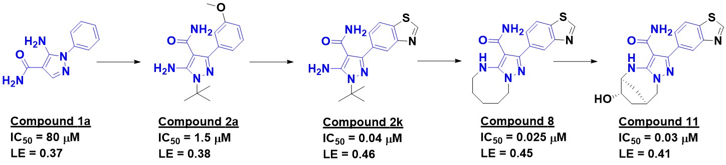
The researchers were interested in receptor interacting protein 2 kinase (RIP2), which is implicated in various inflammatory diseases. A fluorescence polarization screen of 1000 fragments at 400 µM yielded 49 hits with inhibition constants ranging from 5-500 µM. Thirty of these confirmed in a thermal shift assay, and 20 were characterized crystallographically bound to the enzyme. Hit-to-lead chemistry was pursued for five series; the most successful started with compound 1a.


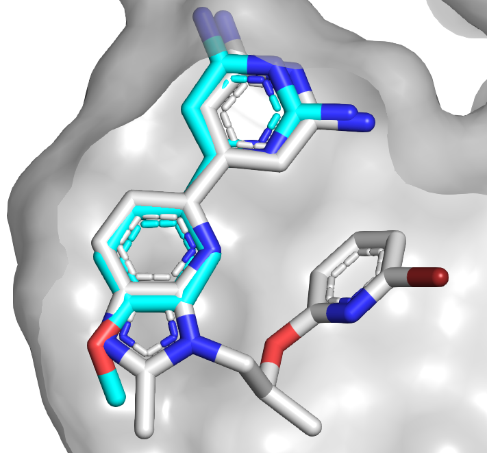
The crystal structure revealed that the carboxamide of compound 1a makes interactions with the hinge region of the kinase, with the phenyl group in the back pocket. A search of related molecules available in-house led to compound 2a, with a satisfying boost in potency. Interestingly, the crystal structure of this molecule bound to RIP2 revealed that the binding mode of the pyrazole moiety had flipped to keep the phenyl ring in the back pocket (compound 1a in cyan, 2a in gray). Enlarging the phenyl group to better fill the pocket led to compound 2k.


This molecule had relatively poor selectivity against several other kinases, but introducing a ring as in compound 8 improved the situation. Crystallography suggested that installing a bridged ring would pick up further interactions with the protein, and although the resulting molecule did not have better affinity, selectivity improved. Finally, a hydroxyl group was introduced (compound 11) to try to pick up interactions with a non-conserved serine residue. This addition did not improve biochemical activity, and in fact a crystal structure revealed that the hydroxyl group was pointing towards solvent, but the activity in human whole blood improved. Importantly, compound 11 was remarkably selective for RIP2: just 1 of 366 other kinases tested at 1 µM showed >70% inhibition.

This is a lovely fragment-to-lead success story that reiterates several important lessons. First, a generic (in this case commercial) and nonselective fragment can lead to novel, selective series. Second, as has been seen multiple times, fragment binding modes can flip unexpectedly, especially during early optimization. Finally, despite the relative flatness of fragment 1a (Fsp3 = 0, though the two aromatic rings are slightly twisted), it could be optimized to a more shapely lead, and the increased complexity is likely responsible for the impressive selectivity. Left unreported is the stability and pharmacokinetics of compound 11: the hydroxyl and all those sp3-hybridized carbons are likely metabolic hotspots. As is so often the case in lead discovery, what solves one problem can too often create another.

27 May 2019

Fragments vs PKC-ι: A*STAR’s second series

Just over a year ago we highlighted work out of A*STAR describing a series of inhibitors for the cancer target protein kinase C iota (PKC-ι). We ended by mentioning that the group had a second undisclosed series. This has now been described in ACS Med. Chem. Lett. by Jacek Kwiatkowski, Alvin Hung, and colleagues.

Compound 1 was among the fragment hits from the high-concentration biochemical screen previously mentioned. Although the researchers did not have a crystal structure, they assumed that the aminopyridine moiety was acting as a hinge binder, which helped them produce a computational model. A simple replacement of the phenyl ring with a pyridyl ring led to compound 2, with a satisfying improvement in potency and ligand efficiency.


As it turned out lots of diverse moieties could be substituted in place of the phenyl, including indoles and phenols. This promiscuity led the researchers to propose that the added heteroatom was making a water-mediated hydrogen bond to the protein; the water could rotate to either accept or donate a hydrogen bond to the ligand. Unfortunately, further growing from this ring did not improve potency.

Returning to their model, the researchers sought to grow from the aminopyridine ring towards a hydrophobic region of the protein. Adding a phenyl group (compound 16) was tolerated, though did not improve the affinity. However, the model suggested that an aspartic acid might be accessible from the phenyl ring, and indeed adding a positively charged piperazine as in compound 19 led to a nearly 100-fold boost in affinity. Unfortunately, the compound’s permeability (measured in a Caco-2 assay) was low, and perhaps because of this it showed only weak antiproliferative activity against hepatocellular carcinoma cells.

Ultimately the researchers were able to solve the cocrystal structure of compound 19 with PKC-ι, which mostly confirmed the model: the aminopyridine interacts with the hinge region, and the second pyridyl moiety likely makes a water-mediated hydrogen-bond with the protein, although the low resolution of the structure makes this somewhat ambiguous. The added piperazine appears to interact with a different aspartic acid than the one targeted.

Although there is more work to be done, it is notable that the researchers were able to optimize a fairly weak fragment to a sub-micromolar compound in the absence of experimental structural information. As they note, “while the empirical SAR remained our ultimate guide in fragment optimization, the model aided the successful design of potent inhibitors.” This is another nice example supporting our 2017 poll results, and recent review, that drug hunters can successfully advance fragments without NMR or crystallography.

18 March 2019

Better properties from fragments: c-Abl kinase activators

Last year we described the discovery of asciminib, an allosteric inhibitor of the kinase BCR-Abl that binds in the enzyme’s myristoyl-binding pocket. As we also highlighted nearly a decade ago, molecules that bind in this pocket can either inhibit or activate the enzyme. Although inhibitors have the most obvious therapeutic potential as anti-cancer agents, activators of the ubiquitously expressed c-Abl protein could potentially treat chemotherapy-induced neutropenia. In a recent J. Med. Chem. paper, Sophie Bertrand and coworkers at GlaxoSmithKline describe their efforts in this area.

The researchers started with a high-throughput screen of 1.3 million compounds. Among the hits was fragment-sized compound 2, which showed good binding and activation in biochemical assays but only modest activity in cells. Building off the left side of the molecule improved biochemical potency, but cell activity still lagged. SAR studies on the dichlorophenyl moiety suggested that this hydrophobic group was probably optimal, and a crystal structure of an analog bound to the enzyme confirmed this. Replacing the central thiazole with other aromatic rings also did little to improve cell activity.

The researchers acknowledge “that the chemistry strategy was largely pursuing compounds with rather poor physical properties,” notably low solubility, high lipophilicity, and high aromatic character. As co-author Robert Young has noted previously, physical properties matter. Happily, a fragment screen identified compound 28.


Adding the acetyl group from the HTS hit generated compound 29, with improved activity compared to the fragment. Moreover, this molecule had better solubility and permeability compared to the more lipohilic, thiazole-containing compound 2. Compound 29 also showed significantly improved activation of c-Abl in a cellular assay. Crystallography revealed that it bound in a similar fashion as compound 2, but with a twisted, more “three-dimensional” shape.

Further optimization, in part informed by previous work done on the thiazole series, ultimately led to compound 52, the most active compound synthesized. Another molecule in the pyrazoline series showed good pharmacokinetic properties in mice. Unfortunately, in vivo efficacy studies had to be halted early due to unexpected (and not clearly understood) toxicity.

This paper nicely illustrates several points. First, the power of fragment-assisted drug discovery, in which information from both HTS and FBLD is combined for lead optimization. Second, the inherently fuzzy line between FBLD and other discovery approaches: had compound 28 been tested in the HTS collection, it likely would have been a hit. Third, the importance of physicochemical properties. And finally, the inadequacy of potency and physicochemical properties alone to produce a developable compound. You can optimize your molecule to the best of your ability but still be sideswiped by nasty surprises such as toxicity. It is helpful to be clever in drug discovery, but you need to be lucky too.