Showing posts with label WaterLOGSY. Show all posts

29 July 2019

SAR by WaterLOGSY?

Among ligand-based NMR methods, WaterLOGSY is nearly as popular as STD NMR. Normally the information obtained is limited: does a given small molecule bind to a protein or not? In a new paper in J. Enzyme Inhib. Med. Chem., Isabelle Krimm and collaborators at the Université de Lyon and University of York try to wring more data from this common experiment.

In WaterLOGSY, magnetization is transferred from water, to protein, and then to bound ligand. This can happen through multiple mechanisms, and even talented NMR spectroscopists have told me they have trouble understanding exactly what is going on. In short, the WaterLOGSY spectra of molecules bound to proteins show a change in sign compared to molecules that don’t bind. Examining ligands in the presence and absence of protein can thus provide evidence for whether a ligand binds.

The researchers go beyond this simple qualitative approach and look at changes in peaks corresponding to specific hydrogen atoms in each ligand. They define a “WLOGSY factor,” which shows an inverse correlation to solvent exposure. In other words, a smaller WLOGSY factor means that a given hydrogen atom in a ligand is more exposed to water, and thus less exposed to protein. If all the hydrogen atoms in a bound ligand have the same WLOGSY factor, this suggests either multiple binding modes, or that the ligand is completely enclosed by the protein. If, on the other hand, different hydrogen atoms in a bound ligand have different WLOGSY factors, this could provide information on the binding mode. This analysis is conceptually similar to the STD epitope mapping the Krimm lab described several years ago, and STD experiments were also run on the proteins here for comparison.

To validate the approach, the researchers tested six proteins (with molecular weights ranging from 22 to 180 kDa) for which fragment ligands had been previously identified with affinities from 50 µM to worse than 1 mM. Screens were done using 400 µM fragment and 5 to 20 µM protein. (NMR aficionados, please see the paper for details on the effects of mixing times and ligand exchangeable protons.)

The results look pretty impressive: for PRDX5, HSP90, Bcl-xL, Mcl-1, and glycogen phosphorylase, the ligand hydrogen atoms previously shown to be solvent exposed from crystallographic or two-dimensional NMR structures do in fact show reduced WLOGSY factors. In the case of human serum albumin, a ligand showed uniform WLOGSY factors, suggesting multiple binding modes, as expected given the multiple promiscuous binding sites on this protein.

To a non-NMR spectroscopist such as myself, this seems like a useful approach for obtaining binding information in the absence of crystallographic data. It also seems easier to run than the LOGSY titration we highlighted a couple years ago. But the first word of this blog is “Practical.” We recently discussed work demonstrating that STD NMR data is perhaps not as easily interpretable as many assume. Have you tried anything like this yourself, and if so how well does it actually work?

18 December 2017

New tools for NMR: 2017 edition

NMR was the first practical fragment-finding method, and continues to be popular. Just over the past year we’ve discussed several new techniques, (here, here, and here), and this post highlights three more.

In Angew. Chem. Int. Ed., Jesus Angulo and colleagues at the University of East Anglia describe differential epitope mapping by STD NMR (DEEP-STD NMR). STD NMR, the most popular of ligand-detected methods according to our poll, can provide some information as to which portions of a ligand are close to a protein, but doesn’t show where on a protein the ligand binds. In DEEP-STD NMR, two separate NMR experiments are conducted and the results compared to provide this information.

The researchers provide two implementation of the technique. In the first, the protein is “irradiated” at two different frequencies; for example, the aliphatic and aromatic regions. Protein residues that are directly irradiated will show a stronger STD to ligand protons than those that are indirectly irradiated, thus revealing whether one region of the ligand is closer to an aromatic or an aliphatic amino acid side chain. If the structure of the protein is known, this can then reveal the orientation of the ligand within the binding site. A similar experiment can be done using H2O vs D2O to determine whether a portion of a ligand is in close proximity to polar residues in the protein.

Water is the subject of the second paper, in J. Med. Chem., by Robert Konrat and colleagues at the University of Vienna and Boehringer Ingelheim. As we’ve previously noted, water often plays a critical role in protein-ligand interactions. The new method, called LOGSY titration, involves doing a series of WaterLOGSY experiments at different protein concentrations and plotting the signals for each proton in the ligand as a function of protein concentration; ligand protons close to the protein show steeper slopes. The researchers examine pairs of bromodomain ligands and demonstrate that LOGSY titration can confirm changes in binding mode previously seen by crystallography. The technique could also reveal what portions of the ligands make interactions with disordered water molecules, which are more difficult to detect in crystal structures.

Both of these techniques provide useful but incomplete information about ligand binding modes. A paper in J. Am. Chem. Soc. by Andreas Lingel and his Novartis colleagues describes how to generate more detailed models. The researchers used a deuterated protein in which all methyl groups (in methionine, isoleucine, leucine, valine, alanine, and threonine) were 13C-labeled. Multiple intermolecular NOEs between the protein and several previously characterized ligands were collected and the resulting distances fed into modeling software to produce good agreement with the known structures. More significantly, the researchers were able to use the method prospectively with two weak (0.9 and 2.8 mM) fragments. The binding models were sufficiently accurate to guide chemical optimization, resulting in molecules with 30-50 µM affinities. Subsequent crystal structures revealed that these bound as predicted. Impressively, this was done on a protein that forms 115 kD hexamers – larger than those typically tackled by NMR.

Teddy would normally close his NMR posts by stating – usually quite forcefully – whether he felt the technique was practical or not. I’m no NMR spectroscopist, so I’ll throw this question out to readers – do you plan to try any of these approaches?

02 November 2015

NMR poll results

The results of our latest poll are in – thanks to all who participated! Of the 119 people who responded to the first question, 87% said they use NMR for finding or validating fragments. Even if we assume that responses were biased towards NMR aficionados, big magnets are clearly popular.

The second question asked about specific NMR techniques. If everyone who said they used NMR in the first question also answered the second, this means the average user applies more than 3 different techniques; I’ll let Teddy weigh in to see whether this matches his experience.
One surprise for me was that, although many techniques are widely used, none are nearly universal; even the most popular methods seem to be used by just over half of respondents.

Among ligand-detected methods (blue in the figure), STD ranks at the top, with line-broadening, WaterLOGSY, and fluorine-based techniques all tied for second place.

Protein-detected methods (red in the figure) also appear quite healthy, with nearly as many respondents using 15N-HSQC/HMQC as STD.

Finally, 11 of you said you use "other" techniques. We didn't include TINS, even though it seems quite useful, because it is only available through the services of ZoBio. But what else is out there?

28 September 2015

NMR poll!

Among fragment-finding techniques, nuclear magnetic resonance (NMR) ranks near the top. Protein-detected methods, like HSQC/HMQC-based SAR by NMR, helped usher in fragment-based drug discovery as a practical endeavor. More recently, ligand-detected methods such as line broadening (or CPMG), STD, and WaterLOGSY appear to have gained the edge. There are also more boutique methods, such as ILOE and spin labeling. And of course, some people proudly embrace their fluorine fetishism.

So what’s your favorite flavor? Now's your chance to weigh in on our latest poll (on the right). The first question asks whether you use NMR, and the second asks which methods you use. PLEASE ANSWER BOTH QUESTIONS - the free version of Polldaddy doesn't track individuals, so we need the answer to the first question to know the total number of respondents.

And, as always, your comments are welcome.

21 October 2014

Benchmark Your Process


So, not everybody agrees with me on what a fragment is.  As has been pointed out years ago, FBDD can be a FADD.  In this paper, from earlier this year, a group from AZ discusses how FBDD was implemented within the infectious disease group. Of course, because of the journal, it emphasizes how computational data is used, but you skim over that and still enjoy the paper :-). They break their process into several steps.
Hot Spots: This is a subject of much work, particularly from the in silico side.  In short, a small number of target residues provide the majority of energy for interaction with ligands.  Identifying these, especially for non-active site targets (read PPI), is highly enabling, for both FBDD and SBDD. To this end, the authors discuss various in silico approches to screening fragments.  They admit they are not as robust as would be desired (putting it kindly).  As I am wont to say, your computation is only as good as your experimental follow up.  The authors indicate that the results of virtual screens must be experimentally tested.  YAY!  They also state that NMR is the preferred method; 1D NMR in particular being the AZ preferred method.  [This is something (NMR as the first choice for screening) that I think has become true only recently.  Its something I have been saying for more than a decade, but I guarantee my cheerleading is not why.] They do note that of the two main ligand-based experiments, STD is far less sensitive than WaterLOGSY.  There is no citation, so I would like to put it out there, is this the general consensus of the community?  Has anyone presented data to this effect?  Specifically, they screen fragments 5-10 per pool with WaterLOGSY and relaxation-edited techniques.  2D screening is only done for small proteins (this is in Infection) and where a gram or more of protein is available.

Biophysics:  They have SPR, ITC, EPIC, MS, and X-ray.  They mention that SPR and MS require high protein concentrations to detect weak binders and thus are prone to artifacts.  They single out the EPIC instrument as being the highest throughput.  [As an aside, I have heard a lot of complaints about the EPIC and wonder if this machine is still the frontline machine at AZ.]  60% of targets they tried to immobilize were successful.  They also use "Inverse" SPR, putting the compounds down; the same technology NovAliX has in their Chemical Microarray SPR.  In their experience, 25% of these "Target Definition Compounds" still bind to their targets. 

They utilize a fragment-based crystallography proof of principle (fxPOP).  Substrate-like fragments (kinda like this?) are screened in the HTS, hits [not defined] are then soaked into the crystal system, and at least one structure of a fragment is solved.  This fragment is then used for in silico screening, pharmacophore models, and the like.  So, this would seem to indicate that crystals are required before FBDD starts.  They cite the Astex Pyramid where fragments of diverse shape are screened and the approach used at JnJ where they screen similar shaped fragments and use the electron density to design a second library to screen.

As I have always said, there are non-X-ray methods to obtain structural information.  AZ notes that SOS-NMR, INPHARMA, and iLOE are three ways.  These are three of the most resource intensive methods: SOS-NMR requires labeled protein (and not of the 15N kind), INPHARMA requires NOEs between weakly competitive ligands (and a boatload of computation), while iLOE requires NOEs of simultaneously binding ligands.  I think there are far better methods, read as requiring fewer resources, to give structural information more quickly (albeit at lower resolution).

The Library:  The describe in detail how they generated their fragment libraries.  They have a 20,000 fragment HCS library.  The only hard filter is to restrict HA less than 18.  I fully support that.  They also generated a 1200 fragment NMR library biased towards infection targets.

The Process:   The authors list three ways to tie these methods together:
  1. Chemical Biology: Exploration of binding sites/development of pharmacophores.  I would add that this is also for target validation.  As shown by Hajduk et al. and Edfeldt et al., fragment binding is highly correlated to advancement of the project. 
  2. Complementary to HTS.  At the conference I am at today, one speaker (from Pfizer) said that HTS was for selectivity, FBDD was for efficiency (or Lord, here comes Pete with that one).  I really like that approach.
  3. Lastly, stand alone hit generation.  
I think this paper is a nice reference for those looking to see how one company put their FBDD process in place. Not every company will do it the same, nor should they.  But there is a FBDD process for every company.

05 May 2014

Biofragments: extracting signal from noise, and the limits of three-dimensionality

What does this protein do? Now that any genome can be sequenced, this question gets raised quite often. In many cases it is possible to give a rough answer based on protein sequence: this protein is a serine protease, that one is a protein tyrosine kinase, but figuring out the specific substrates can be more of a challenge. In a recent paper in ChemBioChem, Chris Abell and collaborators at the University of Cambridge and the University of Manchester attempt to answer this question with fragments.

The bacterium Mycobacterium tuberculosis (Mtb), which causes tuberculosis, has 20 cytochrome P450 proteins (CYPs), heme-containing enzymes that usually oxidize small molecules. Although some are essential for the pathogen, it is not clear what many of them do. The researchers used an approach called “biofragments” to try to pin down the substrate of CYP126.

The biofragments approach starts by selecting a collection of fragments based on known substrates. Of course, the specific substrates are not known, so in this case the researchers started with a set of several dozen natural (ie, non-synthetic) substrates of various other CYPs, both bacterial and eukaryotic. They then computationally screened the ZINC database of commercial molecules for fragments most similar to these substrates and purchased 63 of them. Perhaps not surprisingly given their similarity to natural products, these turned out to be more “three-dimensional” than conventional fragment libraries, as assessed both by the fraction of sp3 hybridized carbons and by principal moment-of-inertia.

Next, the researchers screened their fragments against CYP126 using three different NMR techniques (CPMG, STD, and WaterLOGSY). Since they were primarily interested in hits that bind at the active site, they also used a displacement assay in which the synthetic heme-binding drug ketoconazole was competed against fragments. This exercise yielded 9 hits – a relatively high 14% hit rate.

Strikingly, all of the hits are aromatic, and 7 of them could reasonably be described as planar. In other words, even though the biofragment library was relatively 3-dimensional, the confirmed hits were some of the flattest in the library! The researchers interpreted this to mean that “CYP126 might preferentially recognize aromatic moieties within its catalytic site,” but there could be something more general going on – perhaps aromatics are simply less complex, and thus more promiscuous.

Examining the fragment hits more closely, the researchers found that one of them – a dichlorophenol – produced a spectrophotometric shift similar to that produced by substrates when bound to the enzyme. This led them to look for similar structures among proposed Mtb metabolites. Weirdly, pentachlorophenol came up as a possible hit, and a spectrophotometric shift assay reveals that this molecule does have relatively high affinity for CYP126. Whether this is a biologically relevant substrate for the enzyme remains to be seen.

This is an intriguing approach, but I do have reservations. First, in constructing fragment libraries based on natural products, it is essential to avoid anything too “funky”. The Abell lab is one of the top fragment groups out there, well aware of potential artifacts, and has a long history of studying CYPs, but researchers with less experience could easily populate a library with dubious compounds.

More fundamentally though, I wonder about the basic premise of biofragments. The whole point of fragments is that they have low molecular complexity and are thus likely to bind to many targets, so is it realistic to try to extract selectivity data from them? Indeed, as we’ve seen (here and here), fragment selectivity is not necessarily predictive of larger molecules.

That said, the approach is worth trying. Even if it doesn’t ultimately lead to new insights into proteins’ natural substrates, it could lead to new inhibitors.

17 March 2014

This is another way to do it.

The key to doing something right is to following the directions.  How closely you follow the directions, or don't follow, can be the difference between brilliance and just a good performance, e.g. cooking.  Sometimes, directions are meant as guidelines, like the Pirate Code or the Voldemort Rule.  Late last year, and blogged about here, I published a paper in Current Protocols on how to prosecute an STD screen.  A recent paper in PLOSOne, shows how someone else runs their screens, but with details on library construction, solubility testing, and more.  What makes this paper of interest is the level of detail that they provide.

Library Design: They assembled a diverse fragment library with the following rules: 110≤ molecular weight ≤350, clogP≤3, number of rotable bonds ≤3, number of hydrogen bond doners ≤3, number of hydrogen bond acceptors ≤3, total polar surface area ≤110, and logSw (aqueous solubility) ≥ −4.5. 
I am little confused by the figure and what the text says.  In the text, they seem to have relaxed the MW cutoff, but the figure shows that anything not Voldemort Rule compliant is tossed.  They also preferred that the compound has at least one aromatic peak (for easier NMR detection).  They purchased 1008 from Chembridge, solubilized at 200 mM in DMSO-d6 (ease of NMR detection, again) and then tested the solubility at 1 mM in water.  I would have added some salt here, 50 mM, but that is a quibble.  For purity, they claim a low level of impurity (< 15%)!!!  To me, this is a whole lot of impurity.  But, as has been noted here, purity levels vary from library to library.
Solubility Testing:  They then made sure to experimentally test every fragment for solubility.  I can agree more emphatically with this approach.  Bravo!  They go into great detail, which I will not attempt to replicate here, but thanks to open access, they have included the scripts in the supplemental.  Acceptable compounds had > 0.1 mM aqueous solubility.  For me, this is too low, but to each their own.  They ended up with 893 total fragments (89% passed).  The real data I would like to see is how many fail if the cutoff is set at 0.5 mM or higher.  
Pooling: They then describe their pooling strategy.  I like open access articles for a lot of reasons, and tend to overlook small editorial problems (typos, grammar, etc.), but in this case, let me rant.  The authors state in the text that a random mixing of compounds would lead to severe overlap, exemplified in 3a.  To me, it does no such thing. 

Their approach is very similar to the Monte Carlo-based one that has previously been discussed on this blog.  Their final pools contain 10 fragments at 20 mM (I assume in 100 % DMSO-d6). 
Screening: They also acquired the 1H spectrum, STD (-0.7 ppm, > 1 ppm from any methyl), and WaterLOGSY spectrum of every pool for future reference.  This is a very clever approach as the STD should give no signal while the WaterLOGSY should give inverted peaks for all compounds in the pool (when interacting with a target they will be "right-side up").  Again, the figure may show that (I think if you blow up the figure the WaterLOGSY spectra does have peaks) but it is very difficult to see. 
Three of the 90 pools (3.3%) showed peaks in the aromatic region, most likely due to aggregation (they observed precipitation).  I would like to know if those compounds showed STD peaks also had those methyl groups within 1 ppm of the saturation frequency.  I would also like to know if they removed those compounds from the library, or just dealt with it.  For a paper with a great level of detail, it falls flat in this respect.  
Screening is performed at 10uM Target: 500uM ligand and the following parameters: acquisition time of 1 s, 32 dummy scans, and relaxation delay of 0.1 s, followed by a 2 s Gauss pulse train with the irradiation frequency at −0.7 ppm or −50 ppm alternatively. The total acquisition time was 15 minutes with 256 scans.
Screen Analysis: One of the first things they noticed was that there were difference between the reference spectra (plain water) and the screening sample (protein buffer).  They decided they could not automate the entire process and instead just scripted the data processing and display.  Then they confirmed each putative active as a singleton. 
What they are putting together is a "One Size Fits All" process.  I give them credit for doing this, but I think that you cannot find a single NMR-based process for all targets.  In particular, I think they could have used more typical conditions for the reference spectra.  The paper then goes on and discusses their application to targets of interest.  For me, that is irrelevant.  This paper is an excellent companion to the Current Protocol paper, and due to open access, most likely to get far more citations.

14 September 2012

Fragments vs CYPs – on purpose

The cytochrome P450 enzymes, or CYPs, are a huge class of oxidizing enzymes found across all kingdoms of life. In humans these enzymes metabolize many drugs, and to avoid drug-drug interactions, drug hunters generally shun or re-engineer molecules that inhibit CYPs. But microorganisms such as Mycobacterium tuberculosis (Mtb), the causative agent of tuberculosis, also contain CYPs, and targeting these could lead to a sorely needed new treatment for this disease. A team led by Chris Abell at the University of Cambridge has published just such a strategy in Angew. Chem. Int. Ed.

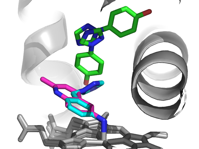
The researchers were interested in CYP121, which is unique to Mtb and important for its viability. They used a thermal-shift assay to screen 665 commercial fragments at 5 mM, of which 66 increased the melting point by at least 0.8 ˚C. 56 of these were further characterized by STD and WaterLOGSY NMR, and 26 showed interactions with the protein and could also be competed with a known substrate. Eight of the most soluble of these were then soaked into crystals of CYP121, leading to four high-resolution structures, three of which are shown here. Isothermal titration calorimetry was used to determine dissociation constants.


 Interestingly, two of these fragments (1 and 2 in blue and red, respectively) coordinate to the heme iron, while the other two do not. Fragment 4 (green in figure above) showed two binding modes in the crystal structure, leading the researchers to make molecules that merge both binding modes. Although the resulting molecules bind in a similar fashion as compound 4 as judged crystallographically, they show at best marginal improvements in affinity and sizable losses in ligand efficiency. Quantum mechanical calculations suggested that this lack of improvement was due to conformational strain within the molecules.

Happily, merging compounds 1 and 2 was much more successful, leading to compound 14, which maintained ligand efficiency and improved affinity. A crystal structure revealed that, as designed, compound 14 binds in a very similar manner as the initial fragments. The molecule was also selective for CYP121 over a different CYP from Mtb as well as several human CYPs.


This is a nice paper not only because it reports a successful example of fragment merging on a new class of targets, but because it also describes several approaches that didn’t work. Fragment merging and fragment linking probably fail more often than they succeed, and this report really digs into the SAR and addresses why merging can be so challenging.

Of course, what would be really cool would be to link compound 14 with compound 4 (ie, link all of the fragments in the top figure), and the paper ends with the statement that this is currently ongoing. It will be fun to see the results.

20 March 2012

Practical Application of NMR

I am sometimes harsh on academic papers, especially those that purport to describe drug discovery. However, Isabelle Krimm and colleagues have continued their excellent work, previously discussed on this blog in a this paper. This paper reads like a "How To" on prosecuting an NMR-based screen. In this work, they have two goals in mind: studying fragments interacting with targets with multiple hotspots and determining the utility of fragments for allostery.

Glycogen phosphorylase is an interesting system to work in: it has an active site and six regulatory sites, including an allosteric site with a variety of positive homotropic and negative heterotropic effects between the various sites. They took 19 known inhibitors that bind to the active site (1-6), inhibitor site (7-9), allosteric site (10-12), and the "new" allosteric site (13-15) and deconstructed them.
Then they screened these fragments against GPa and GPb using both STD and WaterLOGSY. A compound was only deemed a hit if it was observed to bind via both methods. I find this approach very interesting. STD works via NOE from the saturated target to the bound compound. WaterLOGSY works via NOE from bound water to the compound. Each experiment has advantages and disadvantages, but are they truly orthogonal experiments. As a third experiment, the authors use transfer NOE to confirm binding. I would expect to see at least one truly orthogonal method to confirm binding, such as SPR. They then used competition screening against known inhibitors to bucket their fragments based upon the site they are binding to.

While I don't think the results here are not similar to results achieved in industry many times over, this is an excellent paper that shows the power of NMR in screening and how to apply that to drive answers to target validation and compound bucketing.

This paper leads I think to interesting academic musings. Does ontogeny recapitulate phylogeny for compounds derived from fragments? Does it matter if the fragment is 3D or not? Is there a floor below which a fragment will not bind? Does this floor move if you are using 3D fragments vs. highly planar ones?

09 August 2010

Fragment specificity

A frequent topic in fragment roundtable discussions concerns specificity: do fragments hit lots of targets, or just a few? Isabelle Krimm and colleagues at the Université de Lyon in France studied this question experimentally and report their results in a recent issue of J. Med. Chem. The paper provides data for the ongoing debate of whether and how much specificity a fragment should exhibit before being pursued for further lead development.

The researchers assembled a diverse set of 150 fragments and used NMR techniques to determine whether they bind to five different proteins. Three of the proteins, Bcl-xL, Bcl-w, and Mcl-1 are related members of the Bcl-2 family of antiapoptotic proteins, and at least the first of these has been successfully targeted using fragment-based methods. The fourth protein, PRDX5, has proven to be much less yielding to inhibitor discovery, while the fifth, human serum albumin (HSA), binds a wide variety of small molecules.

After applying 1D-NMR techniques (WaterLOGSY and STD) to all of their fragments against each of the five proteins, the researchers used more rigorous but less sensitive 2D-NMR (HSQC) to determine the binding sites of the hits. (This later study revealed, in agreement with previous results from the same lab, that the fragments all bind in the “hot spots” or active sites of the proteins.)

More than two-thirds of the fragments bound to at least one protein, a rather high hit rate. However, the hit rates for each protein varied considerably, with only 7 hits for PRDX5 and 72 for HSA (with a close second of 71 for Bcl-xL). Within the Bcl-2 family there was little specificity observed: Mcl-1, with 29 hits, shared all but one hit with either Bcl-xL or Bcl-2 or both; such non-specificity among related proteins has been discussed previously. In the case of HSA and Bcl-xL, although both proteins had similar numbers of hits, just over half of these were in common, demonstrating that fragment specificity is not difficult even with small-molecule sponges such as HSA. That said, many fragments were remarkably nonspecific, with 22 hitting four of the 5 proteins. Amazingly, all 7 of the hits against PRDX5 also hit all four other proteins.

The physicochemical properties of the fragments that hit one or more proteins were compared with those of the library as a whole, and although most of the parameters were similar, the ClogP values (a measure of hydrophobicity) were considerably higher for hits, and highest of all for the non-specific hits.

These findings are more evidence that, as predicted almost a decade ago, fragments can bind to more proteins than can larger, more complex molecules. The follow-up question, how much does this matter, is still up for debate. There are plenty of examples of developing specific inhibitors from non-specific starting points during the course of fragment optimization. But how non-specific is too non-specific? Would you feel comfortable pursuing any of the fragments that hit all of the proteins?