29 December 2014

Review of 2014 reviews

The year is spinning to an end, and as we did in 2013 and 2012, Practical Fragments is looking back on notable events as well as reviews we didn’t cover previously.

2014 was full of conferences, starting with the CHI meeting in San Diego (here and here), moving to the Zing conference in the Dominican Republic, on to the Fall ACS meeting in beautiful San Francisco, and ending with FBLD 2014 in Basel.

In terms of reviews relevant to the fragment community, John Christopher and colleagues at Heptares published an extensive analysis of “Structure-based and fragment-based GPCR drug discovery” in ChemMedChem early in 2014. The last few years have seen an efflorescence of new structural information on G protein-coupled receptors, and this paper provides a thorough compilation of crystal structures and small molecule ligands. The review also discusses methods that have been used to discover fragments that bind to GPCRs, including TINS, SPR, CEfrag, radioligand binding, and fluorescence assays, and ends with case studies on A2A antagonists and β1AR ligands.

In contrast to GPCRs, kinases represent a well-established target class for fragment-based drug discovery, as exemplified by the first approved drug, vemurafenib. Structural biology has played a major role in this success; more than 200 of the 518 human kinases have had their X-ray crystal structures determined, and more than 3000 protein kinase structures have been deposited in the protein data bank. Astex has put several kinase inhibitors into the clinic, and in Methods in Enzymology Paul Mortenson and colleagues from the company discuss the state of the art. This is a clear and concise review of fragment-based drug discovery in general and as specifically applied to kinases. It serves as an excellent introduction to the topic.

Any chemist who has worked on kinases will be familiar with azaindoles, and in Molecules, Sylvain Routier and colleagues at Université d’Orléans discuss “the azaindole framework in the design of kinase inhibitors.” This provides a thorough compilation of azaindole inhibitors against ALK, Aurora, Cdc7, CHK1, C-Met, DYRK1A, FAK, IKK2, JAK2, KIT/FMS, PAK1, p38α, PIM1, B-Raf, ROCK, m-TOR, and TrkA, replete with synthetic methods. The paper also includes a nice analysis of binding modes. Of the 58 crystal structures of azaindoles bound to kinases in the protein data bank, the majority (48) are with 7-azaindole rather than the three other positional isomers. This isomer (found in vemurafenib) is also over-represented in the patent literature and among commercial compounds.

Another target that has yielded to FBLD is BACE1, a hot but still controversial target for Alzheimer’s disease, and in Bioorg. Med. Chem. Lett. Daniel Oehlrich and colleagues at Janssen review “the evolution of amidine-based brain penetrant BACE1 inhibitors”. This is very much a medicinal chemist’s review, with over 100 chemical structures, including a nice summary of the various chemotypes used by different companies. The authors do an excellent job synthesizing a tremendous amount of data, much of it reported only in the patent literature, and engage in some intriguing chemical sleuthing to guess at the identity of clinical candidates whose structures have not been publicly disclosed, such as MK-8931.

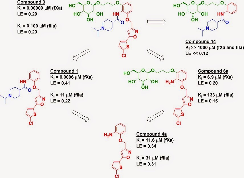
Jia Zhou and collaborators at the University of Texas Galveston and Fuzhou University discuss “Evolutions in fragment-based drug design: the deconstruction-reconstruction approach” in Drug Discovery Today. After briefly describing fragment-finding methods and library design, the review focuses on deconstruction of known ligands to generate “privileged” fragments that are then reassembled into new molecules. Although this approach can be productive, if one doesn’t exclude PAINS the result can be garbage-in, garbage-out.

Finally, in Methods in Enzymology, Katherine Warner (National Heart, Lung and Blood Institute) and Adrian Ferré-D’Amaré (University of Cambridge) review the crystallographic analysis of fragments binding to the TPP riboswitch. This is a concise how-to guide, and the methodology could be applicable to other RNA targets.

And with that, Practical Fragments says farewell to 2014. Thanks for reading, and may the New Year bring wonderful new discoveries!

22 December 2014

Progress in Biophysics and Molecular Biology special issue

The latest issue of Prog. Biophys. Mol. Biol. includes five articles on fragment-related topics. We already discussed one from Astex; brief summaries of the rest follow.

Eddy Arnold (who has an editorial introducing the articles) and colleagues from Rutgers University discuss the advantages of screening fragments crystallographically. Regular readers of this blog will likely be familiar with some of the material, but there is lots of practical advice on fragment cocktail design (that is, choosing which fragments to mix together), optimization of soaking, high-throughput crystallography, and related topics. There is also a nice example of an “unknown known,” where the apparent activity of a compound turned out to be due to contaminating metal.

David Dias (University of Cambridge) and Alessio Ciulli (University of Dundee) have a piece on using NMR in structure-based lead discovery, with a heavy focus on large multi-protein complexes. They succinctly review both ligand-based and protein-based NMR methods and then discuss how these techniques can help determine ligand conformations and binding sites. Next, they discuss how to tackle high molecular weight protein assemblies or protein-protein complexes, often by using clever isotopic labeling strategies. The figures throughout are particularly effective at showing what kinds of information can be obtained from the various techniques.

Andrew Hopkins (University of Dundee) and colleagues are up next with “Fragment screening by SPR and advanced application to GPCRs”. Surface plasmon resonance, of course, is a mainstay of fragment screening, and this is a timely how-to guide by some of the experts in the field. As the title suggests, a major focus is on GPCRs, a class of membrane proteins only recently targeted by fragments. There are some good practical tips on protein immobilization, screening, and weeding out false positives. My sense is that screening GPCRs by SPR remains challenging; most of the fragment libraries screened tend to be small (no more than a few hundred compounds), and sensitivity seems to be an issue, with most of the hits being quite potent by the standards of FBLD (low micromolar or better).

Finally, Theresa Tiefenbrunn and C. David Stout (Scripps) lead us “Towards novel therapeutics for HIV through fragment-based screening and drug design.” Practical Fragments has highlighted fragment efforts against several targets for this virus, including HIV protease, HIV reverse transcriptase, HIV integrase, and TAR RNA; this paper discusses these and more. This is a thorough compilation of copious data and focuses heavily on fragment screening. Crystallography plays a starring role, but SPR and NMR are also prominent. In short, it shows practical applications of the prior papers, and so makes a nice conclusion to this series.

15 December 2014

Fragments vs CDC25B phosphatase – from behind

Protein phosphatases, which remove phosphate groups from proteins, fall into the category of low-hanging but firmly attached fruit: many make great targets, but getting lead-like inhibitors is tough. Indeed, the enzymes seem to be particularly susceptible to PAINS (see for example here and here). A major challenge is the phosphate-binding site, which has a predilection for highly negatively charged (and non-druglike) moieties. In a paper just published in ACS Chem. Biol., Tomasz Cierpicki and his group at the University of Michigan neatly sidestep this issue.

The researchers were interested in the dual-specificity protein phosphatase CDC25B, which is important in cell cycle regulation and thus a potential anti-cancer target. They started with a 1H–15N HSC NMR screen of 1500 fragments in pools of 20, with each fragment present at 0.25 mM. This yielded a single hit: 2-fluoro-4-hydroxybenzonitrile.

Because the researchers were using protein-observed NMR and had previously assigned the backbone resonances, they were able to use chemical shift perturbations to identify the binding site. Surprisingly, this turned out to be not the active site at all, but rather a region about 15 Å away. They were able to confirm this site using X-ray crystallography, which further revealed that the fragment binds in a small pocket near where the substrate protein CDK2 binds.

The researchers noticed a nearby sulfate ion (from the crystallization buffer) and, after first doing a brief SAR by catalog survey, they tried to link this to their hit. Although this certainly didn’t improve physicochemical properties, it did result in tighter binding, and crystallography confirmed that the new molecule bound as designed. This molecule also inhibited the phosphatase, albeit modestly (IC50 1-2 mM). The result suggests that blocking this protein-protein interaction is effective at blocking activity.

It remains to be seen how much affinity there is to be had at this site. Still, I do have a soft spot for phosphatase inhibitors that bind outside the active site. At the very least this paper provides a new direction for an old – and very difficult – class of targets.

10 December 2014

How much information can NMR provide?

A frequent assumption in fragment-based lead discovery is that similar fragments have similar binding modes, which are conserved as the fragments are elaborated. However, this isn’t always the case, a fact that can complicate optimization. Ideally multiple crystal structures help guide the chemistry, but in the real world crystal structures can be difficult to obtain.

One of the seminal papers in FBLD used NMR rather than crystallography to guide design, a strategy still used today. But how effective is NMR at assessing the binding modes of related fragments? This is the question that Isabelle Krimm and colleagues at the Université de Lyon sought to answer in a paper published a few months ago in PLOS ONE.

The researchers were interested in the inflammatory enzyme peroxiredoxin 5 (PRDX5), and they examined its interactions with five catechols: the parent unsubstituted molecule and four derivatives with substituents ranging from methyl to phenyl. Although catechols are PAINS, the researchers took pains to carefully examine the NMR spectra to look for signs of misbehavior.

Two NMR techniques were used, saturation transfer difference (STD) NMR and chemical shift perturbation (CSP). STD is nice because it is a ligand-detected method: you don’t need to go to all the work of assigning the chemical shifts of the protein. One piece of information from an STD experiment is whether a hydrogen atom is exposed to solvent or buried close to the protein, and in this case three of the catechols showed one particular hydrogen atom was exposed to solvent. The unsubstituted catechol provided only a single NMR peak and thus no information, and the fifth catechol was also not very informative, though it did seem to bind. Repeating this “epitope mapping” of all the catechols with human serum albumin instead of PRDX5 gave different results, suggesting a different binding mode.

Of course, there is only so much information you can get from ligand-detected NMR, so the researchers turned to protein-detected NMR and examined the CSPs of proton-nitrogen cross peaks using 15N-HSQC experiments. They also calculated CSPs for various potential binding modes and compared these with the experimentally observed CSPs to generate models. These suggested a common binding mode for the same three catechols that STD revealed as having a single solvent-exposed hydrogen atom each. Combining all this information led to specific binding models for these three fragments.

But how good are the models? Happily, the researchers were able to obtain crystal structures of four of the catechols bound to PRDX5, and these agree quite well with the NMR-derived structures. Unfortunately, the fifth catechol couldn’t be characterized bound to the protein crystallographically; NMR also suggested that this bound differently than the others.

So in the end, NMR was able to successfully predict that three ligands had similar binding modes, while another likely doesn’t. The process does seem to require a fair bit of effort. Nonetheless, in cases where crystallography is difficult or impossible, it may be the best way to get essential structural information, and this paper provides a good road map.

08 December 2014

PAINS Shaming, part deux

So, as regular readers know, we have declared war on PAINS on the blog.  As part of that effort, I (we?, not sure if Dan wants to be associated directly with it) introduced PAINS Shaming. Well, thanks to Angelo Pugliese and Duncan McArthur at the Beatson we have the latest paper to shame. 
The nice thing is that they come right out and call it like it is: a Rhodanine.  To reiterate, from the comment by Baell and Walters:
Rhodanines exemplify the extent of the problem. A literature search reveals 2,132 rhodanines reported as having biological activity in 410 papers, from some 290 organizations of which only 24 are commercial companies. The academic publications generally paint rhodanines as promising for therapeutic development. In a rare example of good practice, one of these publications (by the drug company Bristol-Myers Squibb) warns researchers that these types of compound undergo light-induced reactions that irreversibly modify proteins. It is hard to imagine how such a mechanism could be optimized to produce a drug or tool. Yet this paper is almost never cited by publications that assume that rhodanines are behaving in a drug-like manner.
And as predicted, this paper does not cite Voss et al.  They cite dose-depedent responses for their compounds.  Does it matter?  Not to me.  To their credit, they call these molecules tools, but also tout them for future therapeutic development.  A PAIN can be a useful tool or even lead to non-PAIN containing compounds, but it requires a higher level of proof.  I don't see that here. 

So, here is your PAINS Shaming (Holiday themed): 

03 December 2014

Huge Library + Tiny Hit Rate = Novel Chemotype

As Dan recently pointed out that I pointed out, epigenetics is big.  Bromodomains get a lot of play on this blog.  One bromodomain that is not mentioned a lot in the literature is ATAD2 (because everyone is actually working on it?).  It is promising because of the diverse cellular activities it is involved in.  However, its bromodomain is quite dissimilar from to "druggable" bromodomains.  [Just an aside, can't we get away from druggable already?] Only 3 of seven residues lining the KAc pocket that interact with peptide are similar (compared to Brd4) (Figure 1)
Figure 1.  View of residues within the KAc binding site of BRD4 that interact with diacetylated residues.  Residues from the peptide are shown in teal.
So, in this paper from the Fesik lab at Vanderbilt, the use fragments to discover chemical matter against this tough target.  They utilized 15N-HSQC, like the previous post on bromodomains, because it can detect millimolar binders and (with resonance assignments) determine where on the protein it is binding.  They screened 13800 fragments (NOT a typo!) as mixtures of 12, or 1150 individual experiments.  Using the SO-FAST pulse sequence allows each experiment to be acquired in 7 minutes (6 days of acquisition).  This required more than 2 grams of labeled material.  Hits were then deconvoluted as singletons, resulting in 65 actives with Kds from 350uM to more than 2 mM (determined by HSQC titration).  12 had affinities of less than 1 mM.  This hit rate of 0.1% is low, especially for a fragment based screen, even against a PPI.  While it may ligandable, a hit rate this low still indicates this will be a very tough nut to crack. 

The assignments of ATAD2 are NOT known, but they observed a consistent cluster of resonances being perturbed (Figure 2).
Figure 2.  A. Fragment 1, B, Fragment 5, C Fragment 12.  Green Circles represent resonances which may report on ligand binding.
They discovered several novel chemotypes, never seen against bromodomains, albeit with a very low hit rate, that could be put in three clusters (Figure 3).  Cluster 1 represents known bromodomain inhibitors, while cluster 2 and cluster 3 are unique to ATAD2.  Interestingly, the Kds only differ by 2-fold, but are still more potent than other recently published ATAD2 compounds.
Figure 3.
One representative from each cluster was crystallized (1, 5, and 12).  All three fragments occupy the same pocket and make a critical contact to the conserved N1064.  They also compare their fragments to work from the SGC that scooped them. 
Figure 4. A. Fragment 1, B, Fragment 5, C Fragment 12.
In the end, this is an unsatisfying paper.  There is speculation as to how these fragments can be progressed and made more potent.  But, this entire paper is about the novel chemotypes for ATAD2.  There is no chemistry in a journal that has Chemistry in its title.  I expect more from this journal and this group.  To summarize, if you throw enough fragments at a target you can find a few that bind. 

01 December 2014

Fragments finger a PHD finger

As Teddy recently observed, epigenetics is big, and fragments have played an important role against several targets. One class of proteins that has received less attention is the group of PHD fingers, which recognize methylated lysine residues. The pygo-BCL9 complex contains a PHD finger that binds to a specific methylated lysine residue on histones, and has been implicated in cancer. Marc Fiedler, Mariann Bienz and colleagues at the MRC Laboratory in the UK describe their efforts against this target in a new paper in ACS Chem. Biol.

The researchers started with a virtual screen of 225,000 commercially available compounds. They purchased 313 of the top hits and tested them for binding with protein-detected NMR (1H-15N-HSQC). This produced only three very weak hits – a hit rate of 0.001%. Three additional virtual screens produced a couple dozen more, but all of these were weak; the best had an affinity around 3.5 mM and a ligand efficiency around 0.12 kcal/mol/atom. Co-crystallography proved unsuccessful, probably in part due to the low solubility of the compounds.

Enter fragments. The researchers screened the Maybridgerule of three” 1000-compound library in pools of 5 compounds, each at 1 mM, under the same protein-detected NMR conditions they used previously. Numerous pools appeared to show binding but deconvolution proved unsuccessful for all but two. Strikingly, the two hits – both benzothiazoles – are almost identical, differing only in a single atom substituent (fluorine vs chlorine).

Although the best fragment hit was also weak (Kd = 3.1 mM), it had a much higher ligand efficiency (0.31 kcal/mol/atom). More importantly, it was sufficiently soluble (20 mM!) that it could be cocrystallized with the protein, resulting in a high resolution structure. This revealed that the fragment binds in a narrow cleft – a conclusion independently reached by examining the NMR chemical shift perturbations (CSPs) of protein amino acid residues in the presence of compound.

Testing various analogs did not identify anything significantly more potent, but changing the benzothiazole core to a benzimidazole changed the pattern of CSPs. Additional NMR studies and modeling suggested that these molecules bind not in the narrow cleft but rather in the pocket where methylated lysine binds, and competition studies with a short peptide supported this hypothesis.

This is a nice example of applying fragments against an important emerging target class. It is also a beautiful illustration of molecular complexity in action: as the authors note, the hit rate from fragment screening was around 200-fold higher than the virtual screen, and provided better hits to boot. As with most fragment screens there is still a long way to go to get to a potent compound, but it looks like this group is on the right path.

25 November 2014

Docking covalent fragments

Most drugs interact non-covalently with their target. The conventional wisdom was that covalent drugs – especially irreversible ones – would have dangerous side effects. Although this is still a concern, the success of drugs such as ibrutinib and dimethyl fumarate has caused a resurgence of interest. In a new paper in Nature Chemical Biology, Brian Shoichet, Jack Taunton, and colleagues at the University of California San Francisco describe how computational chemistry can be used to find new covalent inhibitors.

The researchers created a modified version of the program DOCK called – wait for it – DOCKovalent. Happily, they have made this available for free to anyone. To start, you upload your crystal structure and choose which amino acid residue you are interested in targeting. You can then pick from 9 different libraries of various electrophiles, each covering a different class of covalent “warhead”: epoxides, aldehydes, etc. There are about 650,000 molecules in total, roughly half of which easily qualify as fragments, with the rest being lead-like (still < 350 Da). Each molecule is either commercially available or readily synthesized in one or two steps.

The program then virtually links each molecule with the selected protein residue (typically cysteine or serine) and calculates scores based on predicted van der Waals and electrostatic interactions as well as desolvation. Multiple conformations of each ligand are sampled (with fragments there are not that many) as are different rotamers of the nucleophile. Users then manually inspect and test the top hits.

The researchers first benchmarked the program against four proteins with known covalent inhibitors, where it performed well. In the case of the bacterial protein AmpC β-lactamase (which we previously discussed here), the program retrospectively predicted the correct structure of 15 out of 23 known boronic acid ligands. In one case where the prediction differed from the reported co-crystal structure, the researchers re-determined the co-crystal structure at high resolution and found that DOCKovalent was actually correct.

Thus confident, the researchers docked 23,000 commercial boronic acids against AmpC and selected 6 on the basis of score and structural novelty. Of these, 5 had inhibition constants of 3.55 µM or better, with the best being 40 nM. A crystal structure of this compound bound to the protein led them to purchase 7 additional compounds, one of which had Ki = 10 nM and a ligand efficiency of 0.73 kcal/mol/atom. Most of the molecules were also selective against 4 other proteases and were able to reverse antibiotic resistance in AmpC-expressing bacteria.

Of course, by design all of these molecules have a boronic acid warhead; will any such molecule inhibit this enzyme? To find out, the researchers tested 5 low-scoring molecules and found that 4 of them showed, as hoped, less than 10% inhibition at 10 µM. However, a fifth molecule showed reasonable inhibition, with Ki = 3.2 µM. To understand this false-negative, the team solved the crystal structure of the molecule bound to AmpC. Interestingly, the molecule bound in a conformation different than had been predicted – one that also required conformational changes in the protein, which are not allowed in DOCKovalent.

The researchers took a similar approach to seek novel inhibitors of the kinases RSK2 and MSK1 using reversible cyanoacrylamide-containing molecules (previously highlighted here). Here too the researchers were able to identify selective nanomolar cell-active inhibitors.

This looks like a very nice approach. Of course, it does require a crystal structure (or at least a good model). Also, as mentioned above, the fact that the protein is kept rigid means the program will be unable to detect ligands that bind to cryptic pockets, so there is still plenty of opportunity for empirical surprises. Still, the fact that DOCKovalent is freely available will hopefully encourage people to give it a try on their favorite protein.

17 November 2014

Deconstruction, superadditivity, and selectivity

One of the more exciting phenomena in fragment-based approaches is synergy (or superadditivity), in which the binding energy of linked fragments is greater than the sum of the binding energies of the individual fragments. Extreme cases are relatively rare, and the underlying thermodynamics can be counterintuitive, so it is always fun to see new examples. Cosimo Altomare and collaborators at the University of Bari and Consiglio Nazionale delle Ricerche (Italy) describe one in a recent paper in J. Med. Chem.

The proteases factor Xa (fXa) and thrombin (fIIa) are two heavily-studied anticoagulant targets. The paper characterizes a previously described molecule (compound 3) that is selective for fXa but still potent against fIIa, leading to good anticoagulant activity in human plasma as well as profibrinolytic activity. The researchers took a fragment deconstruction approach to better understand the binding to both targets.

As seen previously for fXa, the chlorothiophene moiety (red) is essential for binding, and removing it (compound 14) obliterates any detectable activity on both enzymes. However, while removing the glucose moiety (green) to give compound 1 reduced affinity for fXa by less than ten-fold, it reduced affinity for fIIa by more than two orders of magnitude. In contrast, removing the piperidine moiety (blue) to give compound 6a reduced affinity to both enzymes by several orders of magnitude.


However, these results are context-dependent. Removing both the piperidine moiety and the glucose moiety gives compound 4a, which has similar activity against fIIa as compounds 1 and 6a, where only a single moiety has been removed. In fact, compound 4a (without the glucose) is actually slightly more potent than compound 6a (with the glucose) against fIIa. But, as mentioned above, adding the glucose to compound 1 gives an impressive 110-fold boost in affinity for fIIa. In comparison, a famous early example of cooperativity in an NMR by SAR study gave only a 14-fold boost.

The researchers solved the crystal structure of compound 3 bound to fIIa, which reveals several hydrogen bond interactions between the glucose moiety and amino acid residues that have been previously implicated in allosteric activation of the protein. Perhaps compound 3 is exploiting this allosteric mechanism to bind more tightly.

This is a careful, thorough study and serves as a useful reminder that cooperativity can be huge, but it is still difficult to explain, much less predict.

12 November 2014

Pleasant Surprise

Epigenetics is bigWicked big.  How big?  This big.  Papers come from everywhere.  In this paper, an academic group from Minnesota, goes after SIRT2 with fragments.  SIRT2 is a type III HDAC that resides primarily in the cytoplasm that uses NAD+ as a co-factor (Figure 1).
Figure 1.  Biochemical Reaction of SIRT2

Sirtuins (there are seven) have a long history aready in pharma.  SIRT2 has been fingered as a potential treatment for Parkinson's Disease (PD) and other pathologies, including bacterial infection.  There are a nice range of available inhibitors for SIRT2.
Figure 2.  Known Sirtuin2 Inhibitors
Suramin, a drug originally made in 1916.  I think it was made by pouring hot sulfuric acid over naptha tar (but my chemistry may be off).  Of course, when I saw this I got my dander up and got ready both barrels.  [It also made me chuckle, because many moons ago I co-authored a paper on suramin against RANK-RANKL.  In that paper suramin blocked a PPI.]  In this paper suramin is highly selective for SIRT1 over SIRT2 and 3.  So, as my wife says, your mind is like a parachute, it only works when open.  So, let see what they did.

One of the known inhibitors is


First, they evaluated a SIRT5-suramin crystal structure, where suramin mainly occupies the peptide binding site and a sulfonate protrudes into the nicontinamide binding site.  With this, they came up with the following generic plan (Figure 3). In this case, the circled napthalene is close to the  nicotinamide binding site.  They proposed to merge/link nicotinamide compounds to the appropriate suramin-like moiety. 
Figure 3.  Merging Strategy for SIRT2 Inhibitors
So, my quibble, and its not a big one, but their molecules are big (20 heavy atoms or more).  Oh well, a fragment is in the eye of the beholder.  Dan would call this FADD maybe.  Well, how did they do, you ask?  Well, they were able to generate sub-micromolar compounds (64 being the best) (50 nM![edited]), with selectivity against SIRT1 and 3.  The MOA was competitive vs. substrate and noncompetitive vs. NAD+ (although they could not rule out uncompetitive).  It had low cytotoxicity.  YAY.  Yet, it only had moderate anti-cancer activtity.  Well, those two outcomes could still allow it to be a good PD compound.  But their data indicated that it would have a low probability for crossing the BBB. 
So, in the end, I was pleasantly surprised.  This ended up being nice work with a good breadth of work.  I don't know if this makes it into the "lead-like" space or will remain in the "tool" space, but I like to see this kind of work, especially from academic groups. 

10 November 2014

Plenty of room at the bottom (of chemical space)

One of the key selling points of fragment-based lead discovery is that small fragments can search chemical space much more efficiently than larger compounds, since there are fewer possibilites. Nonetheless, the numbers are still daunting: more than 166 billion molecules with up to 17 non-hydrogen atoms. The question of how many of these are commercially available has come up before. In a paper just published online in Prog. Biophys. Mol. Biol., Chris Murray and colleagues at Astex take a new look at this – and related – questions.

Rather than considering all possible molecules, the researchers focused on six-membered rings with one or two small substituents of no more than six non-hydrogen atoms. Six-membered rings are found in many drugs, so this is a useful area of chemical space on which to focus. The researchers first considered “topologies,” simple two-dimensional representations of molecules. In the coarsest version, benzene, cyclohexane, pyridine, and piperidine would all have identical topologies: a six-membered ring with no substituents.

The researchers looked at how many topologies having up to 16 atoms were listed in the available chemicals directory (ACD) of 2.7 million commercial molecules. Even using the coarse definition where all non-hydrogen atoms were considered equivalent, less than half of 16-atom topologies are commercially available. At finer resolution (for example, differentiating carbon from nitrogen), the numbers dropped even more: less than 4% of the 2223 16-atom topologies with a pyridazine core were available.

However, things get better the smaller the molecule. When considering only molecules with 11 non-hydrogen atoms, all of the coarsest topologies are available, as are more than 70% of pyridazines. From this, the researchers concluded:
We need to focus on fragments with lower heavy atom counts and… improve the sensitivity of our screening methods to make sure that we can identify the binding of these smaller fragments.
The rest of the paper discusses how they applied this approach, and what lessons they learned.

The researchers assert that X-ray crystallography (upon which Astex was founded) is the most sensitive screening method. That may elicit some debate, but is defensible given the presence of extremely weak binders (water, buffer components, detergents) in many crystal structures. They also argue that while NMR may allow detection of fragments with lower solubilities, this may not be a good thing.

Of the 1633 fragments that were in the Astex library between 2001 and 2007, 22% came up as X-ray hits (ie, they showed up in at least one crystal structure). Strikingly, fragments with 11 or 12 atoms were enriched far above their representation in the overall library, while fragments with 17 or more atoms were underrepresented. This is a beautiful confirmation of the “molecular complexity” hypothesis, the idea that there is a sweet spot where molecules are large enough to make productive interactions with a target but not so complex that negative interactions become dominant.

These results led the researchers to redesign their library to focus on fragments having fewer than 17 non-hydrogen atoms, which entailed considerable custom synthesis. The resulting library has 1371 fragments, of which 47% have shown up as X-ray hits. The average size of hits is the same as that of the overall library (12.2 vs 12.4 non-hydrogen atoms and 172 vs 176 Da, respectively), though the hits are slightly more lipophilic (cLogP = 1.1 vs 0.9).

What about “three-dimensionality?” This is a topic that has been discussed quite a lot (herehere, herehere, and here, for starters), so it is nice to have some solid data. One problem is how to define three-dimensionality: simple metrics such as Fsp3 don’t account for the fact that aromatic compounds such as 2,6-substituted biphenyls can be very non-planar. Many people use PMI, but the Astex researchers chose deviation from planarity (DFP). This method puts a hypothetical plane through the molecule that minimizes the deviation of all non-hydrogen atoms from the plane; the average deviation from the plane for each molecule is calculated in Ångstroms. So, for example, benzene has DFP = 0.0 Å, while cycloleucine has DFP = 0.54 Å. In this study, the researchers used a single conformation for each molecule, but since these fragments have on average only 1.3 rotatable bonds this is probably a reasonable simplification.

Roughly 40% of the Astex library has a DFP < 0.05 Å, but these “flat” fragments were enriched to ~50% among hits. Not surprisingly, kinase hits tended to be even more two-dimensional (>60%), but even protein-protein interaction (PPI) hits were, if anything, slightly more planar than the overall collection, which is consistent with another recent study. Indeed, there seems to be nothing special at all about PPI hits, more than half of which were also found against non-PPI targets. The researchers argue that 3D-fragments are inherently more complex and thus less likely to show up as hits, which supports Teddy’s Safran Zunft challenge.

One of the arguments in favor of three-dimensionality is that such molecules may have better physicochemical properties, and the researchers examine the DFP for fragments and resulting leads. It turns out that there is a weak correlation between the shapeliness of a fragment and that of the resulting lead, but there are many exceptions (such as this one).

Some of these data have been publicly presented, but this paper should broaden the discussion. Coming back to the title of this post, the conclusion is that fragments should be made as small as detectable with your assay. And flat is the new black.

05 November 2014

Still Impractical, but Getting Better...

Dan and I don't see every fragment paper and so it's nice when people point out papers to us.  Its typically their recently published paper and they are looking for some sort of recognition/validation from us.  Sometimes its a paper we have on our radar, sometimes it isn't.  Recently, I received an email pointing out this paper from someone I met at FBLD2014.  Well, this is a follow up paper to a paper I discussed a few years back.  The title of the post sums up my thoughts: "Another (Impractical) NMR Method."  One comment from that post (by a co-author from Astex on the current paper) was
This is a relaxation filtered ligand based method (like T1rho or selective T1). You therefore may consider using it in competition mode if you find one suitable "spy" molecule: this would allow with a single point experiment screening mixtures and/or ranking for low affinity hits(especially if solubility is limiting). I would actually give it a try.
So, it looks like she did.  The advantages of Long Lived States (LLS) NMR is that the the dynamic range is wider and it works at low protein concentrations (3 uM or roughly the same as STD or WaterLOGSY) or with very weak affinities.  Against the workhorse HSP90 system, they screened mixtures using LLS and a spy molecule.
  
I still don't think this is a very practical method because it still requires a lot of tailoring to individual systems; in particular the compounds need pairs of protons which are suitable for excitation to the LLS.  Using it in "spy" mode gets around this. I would still hold that this is an impractical NMR method.  I like to see people developing new methods and trying to improve them.  We also need people doing good comparisons of "standard" experiments to new ones.  (Here is how NOT to do a method comparison.) I won't dismiss this out of hand, but there is still a lot of work to be done here to move it into a "front line" screening technique.