Showing posts with label allosteric. Show all posts

31 August 2026

Fragments find cryptic sites on ARID1B

We’ve written previously about the concept of synthetic lethality, in which changes to one protein make cancer cells more dependent on another. Inhibiting this second protein allows selective targeting of cancer cells while sparing normal cells. One such opportunity is the pair of proteins ARID1A and ARID1B, which assemble into a large complex that helps remodel chromatin to regulate transcription. ARID1A is mutated in some 9% of cancers, and ARID1B can partially compensate. CRISPR knockdown studies show that these cancer cells are dependent on ARID1B. ARID1B is huge (2236 amino acids!) and predicted to be mostly disordered – not the easiest target for small molecule drugs. However, a small 109-residue AT-Rich Interactive Domain (ARID), which binds to DNA, had previously been crystallized. This domain is the focus of a new Proc. Nat. Acad. Sci. USA paper by Michael Holliday and colleagues at Relay Therapeutics.
 
Computational assessment using Schrödinger’s SiteMap did not show any ligandable pockets, but molecular dynamics revealed that the N-terminal helix might open to reveal a shallow hydrophobic groove beneath it. Thus encouraged, the researchers performed a 2D NMR (15N-SOFAST-HMQC) screen of 480 fragments and obtained two hits, both with weak but measurable affinities. Interestingly though, the NMR signals are consistent with slow kinetics, suggesting that the molecules bind to rare conformations of the protein. Chemical shift perturbation (CSP) mapping showed that the two fragments bind near one another but not exactly at the same site.
 
Crystallography revealed that compound A-1 bound to a previously cryptic tunnel of ARID that forms by the displacement of a tryptophan side chain – a motion not seen in the molecular dynamics simulation. Preliminary medicinal chemistry improved the potency to mid-micromolar affinity.

 
Compound B-1 proved recalcitrant to crystallography, but cocrystallization with the more potent compound B-2 yielded a high resolution structure. This molecule did in fact bind where the N-terminal helix normally binds, as predicted by molecular dynamics; the helix itself became disordered.
 
But wait, there’s more! To find additional chemical matter, the researchers conducted a virtual screen of 150,000 compounds against ARID. Of the several dozen molecules tested by 2D-NMR, two showed CSPs similar to those caused by compound A-1, and their binding modes were confirmed by crystallography. But one of the molecules that showed CSPs similar to those caused by compound B-1 also caused CSPs in a different region of the protein, and crystallography confirmed binding to yet a third site.
 
In order for the compounds to be selective for cancer cells they need to be selective for ARID1B over ARID1A, which is 83% identical in the ARID domain. NMR experiments revealed that the molecule that binds to the third site binds with similar affinity to ARID1A, while compound A-2 binds about 5-fold less tightly. Happily, compound B-2 showed no binding to ARID1A even at 10 mM. Thus, the researchers have found three starting points for optimization, and evidence that selectivity is possible.
 
As the researchers acknowledge, the compounds will require “substantial additional optimization,” not least for affinity. Intriguingly though, compounds A-2 and B-2 do bind sufficiently close to one another that a fragment linking or merging approach may be warranted.
 
So in the end, ARID1B ARID presents two cryptic sites and a third more conventional (albeit challenging) site. The presence of multiple small-molecule binding sites is consistent with this paper from 2016, but still surprising in such a small protein. As we noted then, just because a site binds fragments does not mean it will support high-affinity ligands. Will ARID1B turn out to be like KRAS, which ended up being druggable, or like IL-1β, which has resisted high-affinity small molecules? The current paper provides good starting points toward answering this question.

25 May 2026

Fragments in the clinic: VVD-214

Just over two years ago we highlighted a new clinical candidate targeting WRN, a covalent inhibitor then called VVD-133214. An open-access paper published near the end of last year in J. Med. Chem. from Shota Kikuchi, David Weinstein, and colleagues at Vividion describes in detail the optimization of the covalent fragment hit to the clinical compound. (Shota presented some of this work at the 2024 DDC meeting.) This paper is also an interesting contrast to non-covalent fragment-finding approaches against this target we wrote about earlier this month.
 
The 2024 post described the chemoproteomic screening that identified compound 1a, which covalently binds to C727 in WRN. An early observation was that some molecules were cooperative with ATP while others were competitive. Given the high concentration of ATP in cells, the researchers prioritized the former category, which led to compound 1f. (Note that while I’m showing only the kinact/KI values, the researchers used biochemical and cell-based assays to drive SAR).
 

The vinyl sulfone warhead is unusual amongst covalent clinical compounds, so the researchers sought to characterize it. The rate of reaction with glutathione for compound 1f is comparable to the approved drug osimertinib: reactive, though acceptable. To try to lower the reactivity and also prevent isomerization of the double bond, the researchers introduced a methyl group. Compound 2a not only showed increased stability but also improved activity against WRN and sub-micromolar activity in a cell-based assay. A crystal structure of a later molecule revealed interactions with a hydrophobic patch on the protein, explaining the improvement in potency. Importantly, the other enantiomer was much less potent.
 
Other rings were tried, unsuccessfully, to replace the pyrimidine and the phenyl moieties. However, changing the cyclopentyl ring to a tert-butyl moiety (compound 5d) further improved the potency to the point where the compound could be tested in vivo, where it proved to be active in a mouse xenograft study. Mass spectrometry experiments revealed prolonged occupancy of C727 out to 24 hours after compound dosing even though the compound itself had been cleared, consistent with a long half-life of the WRN protein. The researchers note that high target engagement (TE) at 24 hours was predictive of tumor growth inhibition, which streamlines optimization since it is easier to run a one-day TE experiment than a multi-week efficacy study.
 
Further optimization of ADME properties ultimately led to VVD-214, which was active in a mouse xenograft study and showed good oral bioavailability and pharmacokinetics in mouse, rat, dog, and monkey. This compound was also profiled in a chemoproteomic assay and found to be quite selective for the C727 of WRN.
 
There are several important lessons in this paper. First, the initial fragment is larger than prescribed by the rule of three, consistent with an analysis of covalent inhibitors last year. Second, much of the SAR was empirical; crystallography was not used until relatively late in the campaign. When a crystal structure was finally solved of VVD-214 bound to WRN it revealed no polar contacts between the ligand and the protein, only hydrophobic interactions, which is rare for fragments, let alone drugs. Perhaps because of the lack of polar interactions, it was impossible to measure the inhibition constant (KI), and saturating the warhead to make it unreactive completely abolished activity. In other words, the binding is largely driven by reactivity, but specific reactivity for WRN rather than generic chemical reactivity.
 
In 2024 just two WRN inhibitors had entered the clinic, the other being a noncovalent molecule called HRO761. We quoted the Vividion team as saying that “this presents a rare opportunity to compare two small molecule oncology drugs targeting the same protein by different mechanisms.” Since then, HRO761 has been quietly discontinued, as has another noncovalent drug, IDE275. Meanwhile, development of VVD-214 is ongoing, and another covalent compound, MOMA-341, which we mentioned here, has also begun human testing. (To be fair, so has yet another non-covalent molecule NDI-219216. And the binding mechanism of a sixth WRN drug, EIK1005, is undisclosed.) While it’s still early in the match, covalent drugs seem to be punching above their weight. May the best drug(s) prevail - Practical Fragments is rooting for them all.

18 May 2026

From noncovalent fragments to covalent WRN inhibitors

Last week we described the discovery and early optimization of noncovalent inhibitors against the the DNA helicase WRN, an interesting oncology target both for its conformational flexibility as well as a ‘synthetic lethal’ approach to cancer drugs. Today we continue the theme with a Bioorg. Med. Chem. Lett. paper from Geoffrey Smith and colleagues at CHARM Therapeutics.
 
At the time the project began, no specific WRN inhibitors had been reported, but a crystal structure of the ADP-bound form of the protein had been published. The computational tool Fpocket, which we wrote about here, was used to identify several ligandable pockets.
 
To find actual ligands, the researchers crystallographically screened a library of 860 "poised" fragments from Enamine and used PanDDA (see here) to identify binders, even those with low occupancies. Several ligands occupied pockets that had been identified by Fpocket. More interestingly, five fragments bound in a previously cryptic site that had not been predicted. This pocket was formed by rotation of two phenylalanine residues as well as peptide backbone movements, consistent with sites able to support high affinity ligands, as we discussed in 2024. Thus, the focus turned to ligands that bind here.
 
Because of its low occupancy in the crystal, the orientation of compound 3 was ambiguous, so the researchers turned to a machine-learning-based protein-ligand co-folding algorithm called DragonFold. This revealed that the fragment binds in close proximity to a cysteine residue, C727, known to be reactive. Scaffold hopping and addition of a covalent warhead led to compound 4d. While the initial compound 3 showed no activity in a WRN helicase assay, compound 4d showed micromolar activity. Moreover, a crystal structure revealed binding to C727. Further SAR led to molecules such as compound 9b, the most potent WRN inhibitor reported in the paper.
 

Compounds were also tested against the closely related helicase Bloom syndrome protein, or BLM, and most of them were active, though the SAR varied between WRN and BLM. The activity against BLM is odd given that the residue corresponding to C727 is a serine, but the researchers note that the molecules might bind to other cysteine residues in BLM. Although no chemical reactivity data are provided for the ligands, I suspect they are somewhat reactive.
 
Several important lessons can be drawn from this brief paper. The fact that an experimental screen was able to identify cryptic pockets missed in a computational screen justifies empirical approaches. The identification of these pockets is all the more impressive given that pre-formed crystals were sufficiently flexible to undergo significant conformational changes. But computational approaches did prove their utility in refining the binding mode of the ligand. And finally, this is another example of appending a covalent warhead onto a non-covalent ligand.
 
Next week we’ll conclude this WRN trilogy with a covalent-first example.

29 December 2025

Review of 2025 reviews

Turning and turning in the widening gyre
The falcon cannot hear the falconer…
 
In our small, annual counterweight to Yeats’ “mere anarchy," Practical Fragments looks back on 2025.
 
This year marked the thousandth post on Practical Fragments, a milestone neither Teddy nor I imagined when the blog launched back in 2008. In terms of conferences, I wrote about CHI’s Drug Discovery Chemistry in San Diego here and Discovery on Target in Boston here.
 
For the past decade I’ve participated in annual J. Med. Chem. perspectives covering fragment-to-lead success stories, two of which published this year. The first, spearhead by Rhian Holvey at Astex, covers the year 2023, while Astex’s David Twigg took the lead on covering the year 2024. In addition to the tabular summaries for which these reviews are best known, both also include tables of “near misses,” none of which made the main tables because the starting points were (sometimes just slightly) too large. Four out of six of these heavyweights are covalent fragments, suggesting that the rule of three may need to be relaxed for these. The most recent paper also includes a table showcasing the eight approved FBLD-derived drugs.
 
Two more general publications are also of interest. In a brief (open-access) editorial in J. Med. Chem. Weijun Xu and Congbao Kang at A*STAR summarize fragment-finding methods and approved drugs and discuss future applications of FBLD in PROTACs and targeting RNA. And in Curr. Res. Pharm. Drug Discov., Geoffrey Wells, Exequiel Porta, and colleagues at University College London present a “graphical review” which covers library design, screening strategies, hit validation, fragment optimization, and a few case studies of approved drugs, along with current challenges.
 
In Drug Des. Devel. Ther., Bangjiang Fang and colleagues at Shanghai University of Traditional Chinese Medicine present an open-access bibliometrics analysis of 1301 fragment-based drug design papers published between 2015 and the end of 2024, which includes top ten lists of institutions, authors, and papers as well as keyword trends and analyses. Annual growth has averaged 1.4%, and the field is both global and collaborative, with 35% of publications involving more than one country.
 
Targets
Two reviews focus on oncology. The first, in Bioorg. Chem. by Milind Sindkhedkar and collaborators at Manipal College of Pharmaceutical Sciences and Lupin Ltd., briefly covers the history and practice of FBDD before providing short summaries of seven of the eight approved drugs to come from it. The second, published open-access in Chem. Rev. by Vanderbilt’s Steve Fesik, is a concise and highly readable introduction and account of the author’s groundbreaking work on BCL-2 family proteins, KRAS, and WDR5.
 
A much longer open-access review in Chem. Rev. by Paramjit Arora and colleagues at New York University covers protein-protein interactions (PPIs). Much of the focus is on larger molecules such as cyclic peptides, peptide mimetics, and other macrocycles, but there are summaries of fragment-based approaches against KRAS and 14-3-3 proteins.
 
The topic of 14-3-3 proteins is treated more fully in an open-access Acc. Chem. Res. paper by Michelle Arkin and colleagues at UCSF. While the focus of most efforts against PPIs is to find inhibitors, for 14-3-3 the goal is to find stabilizers, or molecular glues. The Arkin lab and others have been succeeding using various approaches, particularly disulfide tethering. We wrote about these efforts most recently in 2023, and the new review provides a nice update.
 
Fragment finding methods and libraries
Sahra St. John-Campbell and Gurdip Bhalay, both at The Institute of Cancer Research, published a massive open-access perspective on “target engagement assays in early drug discovery” in J. Med. Chem., covering a host of biochemical, biophysical, and cell-based assays. A table lists more than 50 different techniques, almost half of which are applicable to FBLD. Each row shows what characteristic(s) are measured as well as critical requirements for  protein, sample, and equipment. The paper is also beautifully illustrated with dozens of figures: one shows which techniques are most useful for different types of targets, and each method gets its own diagram.
 
A more focused open-access review is provided by Stefanie Freitag-Pohl and colleagues at Durham University in Biophys. Rev. After surveying various biophysical techniques, the researchers focus on spectral shift analysis, and in particular the Dianthus instrument from NanoTemper Instruments. This plate-based, high-throughput microfluidics-free instrument can detect changes in fluorescence caused by environment or temperature. Examples demonstrate affinity measurements across several orders of magnitude, up to double-digit millimolar, and a nice scheme shows use of the Dianthus in a fragment-screening workflow.
 
Moving to specific techniques, Jia Gao, Ke Ruan, and colleagues at University of Science and Technology of China Hefei provide an open-access survey of “the rise of NMR-integrated fragment-based drug discovery in China” in Mag. Res. Lett. After a brief overview of NMR approaches, they cover case studies from China, most of which are focused on fragment screening rather than optimization.
 
A less common biophysical method is native mass spectrometry (nMS), the subject of an open-access opinion in RSC Med. Chem. by Louise Sternicki and Sally-Ann Poulsen at Griffith University. This is a good survey of the approach; we highlighted a more fragment-focused review by the same authors last year.
 
The most common fragment-finding approach, X-ray crystallography, is covered in two open-access reviews. The first, in Acta Cryst. F by Sarah Bowman and collaborators at University of Buffalo and Brookhaven National Laboratory, focuses on critical early stages, from protein characterization to sample preparation and various crystallization approaches. The second, in Curr. Opin. Strut. Biol. by Martin Noble and colleagues at Newcastle University, starts by briefly reviewing crystallographic fragment screening before turning to fragment libraries. The paper includes a nice table summarizing publicly available libraries at major synchrotrons, with the text describing these in more detail.
 
The provider of one of these libraries, EU-OPENSCREEN, is the subject of an open-access review in SLAS Discov. by Robert Harmel and collaborators at EU-OPENSCREEN ERIC and Fraunhover ITMP. As we wrote last year, EU-OPENSCREEN is a broad consortium whose mission is to advance early drug discovery by providing access to technology and expertise. The new paper summarizes the four compound collections, including the European Fragment Screening Library (EFSL), and surveys progress to date. It also lays out ambitious plans, including expanding to >30 sites in nine countries.
 
Computational approaches and cryptic sites
Despite the hype about artificial intelligence in the broader world, AI in fragment-based drug discovery has been less common. In Curr. Opin. Struct. Biol., Woong-Hee Shin and colleagues at Korea University College of Medicine summarize applications to fragment growing, merging, and linking. The open-access paper includes a handy table of 13 programs, and includes GitHub links where available.
 
Cryptic binding sites, defined by Ehmke Pohl and collaborators at Durham University and Cambridge Crystallographic Data Centre “as binding pockets that exist in the ligand-bound state of a protein but not in its apo form,” are the focus of an open-access review in Bioinform. Adv. The researchers cover earlier computational approaches for finding these, especially molecular dynamics (MD) and machine learning (ML). They note that a key challenge for ML is the limited quantity and quality of experimental data: undiscovered cryptic sites would be misclassified as non-binding sites.
 
Yowen Dong, Ge-Fei Hao, and colleagues at Guizhou University review “computational methods for identifying cryptic pockets” in Drug Discov. Today. As with the previous review, these are divided between molecular dynamics and AI-based techniques, which are discussed individually and then compared. The researchers apply six approaches to the model bacterial protein TEM-1 β-lactamase and find that, for this highly studied single protein, the AI-based methods are much faster (seconds instead of days) and just as accurate, though MD-based methods provide more insight into formation mechanisms of cryptic pockets.
 
Covalent ligands
Allosteric sites are an important sub-class of cryptic pockets, and in J. Med. Chem. Jianing Li and colleagues at Purdue University discuss covalent allosteric inhibitors. After briefly discussing advantages of covalent molecules, they review examples targeting protein phosphatases, kinases, and GTPases, such as KRAS.
 
Of course, covalent molecules are not limited to allosteric sites. An open-access review in Bioorg. Med. Chem. Lett. by Walaa Bedewy, John Mulawka, and Marc Adler at Toronto Metropolitan University summarizes published covalent protein ligands, grouping them by target site:  active sites, residues adjacent to an active site, protein-protein interfaces, cofactor binding sites, and allosteric sites.
 
Chem. Rev. published two massive reviews on covalent ligands, each with more than 300 references. The first, by Tomonori Tamura, Masaharu Kawano, and Itaru Hamachi at Kyoto University, covers a wide range of topics, from covalent drugs, to peptide- and protein-based covalent inhibitors, to chemical biology labeling and target engagement strategies, to covalent bifunctional molecules such as PROTACs and radionucleotide-based molecules, and even covalent modification of DNA and RNA. The paper includes 68 figures, many reproduced from the original publications.
 
The second (open-access) Chem. Rev. paper, by Ku-Lung Hsu and colleagues at University of Texas at Austin, focuses on covalent ligands targeting protein residues other than cysteine, particularly lysine and tyrosine; we highlighted some of Hsu’s work recently. The paper also discusses naturally occurring molecules that bind to lysine, such as pyridoxal phosphate and aldose sugars.
 
Methods for finding covalent ligands are the focus on an open-access review in JACS Au by Mengke You, Hong Liu, and Chunpu Li at Shanghai Institute of Materia Medica. Specifically, they review disulfide tethering, activity-based protein profiling (ABPP), covalent DEL, phage and mRNA display, and sulfur(IV) fluoride exchange (SuFEx), with examples for each.
 
The last paper on this topic, in J. Med. Chem., offers a brief but important overview of all covalent FDA-approved small molecule drugs through 2023. Samuel Dalton and collaborators at Isomorphic Laboratories and Merck counted 128 covalent drugs, about 7% of all small molecule drugs. More than half are antibiotics, and more than 85% target serine or cysteine. Only 10% are reversible, but this number is rapidly increasing, with 11 of the 13 reversible covalent drugs approved since 2010. Importantly, the names, chemical structures, indication, target and target residue, warhead, and key references for all the drugs are provided in the supporting information.
 
Miscellaneous
Deconstruction of ligands to smaller fragments that are then “reconstructed” into new leads is a venerable approach in FBLD and the subject of an open-access perspective in J. Med. Chem. by J. Henry Blackwell, Iacovos Michaelidies, and Floriane Gibault at AstraZeneca. Multiple examples dating as far back as the late 1990s are provided, along with appropriate caveats about potential changes in fragment binding modes and protein conformations.
 
Finally, an open-access perspective in J. Med. Chem. by Dean Brown (Jnana Therapeutics) examines the 104 oral drugs approved from 2020 through 2024, including structures, dosing, pharmacokinetics, and safety. Roughly a third of these drugs are dosed more than once per day, and almost a quarter have a black box warning, while 42% have at least one contraindication. Dean warns that “overly prescriptive [development candidate] criteria may inadvertently stifle the development of innovative drugs,” and that it is difficult but important “to be the champion for a compound that others perceive as ‘un-drug like.’” The growing success of covalent drugs illustrates that some organizations are taking this to heart.
 
And that’s it for 2025. Thanks for reading and special thanks for commenting. And in 2026, may the best of us be filled with passionate intensity.

02 December 2024

Mapping protein conformations with fragments

Proteins can be remarkably dynamic, and, as we noted recently, different conformational states can reveal different pockets for small molecule ligands. But how can one survey and categorize all the possibilities? In a recent J. Chem. Inf. Model. paper, Doeke Hekstra and colleagues at Harvard University present a new tool for doing so.
 
High-throughput crystallographic fragment screens are becoming faster and more widely accessible, and the researchers wondered whether the information from these screens could be used to map protein conformational landscapes. To do so, they built a Python program called COLAV, short for COnformational LAndscape Visualization. This open-source tool can compile data from hundreds of protein coordinate files and then, for each protein, calculate the dihedral angles between backbone atoms, the pairwise distances between the alpha-carbon atoms, and the strain.
 
To a first approximation, dihedral angles capture local movements, while distances between alpha-carbons capture global movements, such as the distance between the N-terminus and C-terminus. Strain measurements are also local but can reveal particularly important features such as hinge movements. Also, while dihedral and pairwise distances can be calculated for single proteins, strain measurements are calculated after first aligning multiple structures.
 
Having calculated these three parameters for individual protein structures, COLAV can compare them across the selected set of structures using principal component analysis (PCA). These comparisons can reveal clusters with similar dihedral angles, pairwise distances, or strain.
 
The researchers provide two case studies. The first is the metabolic disease target PTP1B, which we recently wrote about here. This enzyme has been pursued intensively for decades, so the researchers were able to draw on 163 individual protein structures deposited in the protein data bank (PDB) as well as 187 structures from a high-throughput crystallographic fragment screen. PTP1B contains two flexible loops, each of which adopts one of two conformations, and COLAV successfully segregated all 350 structures into four clusters. Importantly, these four clusters were found whether the structures were pulled from the PDB (representing experiments conducted across multiple labs and years) or from the fragment screen, suggesting that a single crystallographic fragment screen can identify most or all of the conformational states available to a protein. This is particularly impressive given that most of the fragments bound in allosteric sites while most of the ligands found in the PDB bound in the active site.
 
Next, the researchers turned to the main protease (MPro) of SARS-CoV-2, the subject of intense and successful drug discovery efforts. They used 656 structures from the PDB and 631 structures from high-throughput crystallographic screens to perform COLAV analyses. Unlike PTP1B, discrete conformational clusters were not observed; rather a continuous band was seen, suggesting that the protein can assume myriad conformations. Here too though, the fragment screens were able to sample most of the conformations observed in the PDB.
 
The fact that a single high-throughput crystallographic screen can capture the conformations seen in hundreds of hard-won discrete protein-ligand crystal structures is encouraging, though of course the paper only describes two case studies. Also, as the researchers note, any structure that cannot be crystallized is not sampled. Since COLAV is free to use, it will be fun to see it applied to other proteins.

28 October 2024

Which cryptic sites are ligandable, and why?

Many interesting proteins have flat, featureless surfaces, lacking the deep pockets in which small molecules usually bind. But structures can be deceptive: crevasses can open unexpectedly, revealing “cryptic sites” for ligands. Or not – just because a site is available does not mean it is ligandable (able to bind to ligands with high affinity). A new (open accesspaper in Drug Disc. Today by Sandor Vajda and collaborators at Boston University and Stony Brook University asks “which cryptic sites are feasible for drug targets?” (Sandor presented some of this at FBLD 2024 last month.)
 
To get started, the researchers turned to the aptly named CryptoSite, a previously published list of 93 proteins where unexpected pockets had been found. Each protein has at least two published crystal structures, one in the apo form and one with a ligand bound to the (no longer) cryptic pocket. Cryptic sites form primarily through two mechanisms. In the first, amino acid side chains move aside, opening a pocket. In the second, larger motions occur in protein loops or secondary structural elements, such as alpha helices, creating pockets.
 
Of the 18 cases for which cryptic sites formed primarily through the movement of side chains, ten had published affinities for the ligands, and all of these were weak, with the best being low micromolar. In contrast, of the 27 cryptic sites created by loop movements for which affinity information was available, all but two were nanomolar binders. From this evidence, the researchers suggest that cryptic sites formed only by the motion of side chains are not sufficient to support high affinity ligands. Why?
 
The researchers note that side chain motions occur very rapidly, on a timescale of 10-11 to 10-10 seconds, much faster than ligand binding, which at its fastest is 10-8 seconds. Thus, “a fast-moving side chain that spends a substantial fraction of time in the pocket interacting with the other residues competes with ligands for binding and, hence, acts as a competitive inhibitor.” This intuitive picture is supported in the paper by mathematical simulations.
 
In contrast, loop movements occur on 10-9 to 10-6 second timescales, while the movements of secondary structure elements are even slower. Thus, a ligand could bind while the cryptic site is open, and, like a wrench in a machine, keep it open.
 
This finding is important. As the researchers point out, the molecular dynamics calculations frequently used to find cryptic pockets are typically run at short timescales likely to miss loop movements. Other computational methods used to assess ligandability may also suffer; the researchers note that their program FTMap, which we’ve written about here and here, overestimates the ligandability of cryptic sites created by side chain movements.
 
Of course, just because a cryptic site is created by loop movements does not mean it is ligandable, as we discussed for interleukin-1β. And the researchers acknowledge that covalent inhibitors might be able to take advantage of less traditionally ligandable sites, cryptic or otherwise. Certainly this has been the case for KRAS. I’m confident that many more examples will be forthcoming.

30 October 2021

Asciminib: the sixth fragment-derived drug approved

Yesterday, on October 29, the US FDA approved asciminib (ABL001, from Novartis) for two subsets of patients with chronic myeloid leukemia (CML), making it the sixth fragment-derived drug to reach the market.
 
In common with the five other approved fragment-based drugs, asciminib is a cancer therapeutic. Like three of them, it is a kinase inhibitor. But there the resemblance ends. As we discussed at length in 2018, asciminib targets not the hinge region of BCR-ABL1, but an allosteric myristoyl-binding pocket on the protein. This unique mechanism of action provides improved selectivity over conventional kinase inhibitors, which could be part of the reason the drug causes fewer side effects than other BCR-ABL1 inhibitors.
 
Another advantage of targeting the allosteric pocket is to sidestep resistance. One group for which asciminib was approved is for patients with the BCR-ABL1 T315I mutation, which causes resistance to other approved therapeutics. Combining asciminib with other drugs might prevent resistance from emerging in the first place.
 
The approval of sotorasib in May was a study in speed, with less than three years spent in the clinic. In contrast, asciminib was first dosed in 2014. Even getting there was far from certain: as Wolfgang Jahnke recounted five years ago, the project started as a grass-roots effort and was halted twice. Imatinib, which targets the hinge region of BCR-ABL1, also faced a fraught journey to the clinic before being approved twenty years ago.
 
These stories of persistence paid off, and today humanity has a new weapon against CML. And this is just the beginning: a dozen clinical trials with asciminib are either announced or in progress. Practical Fragments wishes to offer everyone involved congratulations, luck, and thanks.

25 March 2019

Tiny fragments at high concentrations give massive hit rates

Screening fragments crystallographically is becoming more common, especially as the process becomes increasingly automated. Not only does crystallography reveal detailed molecular contacts, it is unmatched in sensitivity. At the FBLD 2018 meeting last year we highlighted work out of Astex taking this approach to extremes, screening very small fragments at very high concentrations. Harren Jhoti and colleagues have now published details (open access) in Drug Discovery Today.

The researchers assembled a library of 81 diminutive fragments, or “MiniFrags”, each with just 5 to 7 non-hydrogen atoms. Indeed, the fragments adhere more closely to the “rule of 1” than the “rule of 3.” Because the fragments are so small, they are likely to have especially low affinities: a 5 atom fragment with an impressive ligand efficiency of 0.5 kcal mol-1 per heavy atom would have a risibly weak dissociation constant of 14 mM. In order to detect such weak binders, the researchers screen at 1 M fragment concentrations, almost twice the molarity of sugar in soda! Achieving these concentrations is done by dissolving fragments directly in the crystallographic soaking solution and adjusting the pH when necessary. Although this might mean preparing custom fragment stocks for each protein, it avoids organic solvents such as DMSO, which can both damage crystals and compete for ligand binding sites.

As proof of concept, the researchers chose five internal targets they had previously screened crystallographically under more conventional conditions (50-100 mM of larger fragments). All targets diffracted to high resolution, at least 2 Å, and represented a range of protein classes from kinases to protein-protein interactions. The hit rates were enormous, from just under 40% to 60%, compared to an average of 12% using standard conditions.

Astex has previously described how crystallography often identifies secondary binding sites away from the active site, and this turned out to be the case with MiniFrags: an average of 10 ligand binding sites per protein. In some cases protein conformational changes occurred, which is surprising given the small size and (presumably) weak affinities of the MiniFrags.

All this is fascinating from a molecular recognition standpoint, but the question is whether it is useful for drug discovery. The researchers go into some detail around the kinase ERK2, which we previously wrote about here. MiniFrags identified 11 ligand-binding sites, several of which consist of subsites within the active site. Some of the MiniFrags show features previously seen in larger molecules, such as an aromatic ring or a positively charged group, but the MiniFrags also identified new pockets where ligands had not previously been observed. The researchers argue that these “warm spots” could be targeted during lead optimization.

One laudable feature of the paper is that the chemical structures of all library members are provided in the supplementary material. Although it would be easy to recreate by purchasing compounds individually, hopefully one or more library vendors will start selling the set. If MiniFrag screening is standardized across multiple labs, the resulting experimental data could provide useful inputs for further improving computational approaches, as well as providing more information for lead discovery.

18 March 2019

Better properties from fragments: c-Abl kinase activators

Last year we described the discovery of asciminib, an allosteric inhibitor of the kinase BCR-Abl that binds in the enzyme’s myristoyl-binding pocket. As we also highlighted nearly a decade ago, molecules that bind in this pocket can either inhibit or activate the enzyme. Although inhibitors have the most obvious therapeutic potential as anti-cancer agents, activators of the ubiquitously expressed c-Abl protein could potentially treat chemotherapy-induced neutropenia. In a recent J. Med. Chem. paper, Sophie Bertrand and coworkers at GlaxoSmithKline describe their efforts in this area.

The researchers started with a high-throughput screen of 1.3 million compounds. Among the hits was fragment-sized compound 2, which showed good binding and activation in biochemical assays but only modest activity in cells. Building off the left side of the molecule improved biochemical potency, but cell activity still lagged. SAR studies on the dichlorophenyl moiety suggested that this hydrophobic group was probably optimal, and a crystal structure of an analog bound to the enzyme confirmed this. Replacing the central thiazole with other aromatic rings also did little to improve cell activity.

The researchers acknowledge “that the chemistry strategy was largely pursuing compounds with rather poor physical properties,” notably low solubility, high lipophilicity, and high aromatic character. As co-author Robert Young has noted previously, physical properties matter. Happily, a fragment screen identified compound 28.


Adding the acetyl group from the HTS hit generated compound 29, with improved activity compared to the fragment. Moreover, this molecule had better solubility and permeability compared to the more lipohilic, thiazole-containing compound 2. Compound 29 also showed significantly improved activation of c-Abl in a cellular assay. Crystallography revealed that it bound in a similar fashion as compound 2, but with a twisted, more “three-dimensional” shape.

Further optimization, in part informed by previous work done on the thiazole series, ultimately led to compound 52, the most active compound synthesized. Another molecule in the pyrazoline series showed good pharmacokinetic properties in mice. Unfortunately, in vivo efficacy studies had to be halted early due to unexpected (and not clearly understood) toxicity.

This paper nicely illustrates several points. First, the power of fragment-assisted drug discovery, in which information from both HTS and FBLD is combined for lead optimization. Second, the inherently fuzzy line between FBLD and other discovery approaches: had compound 28 been tested in the HTS collection, it likely would have been a hit. Third, the importance of physicochemical properties. And finally, the inadequacy of potency and physicochemical properties alone to produce a developable compound. You can optimize your molecule to the best of your ability but still be sideswiped by nasty surprises such as toxicity. It is helpful to be clever in drug discovery, but you need to be lucky too.

04 February 2019

Taking a step towards STEP activators

Most drugs – and small molecule modulators in general – inhibit something, often an enzyme. Enzyme activation, on the other hand, is rare; we’ve highlighted just a few cases on Practical Fragments over the past decade. A new example is described in J. Med. Chem. by Christofer Tautermann and collaborators at Boehringer Ingelheim and the Beckman Research Institute of the City of Hope.

The researchers were interested in the protein tyrosine phosphatase non-receptor type 5 (PTPN5), also known as striatal-enriched protein tyrosine phosphatase (STEP). As its name suggests, this enzyme is found in the brain, and has been implicated in multiple neuropsychiatric disorders. However, phosphatases are tough targets due to their small, polar active sites. The problem is exacerbated for CNS targets, because negatively charged molecules have a hard time crossing the blood-brain barrier. Thus, the researchers sought allosteric modulators.

They began with a screen of 3083 fragments using STD NMR, differential scanning fluorimetry, and microscale thermophoresis. Validation of the several hundred hits by 2-dimensional NMR confirmed just seven, and comparison of the protein chemical shifts with those caused by a non-specific active site binder (sodium vanadate) suggested that compound 2 bound outside the active site. Crystallography confirmed this, revealing that the compound binds on the “back side” of the protein, about 20 Å from the catalytic pocket. The affinity was extraordinarily weak, with no functional activity, so the researchers used NMR to drive the SAR. Ultimately this led to fragment-sized BI-0314, with measurable affinity by isothermal titration calorimetry (ITC). Crystallography revealed that it binds in the same pocket as compound 2.

Surprisingly, far from being an inhibitor, BI-0314 actually showed activation of the enzyme in functional assays, increasing the activity by up to 60% at 0.5 mM. Careful mechanistic analysis revealed that this was due to an increase of kcat, while the KM for substrate was mostly unchanged. Molecular dynamics simulations suggested that BI-0314 increases the rigidity of the enzyme, and also stabilizes the active conformation. As expected of an allosteric modulator, the molecule was selective for STEP, with no activity (activating or inhibitory) for a couple other phosphatases.

As it turns out, the researchers were actually interested in STEP inhibitors, so they didn’t pursue BI-0314 further. As they note, there is still much to be done to generate a useful chemical probe, in particular improving potency. Laudably, the researchers are making BI-0314 available to other researchers free of charge. Perhaps someone else will be able to take this forward, as we’ve seen for other published fragments. And indeed, as researchers at Novartis have shown with asciminib, the transition from an allosteric binder with no functional activity to an inhibitor is possible – perhaps the same will hold true for an activator. If you are interested in STEP, you now have a new site to explore, and even a well-characterized starting point.

03 December 2018

Fragments vs lectins - allosterically

Carbohydrates are ubiquitous in nature but largely ignored in drug discovery. This is because interactions between carbohydrates and proteins, while important, tend to be quite weak; sugar binding sites in proteins rarely have deep, ligandable binding pockets. The few case studies we’ve highlighted (here, here, and here) have resulted in weak and/or large ligands.

However, you don’t need to target the active site to inhibit a protein: one of the most advanced fragment-derived drugs in the clinic is an allosteric inhibitor. Recognizing that many proteins contain secondary (and potentially allosteric) binding sites, Marc Nazaré (Leibniz Forschungsinstitut für Molekulare Pharmakologie), Christoph Rademacher (Max Planck Institute) and collaborators at Freie Universität Berlin and Berlin Institute of Health set out to find some, as they report in a recent paper in J. Am. Chem. Soc.

The researchers were interested in the protein langerin, a C-type lectin receptor involved in pathogen recognition. They screened the extracellular domain against a total of 871 fragments using a combination of NMR methods: STD, T2-filtered, and 19F NMR. A total of 78 fragments confirmed in at least two of these assays, of which 53 also confirmed by SPR. Three of these fragments inhibited the binding interaction between langerin and the polysaccharide mannan.

Next, the researchers acquired or synthesized more than a hundred derivatives of the active fragments and tested them in their battery of assays. Throughout the process they were careful to look for and exclude compounds that showed bad behavior such as aggregation or instability.

Ultimately, the best compounds showed triple-digit micromolar affinity by SPR and double-digit micromolar inhibition in the mannan-binding assay. Interestingly, these compounds do appear to be allosteric: they reduce the affinity of langerin towards mannan but don’t appear to directly block binding. Moreover, two-dimensional (HSQC) NMR studies suggest that the compounds bind to a different binding site on the protein than the carbohydrate does.

Of course there is still a long way to go: the compounds are far too weak to be useful chemical probes at this point. Still, this is a nice tour-de-force of biophysics. And perhaps – as we’ve seen before – someone else will be able to improve the potency of these molecules.

24 September 2018

Fragments in the clinic: Asciminib

Imatinib is the early poster child of personalized medicine. The drug famously works by binding to the mutant kinase BCR-ABL1, and its approval by the FDA for chronic myelogenous leukemia in 2001 arguably launched hundreds of programs targeting kinases. Although imatininb is remarkably effective, resistance sometimes develops, usually caused by mutations that lead to loss of affinity for the drug. Imatinib and other approved drugs that target BCR-ABL1 all bind in the active (ATP-binding) site of the kinase, and they all have various off-targets that can lead to toxicity. To sidestep these issues, researchers at Novartis have developed an allosteric inhibitor, as described by Andreas Marzinzik and colleagues in a new paper in J. Med. Chem.

The ABL1 kinase is naturally autoinhibited by the binding of a myristoyl group to an allosteric pocket. Although the pocket exists in BCR-ABL1, the site that is normally myristoylated is lost. The researchers wanted to create a molecule that would mimic the function of the myristoyl group and exert its inhibitory effect within the allosteric pocket.

An NMR-based screen of 500 fragments yielded 30 hits – perhaps not a surprisingly high hit rate given the lipophilicity of the pocket. Compound 2, with low micromolar affinity, had a high ligand efficiency. Unfortunately, it and similarly high-affinity fragments showed no cell-based activity. A crystal structure of compound 2 bound to the protein revealed that, although the fragment binds in the myristate pocket, its binding mode would actually prevent the conformational change necessary for allosteric inhibition. Tweaking and growing the fragment led to molecules such as compound 4, which were still inactive.


To determine why, the researchers developed a clever NMR assay based on a specific valine residue located in a disordered region of the protein that becomes helical in the allosterically inhibited state of the protein. This assay allowed them to distinguish which protein conformation molecules bound and revealed that, contrary to design, compound 4 did not in fact bind to the inhibited form of the protein. Other researchers had found a different series of molecules that also bind in the myristate pocket, and these all contained a trifluoromethoxy group. When this moiety was grafted onto compound 4, the resulting compound 5 showed cell-based activity.

Now the medicinal chemistry began in earnest. Crystallography revealed a lipophilic cleft in the allosterically inhibited form of the protein which could be filled with a pyrimidine, and the cationic solubilizing group in compound 5 was replaced by the neutral moiety in compound 7. This compound showed some hERG channel inhibition, which could be fixed by replacing the pyrimidine with a pyrazole. Also, crystallography revealed that there was a little extra space near one of the fluorine atoms, which could be replaced with a chlorine in the clinical compound asciminib (ABL001). A crystal structure of this molecule shows it binding to the inactive conformation of the protein (the helix that forms is in the upper right).


Asciminib effectively inhibits proliferation of cells containing either wild-type or T315I BCR-ABL1, the latter being one of the more pernicious resistance mutations. The compound is also highly selective against > 60 other kinases, and is only active against CML cell lines in a panel of 546 cancer cell lines, suggesting that it should be well tolerated. Mouse xenograft models were also impressive, and the compound is currently in a phase 3 clinical trial.

This is a thorough, clearly written account combining biophysics, modeling, chemistry, and biology to discover a first-in-class drug. It is also a useful reminder that binding alone may not be sufficient to cause desired effects. As with all the clinical-stage programs, Practical Fragments wishes everyone involved the best of luck!

15 January 2018

Fragments vs USP7, two ways, both allosteric

Proteins in cells are constantly synthesized and degraded in a complex, highly regulated manner managed in part by the ubiquitin proteasome system. Simplistically, a ubiquitous small protein called ubiquitin is conjugated to other proteins, targeting them for destruction, and some of the proteins thus targeted control the stability of still other proteins. But ubiquitination is not destiny: ubiquitin can be removed by more than 100 deubiquitinating enzymes, or DUBs.

As I said, this is complex. But complexity has never stopped folks from pursuing drug targets, and multiple groups are interested in a particular DUB called USP7, which is implicated in cancer and other indications. USP7 is one of more than 50 members of a subfamily of DUBs that use cysteine as a catalytic residue. Selectivity is an obvious challenge, and since cysteine is chemically reactive, any screening result carries a high risk of being an artifact. Two recent papers describe how fragment-based approaches led to potent, selective inhibitors.

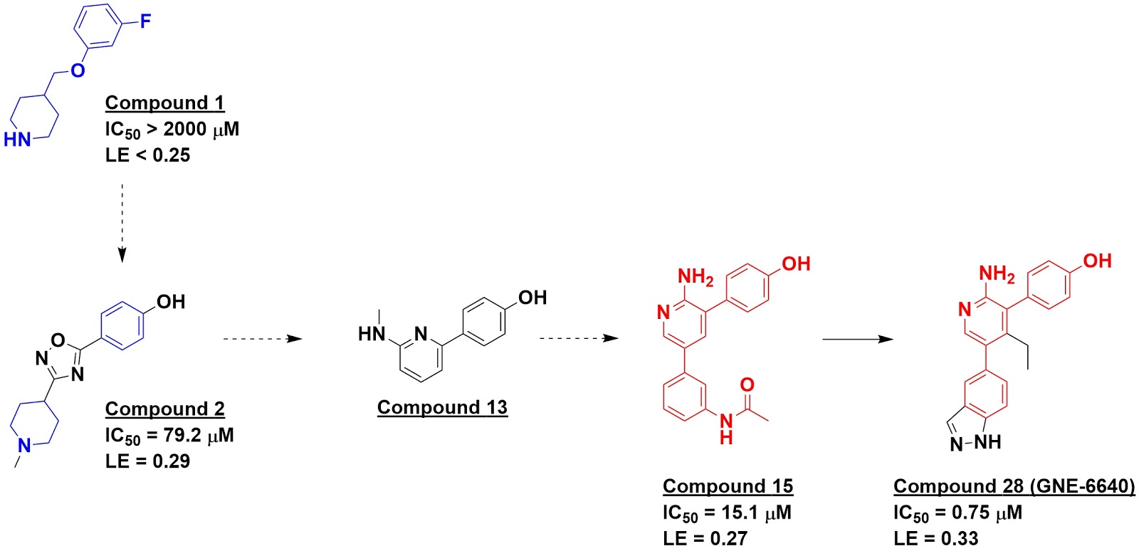
The first, published in J. Med. Chem. by Paola Di Lello, Vicki Tsui, and coworkers at Genentech, started with an NMR fragment screen. This identified molecules such as compound 1, which NMR data suggested bound near the active-site cysteine. This and other fragments were used to conduct virtual screens of the much larger Genentech library, and 21 of these were then tested experimentally. Most of these either didn’t bind, bound to multiple sites, or caused protein aggregation, but four of them, including compound 2, showed clear binding to a specific site on USP7 and also inhibited the enzyme in a biochemical assay.

Surprisingly, protein-detected NMR suggested that these four molecules did not bind in the active site as expected but rather in an adjacent “palm site”, a hypothesis that was confirmed by a crystal structure of compound 2 bound to USP7. This led the researchers to reexamine other hits from the original NMR screen, where they identified several aminopyridinephenols, such as compound 13.


Meanwhile, a biochemical HTS against USP7 had identified 76 hits, but most of these turned out to be artifacts, and none of them yielded co-crystal structures with the enzyme. The fragment findings led the researchers to revisit some of the weaker hits that had been overlooked, such as compound 15. This led to a crystal structure showing binding in the palm site, and further medicinal chemistry ultimately led to molecules such as compound 28 (GNE-6640), with nanomolar activity in both biochemical and cell-based assays. A separate paper in Nature characterizes the biology in more detail, revealing that molecules in this series interfere with ubiquitin binding and are highly selective for USP7.

Another fragment effort on this target was reported by Timothy Harrison and collaborators at Almac and Queen’s University, Belfast in Nat. Chem. Biol. An SPR screen of 1946 fragments against the catalytic domain of USP7 led to compounds such as fragment B. This was combined with molecules from other groups that had been reported in the literature, leading to compound 1. Subsequent medicinal chemistry, informed by crystallography, led to compound 4, with low nanomolar biochemical and cell-based activity and excellent selectivity. The enantiomer is much less active, and compound 4 should be a useful chemical probe to further understand the biology of USP7.


Remarkably, not only do the two series of molecules bind some distance away from the active site cysteine (yellow, upper right), they bind in completely different, non-overlapping sites!

These papers illustrate the importance of allosteric sites for tackling specific members of large protein families. They are also both cases of “fragment-assisted drug discovery.” Unlike many success stories we’ve highlighted, it is difficult or impossible to find the initial fragment in the final molecules. Heck, Genentech’s best molecules bind in a completely different site from where the first fragment hits bound. Being open to such possibilities, and using all available data from every possible source, are keys to success.

26 December 2017

Review of 2017 reviews

The year is done, and the darkness
Falls from the wings of Night.

As we've done since 2012, Practical Fragments is using the last post of the year to highlight conferences as well as reviews not previously discussed.

Significant events included the venerable CHI FBDD meeting in San Diego, the NovAliX Biophysics conference in Strasbourg, and the first-ever fragment conference in Shanghai. We discussed a special issue of Essays in Biochemistry devoted to structure-based drug design, and Teddy came out of retirement to provide an entertaining summary of his experience putting together a book on biophysics in drug discovery - well worth reading if you're ever tempted to edit one yourself.

As in years past, several reviews were devoted to the broad topic of FBDD. Below, I’ll outline the general reviews, followed by those focusing on particular targets, techniques, and other topics.

György Keserű (Hungarian Academy of Sciences) and Mike Hann (GlaxoSmithKline) ask “what is the future for fragment-based drug discovery?” in Fut. Med. Chem. After a concise summary of the topic, they answer that it “includes target discovery and validation, the development of chemical biology probes, pharmacological tools and more importantly drug-like compounds.” In other words, the future looks bright.

FBDD is more comprehensively covered by Ben Davis and Stephen Roughley (Vernalis) in Ann. Reports Med. Chem. This is a complete, self-contained guide to the field, covering everything from history, theory, fragment library design, and fragment-to-lead approaches. It is ideal for a newcomer, but there are enough insights throughout that it makes a rewarding read for experts too.

Of the thirty-plus fragment-derived drugs that have made it to the clinic, none are directed against neglected diseases. Gustavo Henrique Goulart Trossini and colleagues at Universidade de São Paulo review some of the work that has been done in this area in Chem. Biol. Drug Des.

And rounding out general reviews, Christopher Johnson (Astex) and collaborators examined all 28 successful fragment-to-lead programs published in 2016, defined as at least a 100-fold improvement in affinity to a 2 µM or better compound. This is a sequel to our analysis of the 2015 literature, also published in J. Med. Chem., and many of the trends are similar. Interestingly, many leads maintained high ligand efficiencies, and there was no correlation between the “shapeliness” (deviation from planarity) of fragments and that of the resulting leads. Consistent with our recent poll on the importance of structural information, 25 of the 28 examples used crystallography at some point.

Targets
Three of the success stories from 2016 involved bromodomains, the subject of an entire month of Practical Fragments’ posts last year. In Arch. Pharm., Mostafa Radwan and Rabah Serya (Ain Shams University, Cairo) review this target class, with a particular emphasis on the four BET family proteins.

More than 30% of enzymes are metalloenzymes, yet these are targeted by fewer than 70 FDA-approved drugs. One of the first published examples of FBDD involved a metalloenzyme, but most efforts have been focused on a limited set of metal-binding pharmacophores, such as hydroxamic acids. Seth Cohen (University of California, San Diego) has been steadily building libraries of metallophilic fragments, and in Acc. Chem. Res. he describes how this approach can lead to new classes of inhibitors.

Protein-protein interaction inhibitors are another underrepresented class of drugs, though one approved FBDD-derived molecule falls into this category. In Methods, Daisuke Kihara and collaborators at Purdue University look at in silico methods to discover PPI inhibitors, including fragment-based approaches.

Unlike PPIs, kinases have been highly successful drug targets. We recently highlighted one review of cyclin-dependent kinases (CDKs), and in Eur. J. Med. Chem. Marco Tutone and Anna Maria Almerico (Università di Palermo) provide another. Although the main focus is on in silico methods, there is a section on FBDD.

Techniques
As noted above, X-ray crystallography has played a role in most successful fragment to lead programs. In the open-access journal IUCrJ, Sir Tom Blundell (University of Cambridge) provides an engaging and personal view of protein crystallography, a field in which he has played a starring role, starting with his early involvement in determining the crystal structure of insulin. He also notes that the interchange of ideas and techniques between academia and industry has long been a crucial driver of advances.

NMR was the first practical method used for FBDD, so it is not surprising that there are several reviews on the topic. In Arch. Biochem. Biophys., Michael Reily and colleagues at Bristol-Myers Squibb provide a detailed overview of NMR in drug design. This covers not just the ligand- and protein-detection methods often used in fragment screening, but also more intensive techniques to characterize protein-ligand interactions.

A briefer look at many of these topics is provided by Yan Li and Congbao Kang (A*STAR) in Molecules. This review also highlights more unusual approaches such as NMR experiments on living cells.

Artifacts are a fact of life in both FBDD and HTS, and it is always important to recognize these early. In J. Med. Chem. Anamarija Zega (University of Ljubljana) discusses how NMR can help. This includes methods to detect aggregators and covalent modifiers. Of course, NMR methods can introduce their own artifacts, and these are also covered.

Other topics
Speaking of artifacts, PAINS are responsible for quite a few. The term “PAINS” has also been somewhat controversial, and in a new paper in ACS Chem. Biol. Jonathan Baell (Monash University) and J. Willem Nissink (AstraZeneca) examine the “utility and limitations” of the term Jonathan coined seven years ago. As they acknowledge, the PAINS filters were derived from just 100,000 compounds run in a limited set of assays. This means that not every bad actor will be recognized by PAINS filters, and some compounds that are may only be PAINful in certain assay formats. Like Lipinski’s rule of 5, it is important to recognize the limits of applicability. As the authors note, “the key is to remain evidence-based.”

Another sometimes controversial topic is ligand efficiency and associated metrics, the subject of an analysis in Expert Opin. Drug Disc. by Giovanni Lentini and collaborators at the University of Bari Aldo Moro. This includes extensive tables of rules and metrics, both common and obscure. The authors note that, while metrics can be useful, it is important not to use them as a “magic box.” As they quote William Blake, “to generalize is to be an idiot.”

Shawn Johnstone and Jeffrey Albert (IntelliSyn Pharma) discuss pharmacological property optimization for allosteric ligands in a review in Bioorg. Med. Chem. Lett. As we recently noted, fragments are particularly suited for discovering allosteric sites, and this paper discusses how to characterize these.

Finally, Jörg Rademann and collaborators at Freie Universität Berlin discuss protein-templated fragment ligations in Angew. Chem. Int. Ed. Earlier this year we highlighted some of his work, and this review provides a thorough analysis of both reversible and irreversible approaches, with good discussions of detection methods, chemistries, and case studies.

That’s it for the year. Thanks for reading, and especially for commenting.

And may 2018 be filled with music, and light.