30 December 2010

Flattening fragments

If you want to visualize and manipulate crystallographic data there are plenty of options, such as PyMOL and OpenAstexViewer. However, powerful molecular graphics programs such as these have a learning curve associated with them, and sometimes it’s nice to get a simple two-dimensional image. In the December issue of ACS Medicinal Chemistry Letters, Katrin Stierand and Matthias Rarey describe software they’ve developed to do this.

PoseView, which can be used online free of charge, automatically generates a two-dimensional view of a protein-ligand complex and labels five different types of interactions: hydrogen bonds, metal interactions, pi-cation interactions, pi-pi stacking, and undirected hydrophobic contacts.

The researchers tested their software by using it to draw nearly every protein-ligand interaction in the Protein Data Bank (PDB) in which the ligand has 5 to 80 atoms, excluding crystallization buffer components and things like heme or iron-sulfur clusters. For the remaining 200,000+ complexes, PoseView successfully generated plots for 85% of them; interestingly, most of the failures resulted because there were no apparent interactions between the protein and “ligand.” Of the successes, 80% produced high-quality figures in which there were no overlapping features – not bad considering the challenge of reducing a three-dimensional model to a two-dimensional image.

The paper includes some interesting data about protein-ligand complexes in general. For example, over 90% of complexes in the PDB have less than 11 directed interactions (ie, interactions other than hydrophobic contacts), with the largest number having only a single directed interaction. It would be interesting to see how these statistics apply to fragment-sized ligands.

PoseView is incredibly easy to use: just enter a pdb code or upload a structure, and the program spits out an image. I tried it on the very first fragment I discovered, and the result looks quite attractive:

Of course, there is always room for improvement: for example, it would be nice if you could change fonts or colors (the output is a pdf). Still, this is a useful addition to the toolbox of modeling programs.

And with this post, Practical Fragments says goodbye to 2010 and wishes everyone a happy holiday season. We look forward to more exciting developments in 2011 – and if we miss any, please send them our way!

22 December 2010

SAR by Crystallography – minus the “A”

Practical Fragments has highlighted at least a couple papers (here and here) where fragments were identified by crystallography but where functional activity was not reported. Indeed, most experienced fragment hunters will be able to point to cases where they observed well-defined electron density but were unable to measure inhibition. Although this is often a source of frustration, researchers from Johnson and Johnson decided to avoid measuring activity altogether until after a couple cycles of crystallography-guided library design, as they describe in a paper published this month in J. Med. Chem.

The researchers were interested in the protein ketohexokinase, an ATP-dependent enzyme that is a potential target for metabolic diseases. They assembled a primary fragment screening library of 900 compounds with the following characteristics:

• 6-15 non-hydrogen atoms
< 3 hydrogen bond acceptors
< 3 hydrogen bond donors
< 2 rings
• No unspecified chiral centers
• No unpleasant functionalities (peroxides, acyl halides, etc.)
• Prioritization given to molecules found in known pharmaceutical compounds (similar to the Fragments of Life)

This is a fairly standard set of criteria for assembling a fragment library. The researchers broke with convention by pooling fragments into groups of 5, with the members of a given fragment pool as structurally similar as possible. Normally when researchers pool libraries for crystallography-based fragment screening the idea is that each member of a given pool will be structurally unique to facilitate identification. In this case, though, the researchers were interested in observing general features of how fragments bind to the target protein.

Crystals of hexokinase were soaked with the pools, and 60 of these pools yielded electron density in the active site – a very high hit rate of 30%. A number of generalizations could be made about what molecular features were preferred in different parts of the binding site, and this information was used to build a secondary library of about 350 compounds based on 6 scaffolds; the idea was to merge or grow fragments found in the primary screen. These compounds were similarly pooled and screened crystallographically but not functionally, yielding hits from 4 of the scaffolds. The structures of these hits were then used to generate a third library of several hundred compounds around at least 4 separate scaffolds. These compounds were then tested individually to see if they could inhibit ketohexokinase, and were also characterized crystallographically. Gratifyingly, a number of these were active at submicromolar potency, including compound 6.


In addition to showing decent potency against ketohexokinase, compound 6 exhibited respectable drug-like properties as well, including lack of activity against the CYP450 enzymes, high oral bioavailability, and good selectivity against receptors, ion channels, and protein kinases, though other ribokinases were not tested.

This is an interesting and potentially useful approach, but it would be fun to see how it compares with a more “traditional” strategy, where individual fragments are advanced. It should be relatively straightforward to assay fragments from the first and second library for affinity; was fragment 3 (precursor to compound 6) one of the most potent or ligand efficient? Or was the information from fragments with no detectable activity instrumental in delivering the best compounds?

17 December 2010

Fragment-based job listings

The fragment-based community is very diverse, with practitioners coming from biochemistry, biophysics, chemistry, computational chemistry, and many other disciplines. Communicating job openings to such a fragmented community can be difficult, but we think Practical Fragments can provide a common place for employers who want to build up fragment expertise of any flavor. If you have an opening you are trying to fill, please add it as a comment to this post. These comments will then be listed in the “Job Openings” link under the “Links of Utility” heading on the right side of the page, so they will always be easy to find.

(Note: this is for positions in fragment-based drug discovery only. There is also a LinkedIn group that lists jobs with a broader but overlapping focus.)

10 December 2010

Hsp90 and fragment linking

There has been no shortage of fragment-based approaches directed toward the anti-cancer target Hsp90, most of which have relied on growing fragments (see here for some impressive recent examples). Researchers from Abbott published a report providing a couple examples of linking fragments against this target a few years back, but in those cases the ligand efficiencies of the linked molecules were dramatically lower than those of the initial fragments. In a recent paper in ChemMedChem, researchers from Evotec describe an example that maintains the ligand efficiency.

The research group had previously conducted a fragment screen against Hsp90, resulting in a number of hits. In the new paper, fragment hits 1 and 2 (see figure) were both found to have fairly low affinities, but were characterized crystallographically. Interestingly, fragment 2 could adopt at least two very different conformations, depending on whether it was co-crystallized in the presence of fragment 1. In the ternary structure, the two fragments come within about 3 Å of each other, and molecular modeling suggested that four atoms should be able to link them.


Gratifyingly, when such a compound was made and tested, it inhibited the enzyme several hundred-fold more tightly than either of the initial fragments. The crystal structure revealed that the compound binds similarly to the ternary structure of Hsp90 and fragments 1 and 2.

The authors note that “the binding free energy of the linked fragment 3c was found to be exactly the sum of those of the original two fragments.” Of course, this is still a long way from an ideal linking situation: as noted earlier this year a good linker should lead to super-additivity (an improvement of ligand efficiency), not just additivity (maintenance of ligand efficiency). Nonetheless, this example is still better than many attempts at linking, which often are less than additive.

02 December 2010

Fragment-based conferences in 2011

As 2010 winds to a close, it’s time to start planning for 2011. As in previous years, fragment events seem to cluster in the first half of the year, so start planning now!

February 21-22: SMi’s 10th Annual Advances & Progress in Drug Design Conference will be held in London, and has a number of fragment-based talks. There is also a separate half-day post-conference workshop on the topic on February 23.

February 23-25: CHI’s Molecular Medicine Tri-Conference will again be held in my beautiful city of San Francisco, with a program on medicinal chemistry that includes a fragment section. Notes on this past year’s meeting can be found here.

March 7-8: Fragments 2011, the Third RSC-BMCS Fragment-based Drug Discovery meeting, will be held in Stevenage, UK. This is a biennial event; the last was in Alderley Park in 2009 (you can read about it here and here). Registration is now open, as is a call for posters through January 31.

April 12-13: Cambridge Healthtech Institute’s Sixth Annual Fragment-Based Drug Discovery will be held in San Diego, with at least one pre-conference short course on the topic on April 11. You can read impressions of this past year’s meeting here.

Know of anything else? Organizing a fragment event? Let us know and we’ll get the word out.

29 November 2010

More techniques: NovAliX and Graffinity combine MS and SPR

We’ve written previously about Graffinity, which uses surface plasmon resonance (SPR) to find fragments, and NovAliX, which initially focused on mass spectrometry. The two companies have been collaborating since last year, and this has apparently been a fruitful partnership: this month NovAlix acquired a majority ownership stake in Graffinity (click here for press release). Earlier this year NovAlix also purchased an NMR-focused company, and they have already added crystallography expertise. Finding fragments effectively requires using a range of orthogonal technologies, and this latest move gives NovAlix a full suite of biophysical techniques.

12 November 2010

Fragments vs PDK1

Kinases have been a particularly productive target class for fragment-based drug discovery (and drug discovery in general), with nearly half of reported FBDD-derived clinical candidates targeting kinases. The latest dispatch from this field can be found in the November issue of ACS Medicinal Chemistry Letters.

In this paper, Jeffrey Axten and colleagues at GlaxoSmithKline describe their use of fragment screening to identify inhibitors of PDK1, a popular anti-cancer target. They started by assembling a library of fragments biased towards the purine-binding site of kinases, and tested 1065 of these in a biochemical screen at 400 micromolar concentration. Of these, 193 inhibited activity at least 60% and were further characterized; 89 had IC50 values better than 400 micromolar. A set of 36 of these, chosen on the basis of ligand efficiency and chemical tractability, were chosen for follow-up.

Saturation transfer difference (STD) NMR was used to confirm which fragments bound to PDK, which cut the number of hits in half. X-ray crystallography experiments were started before NMR and performed on 7 fragments; only the fragments that were confirmed by NMR gave interpretable data. One of these was the aminoindazole compound 8 (see figure).
A substructure search was conducted to find more elaborated molecules within the corporate screening collection, leading to compound 19, which has sub-micromolar potency. This compound also showed some signs of selectivity for PDK1 over other kinases. Although the paper stops here, Jeffrey Axten gave a nice presentation at FBLD 2010 in which he discussed subsequent medicinal chemistry that ultimately led to novel, high picomolar inhibitors of PDK1.

There are at least two lessons from this story. First, the significant attrition from the biochemical screen again emphasizes the need for orthogonal methods of fragment validation. Second, even though the fragment identified has been around the block with respect to kinases (as of last year, the aminoindazole substructure had appeared in over 70 kinase patents), skillful medicinal chemistry can still get you to novel compounds.

07 November 2010

Pin1 revisited

Earlier this year we highlighted a paper from Vernalis that described the use of NMR methods to discover inhibitors of the anti-cancer target Pin 1. In a recent issue of Bioorg. Med. Chem. Lett. the same team now reports a second series of compounds that inhibit this protein, also discovered and advanced through FBLD. The two papers together provide some interesting lessons.

Rather than using NMR, the researchers identified the second series of compounds with an inhibition assay. After screening 900 fragments at 2 mM, they obtained 40 hits, including 3 compounds previously discovered by NMR. Disturbingly though, follow-up NMR experiments confirmed binding for only 2 of the 37 new hits, suggesting that the remaining compounds may act through pathological mechanisms (see also here). Still, two hits are better than none, and the binding mode of one of the fragments (compound 3 in figure) was determined by X-ray crystallography. Several analogs of this were purchased and tested, and compound 5 was found to have an improved potency and ligand efficiency.


At this point chemistry entered the picture. The researchers synthesized several analogs of compound 5, guided by crystallography and modeling. This led to compound 10e and eventually to compound 20, with sub-micromolar biochemical activity and measurable cell activity.

Ultimately though, as in the previous Pin1 series, even this modest cellular potency was gained at the cost of unacceptable increases in size and hydrophobicity. This brings up an interesting question: at what point do you declare a target undruggable? The authors note that “the nature of the Pin1 active site makes it difficult to optimise hits into drug-like molecules.”

Fragment-based approaches can sometimes deliver inhibitors to challenging targets where HTS has failed. However, if the inhibitors can’t be transformed into drugs, is finding them actually a good thing? Researchers are getting better at improving potency at the same time as ligand efficiency for some targets, but ultimately getting to clinical candidates for harder targets may come down to how many resources you are willing to throw at a project: molecules such as ABT-263, though far from rule-of-5 compliant, are doing well in the clinic, but only after the investment of dozens if not hundreds of people-years.

03 November 2010

Ligand efficiency hot spots

Hot spots are regions of a protein with a particular predilection for binding to small molecules – thermodynamic sinkholes, so to speak. Discovering one of these can get you to potent molecules very quickly. In an effort to better understand hot spots, Iwan de Esch and colleagues at VU University in Amsterdam and collaborators at Beactica have deconstructed a potent ligand for nicotinic acetylcholine binding protein (AChBP), a model protein for ligand-gated ion channels, which are implicated in a variety of neurological diseases. They report their results in a recent issue of J. Med. Chem.

The researchers started with the previously reported quinuclidine compound 6 (see figure) and fragmented this into 20 analogs. They tested these in a surface plasmon resonance (SPR) assay as well as in a more conventional radioligand binding assay; the agreement between these very different assay formats was excellent, further validating the utility of SPR as a useful tool for discovering fragments.


Not surprisingly, some of the fragments have higher ligand efficiencies than the larger, more potent molecule, suggesting that there is a hot-spot that recognizes the core fragment 25 (which is structurally related to nicotine). This concept of “group efficiency” has been described previously, and can be useful for optimizing fragments. For example, compound 22, without a basic nitrogen atom, has the lowest ligand efficiency in the bunch; presumably, simply adding the nitrogen would give a sizable boost in potency.

However, one needs to be cautious. The researchers use computer docking to develop models of how each of these fragments bind, but as we have seen before, isolated fragments do not always recapitulate the binding modes of fully elaborated molecules. Still, particularly in the absence of structure (as is the case with many ion channels), exercises such as this could provide useful ideas for what to do with fragment hits.

29 October 2010

Fragment linking for specific Bcl-2 inhibitors

One of the most well-known examples of a fragment-based program that has yielded a clinical compound is Abbott’s Bcl-2 effort: ABT-263 is currently in over a dozen trials for various cancers. However, this molecule hits several proteins in the Bcl-2 family, and a more specific inhibitor of Bcl-2 alone may have lower toxicity. Phil Hajduk and colleagues have used SAR by NMR to do this, as they report in the latest issue of Bioorg. Med. Chem. Lett.

The researchers started with a protein-detected NMR screen of 17,000 compounds; compound 1 (see figure) was found to be fairly potent, and also roughly 20-fold selective for Bcl-2 over the related protein Bcl-xL. Hajduk’s team did not have a crystal structure at this point, but they were able to use NMR to determine that the compound lies in a large hydrophobic groove. This is the same groove found to bind biaryl acids in earlier work, so the researchers screened a set of 70 of these to see if they could bind in the presence of compound 1. Interestingly, compound 6 was equally potent in the presence or absence of compound 1, suggesting that both fragments could bind simultaneously.
NMR was then used to determine the ternary structure of fragments related to compound 1 and compound 6 bound to Bcl-2, and linking these led to compound 25, with high nanomolar potency. Although this represents a good boost in potency, the binding energies were not additive (let alone synergistic). An NMR structure of one of the linked molecules revealed that, although it binds in the same groove as its component fragments, its position is shifted, and also that one of the protein side chains moves to deepen a hydrophobic pocket. Additional chemistry to fill this pocket led to compound 29, with 40 nM biochemical potency and measurable cell activity, as well as greater than 1000-fold selectivity for Bcl-2 over Bcl-xL and at least 28-fold specificity over other Bcl-2 family members.

This is a nice example of starting with a modestly selective fragment (albeit a jumbo-sized one) and, through fragment linking, increasing both the potency and specificity towards the target protein.

24 October 2010

Small but PAINful

We’ve written previously about the phenomenon of compound aggregation, and how this can lead to false positives in high-concentration screening (see also here). Unfortunately, aggregation is not the only thing that can trip you up. Earlier this year Jonathan Baell and Georgina Holloway published a description and systematic catalog of “Pan Assay Interference Compounds,” or PAINS, and Baell has now followed up on this with a Perspective in the October issue of Future Medicinal Chemistry. (For those of you without journal access, he has also summarized some of this material here.)

PAINS are compounds that frequently show up as screening hits, but that act through non-specific mechanisms such as covalent attachment to proteins or generation of hydrogen peroxide. Sometimes it is not the compound itself that is problematic but a breakdown product or leftover reactant; most readers with experience in hit-to-lead discovery will experience a painful sense of déjà vu reading about hits that did not confirm when the compound was resynthesized or repurified. This problem is exacerbated when trifluoracetic acid (TFA, which is toxic to cells and can accelerate compounds’ decomposition) is used in reverse-phase purification of compounds; apparently Pfizer has systematically repurified hundreds of thousands of their screening compounds to remove traces of TFA.

The problem with PAINS is that they may show convincing biochemical and even cell based activity, but mechanistically be useless for further advancement to drugs or even chemical probes. Unfortunately this does not prevent them from being published, where they are picked up by other researchers and identified as “privileged pharmacophores” by computational chemists. This leads to more (wasted) research and more (useless) publications in a vicious circle Baell terms “pollution of the scientific literature.”

Although the focus of the Perspective is high-throughput screening, fragments can be PAINS too, as the sampling below shows.


Compound 1 is obviously a Michael acceptor and compound 2 is unsurprisingly reactive with cysteine, but the problems with compound 3 are less obvious, and compound 4 generates hydrogen peroxide in the presence of reducing agents and oxygen – a mechanism it likely took some time to track down.

One of the fun aspects of reading this paper is that Baell is not shy about calling out high-profile publications that are likely to report false positives, though he readily admits that he too has been misled. The question is what to do: using computational filters to get rid of the worst offenders only eliminates 5-12% of commercial compounds, but more stringent screens can eliminate upwards of 95%!

Baell calls for better stewardship of the scientific literature:
Journals therefore have a responsibility here because if misleading information is published, unintentional though it may be, it is more likely to propagate and be taken as fact by others who may then initiate flawed research projects. Research resources are too precious for this to be acceptable.
I agree whole-heartedly with this, though given what I see published on a weekly basis I despair of this happening any time soon. Ultimately of course the onus is on researchers to carefully follow up and fully understand the mechanism of their hits, particularly those with dubious structures: don’t contribute to the pollution yourself, and don’t let friends (or papers you review) pollute!

14 October 2010

FBLD 2010

The last major fragment event of this year is over, but it ends on a high note: FBLD 2010 has remained true to its predecessors in bringing together a great group of fragment enthusiasts in a Gordon-Conference-like environment. With 30 talks and even more posters I won’t attempt to be comprehensive or even representative, but will instead just pick out a few themes. Those of you who were there, please chime in with your own observations.

One of the themes was the shape of chemical space, and what makes a good binder. Jean-Louis Reymond, who has been systematically enumerating all stable molecules containing carbon, nitrogen, oxygen, and a few other atoms, has already published up to 13 heavy atoms but is now expanding his analysis to molecules containing up to 17. The issue of whether more attention should be given to three-dimensional fragments was discussed, with Ken Brameld reporting that fragments in crystal structures at Roche and the protein data bank contain fewer “flat” compounds than does the ZINC database of commercial molecules. However, analyzing 150,000 molecules with MW < 300 that had been screened in 40-100 high-throughput screens at Roche did not show any shape differences between the 50,000 molecules that showed up in at least one screen and those that didn’t. Interestingly, this ratio also came up in a talk by Tony Giannetti of Genentech, who said that across 13 screens 36% of their fragments hit at least one protein, while the rest didn’t hit any. Vernalis has found similar results; is there any way to enrich for the productive binders?

While FBLD 2009 had a strong computational theme, a major thrust of this conference was using biophysics to detect and confirm fragment binding. Tony discussed best-practices in SPR, and noted that since small molecules are “brighter” in NMR assays than proteins it is possible to find even very tiny fragments, including a 6 heavy-atom compound with a Kd of 600 micromolar. Tony also described the use of SPR for weeding out badly behaved compounds. Spookily, he noted that promiscuity is a function of compound, protein, and buffer, so it is not possible to weed out bad actors in a library before screening: one compound that was promiscuous against 8 targets bound legitimately and gave a crystal structure with a ninth. Adam Renslo of the University of California San Francisco described how easy it is to be misled by such phenomena. Glyn Williams described how Astex uses biophysical techniques to detect problem compounds, and noted that oxidizers can be particularly insidious – a trend that will likely continue as people explore novel heterocycles.

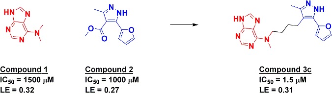
Glyn also presented a fascinating if slightly depressing discussion of ligand efficiency. As many have found, it can be challenging to maintain ligand efficiency during the course of fragment optimization. Yet even this goal is too modest. A fragment pays about 4.2 kcal/mol in binding energy when it binds to a protein due to loss of rotational and translational entropy; since this enropy cost is only paid once, atoms added to this molecule do not have this liability . Thus, merely maintaining ligand efficiency means that the atoms being added are binding less efficiently. This point was also emphasized by Colin Groom of the Cambridge Crystallographic Data Centre.

Membrane proteins are increasingly being targeted by fragment-based methods, as recently discussed on this site, and both Gregg Siegal of ZoBio and Rebecca Rich of the University of Utah presented progress against GPCRs.

There was general agreement that many approaches can find fragments, and that using several orthogonal methods is a good way to separate the true binders from the chaff, but a continuing challenge is what to do next. The last two sessions were devoted to chemical follow-up strategies and success stories. Some of these have been at least partially covered on Practical Fragments (for example here, here, and here) but there were a number of unpublished examples too – we’ll try to discuss these individually as they emerge.

If you missed this or the previous two conferences you’ll have another chance in 2012, when the meeting will be held in my fair city of San Francisco. And if you can’t wait that long, there are at least two fragment conferences scheduled for next year – details to come shortly.

06 October 2010

Commercial fragments – how do they compare?

Earlier this year we updated our list of commercial fragment suppliers. Now Chris Swain at Cambridge MedChem Consulting has analyzed the structures and properties of eleven of these.

A major conclusion, which will be a disappointment to purchasers of fragments but a boon to suppliers, is that there is very little overlap in terms of exact molecules. Given the vastness of chemical space this shouldn’t be too much of a surprise, but it is striking that, of the 40,000+ molecules represented, less than a dozen are sold by four or more companies. Looking at molecular similarity rather than identity increases the amount of overlap, but for the most part each collection is quite differentiated from the others.

Where it gets really interesting is in the analysis of chemical properties, which Swain has calculated for each fragment set. These include cLogP, molecular weight, polar surface area, H-bond donors and acceptors, heavy atom count, and rotatable bond count. Here the collections are dramatically different, with some being strictly Rule-of-3 compliant while others are much less so. There are also interesting differences in distributions: some collections are distributed around a low molecular weight median, while others are biased towards larger molecules. Finally, an analysis of molecular diversity reveals some collections to be very diverse while others have clusters of closely related molecules.

Of course, people differ in how much weight to put on simple molecular properties. Also, an analysis such as this is necessarily a static snapshot: commercial offerings change over time, and new suppliers continue to enter the market. Moreover, some of the companies offer many more fragment-sized molecules as extension sets beyond their core fragment collections. Still, this is a valuable resource for anyone building or expanding a custom fragment collection. The only thing that would make it even more useful would be price per fragment!

02 October 2010

TINS and STD and SPR – oh my!

Following up on our last post on the use of the NMR technique TINS applied to a membrane protein, the same research group has now compared TINS with other techniques on a more conventional target. In addition to TINS, they conducted fragment screens using another commonly used NMR technique as well as surface plasmon resonance (SPR) and high-concentration screening; the results appear in the Journal of Biomolecular Screening.

TINS involves immobilizing a target protein onto a resin, then flowing fragments across the resin and determining whether they bind to the target as assessed by a reduction in their NMR amplitudes. A reference protein is evaluated at the same time; only fragments that bind to the target protein and not the reference are considered hits. In this case, the researchers chose the viral protein RNA-dependent RNA Polymerase (RdRP) as the target and the PH domain from the human protein Akt1 as the reference. Using a total of 4 mg of RdRP, they screened a library of 1270 commercially available fragments in pools of 3 to 5 compounds each, resulting in 74 hits.

One of the most commonly used NMR techniques for fragment screening is saturation transfer difference (STD), in which the magnetization of the protein target is saturated, and so magnetization transfers to any ligands bound to the protein. The researchers tested 133 fragments (both hits and non-hits from the TINS experiments) and found a total of 49 hits, of which 40 had also been found by TINS.

The 83 fragment hits from both TINS and STD were tested for their ability to inhibit polymerase activity at concentrations up to 2 millimolar; 70 of them showed some activity, and a few of these seemed to actually activate the enzyme.

Finally, a selected set of 62 fragments (all of which were hits in at least one of the three assays) were tested in an SPR assay at concentrations up to 0.2 millimolar. Of these, around half showed binding, and these tended to be the fragments that showed the greatest activity in the enzymatic assay.

The authors conclude that TINS picks up more hits than the other assays, though high-concentration screening comes close. This may be true, but it would have been nice if they had run the entire set of 1270 fragments through each of the different methods; it is possible that there were false negatives in the TINS experiments that could have been picked up by the other techniques. Moreover, some of the TINS hits that didn’t confirm in other assays may well have been false positives. Still, there are lots of useful data in this paper, and it demonstrates yet again the importance of using multiple, orthogonal techniques to discover and properly validate fragments.

28 September 2010

Fragments vs membrane proteins with TINS

Fragment-based ligand discovery owes much of its success to the rise of biophysical techniques such as NMR, crystallography, and – more recently – surface plasmon resonance. These have allowed the discovery of fragments against a wide range of proteins, but one notable exception has been membrane proteins, the targets of more than half of marketed drugs. In a recent issue of Chemistry and Biology, Gregg Siegal and colleagues take a crack at this diverse group of proteins.

The researchers, from Leiden University, ZoBio, and elsewhere, use an NMR-based technique called target immobilized NMR screening, or TINS. In this method, a protein is immobilized onto a solid support. A reference protein is also immobilized; this reference is usually a well-characterized protein that does not bind to many small molecules. Each protein is then put into its own compartment of a two-compartment flow-cell, and this is inserted into an NMR spectrometer. Mixtures of fragments are then flowed through both chambers: those that interact with protein show a reduction in the amplitudes of their NMR spectra. By choosing fragments that show such a reduction for the target protein and not the reference protein, fragments that bind to the target can be differentiated from those that bind to proteins in general. After each NMR experiment, the fragments are washed away and replaced with a new set of fragments. TINS has been applied to a number of soluble proteins, as reviewed here. Remarkably, the immobilized protein samples often remain stable through hundreds of screening cycles.

Membrane proteins are notoriously difficult to crystallize or characterize by NMR. Moreover, it is often difficult to obtain enough protein to work with. However, since TINS relies on a decrease in signal from the fragment rather than a signal from the protein itself, Siegal and colleagues tested whether they could use the technology to discover fragments that bind to membrane proteins.

The researchers chose a protein called disulphide bond forming protein B (DsbB), which is found on the inner membrane of E. coli and other Gram-negative bacteria and may be important in virulence factor folding. One of the challenges of membrane proteins is keeping them properly folded, and the researchers used two different approaches to do this, either detergent micelles or “nanodiscs,” lipid bilyaers surrounded by a scaffold protein. Using less than 2 milligrams of DsbB, the researchers used TINS to screen a set of 1071 fragments in groups of about 5 each, with each fragment present at 500 micromolar concentration, a process that took 5 and a half days.

The TINS process led to 93 hits, a respectable hit rate of 8.7%. Each of these was then tested in a functional assay at 250 micromolar concentration, and more than half of the hits inhibited DsbB activity by at least 30%. Eight of these were subsequently characterized using full IC50 curves and kinetic analysis. The potencies were impressive, ranging from 7 micromolar to 193 micromolar, with ligand efficiencies as high as 0.45 kcal/mol/atom. DsbB has the advantage that it has been characterized structurally, and the researchers used chemical shift information from 2-dimensional NMR experiments to show that the fragments could be divided into two groups, with one set competing with a quinone cofactor and the other binding at a different site.

This paper demonstrates that it is possible to find fragments that bind to membrane proteins. Of course, the next question is, what can you do with the fragments? In this case there were structural data about the target, but this will not generally be true for membrane proteins, and in the absence of structure, advancing fragments to leads can be challenging. On the other hand, medicinal chemists have been developing drugs against membrane targets for decades without knowing their precise structures, so perhaps the challenge is as much psychological as scientific.

20 September 2010

Allosteric FPPS inhibitors – not so negative

The protein farnesyl pyrophosphate synthase (FPPS) has long been a target of drugs for osteoporosis, and some data have suggested it could be a productive target for other diseases too. However, approved drugs that target FPPS contain two phosphonates, highly negatively charged moieties that, while nicely directing the drugs to bone, lead to low plasma and soft tissue levels. Researchers at Novartis have now identified fragments that bind to an allosteric site on FPPS and so lack the phosphonate moieties. An article in the September issue of Nature Chemical Biology describes the discovery of these fragments and how they were advanced to nanomolar inhibitors.

The researchers, led to Wolfgang Jahnke in Basel, Switzerland, started by screening a library of only 400 fragments using NMR. Several low affinity (millimolar) hits such as compound 1 were identified, but surprisingly these were not competitive with bisphoshonate drugs, and some even bound synergistically. Crystallography revealed that they were binding in a previously undiscovered allosteric site. (The same group also used fragment screening to explore an allosteric site in another protein.)


To follow up on these observations, the researchers tested 40 related compounds in the Novartis internal compound collection, again using NMR to detect binding. This led to the more potent compound 5, and two rounds of focused library assembly and screening led to low micromolar inhibitors such as compound 7. Structure-based design led to molecules such as compound 11, which has comparable potency to approved drugs that target FPPS. Compound 11 was further characterized using ITC and crystallography, and although its two carboxylic acids likely account for a relatively low cellular permeability, it does not show any affinity for bone.

Interestingly, a high-throughput screen conducted against FPPS did not yield any inhibitors with an IC50 better than 5 micromolar. So in this case not only did a fragment-based approach discover a new series of molecules against a new site on an old target, it succeeded where conventional HTS didn’t.

13 September 2010

Protein-templated click chemistry – just add copper

We’ve written previously about protein-templated chemistry (here and here), in which a protein catalyzes the formation of a more potent inhibitor from two lower affinity fragments. Of course, proteins aren’t the only things that can catalyze reactions: copper is well-known to promote the cycloaddition between azides and alkynes. Like peanut butter and chocolate, it turns out that copper in the context of a protein can be even better than either alone, as reported in a recent issue of Angew. Chem. by an international team of researchers from Japan and the US.

The researchers were interested in using in situ click chemistry to discover inhibitors of histone deacetylases (HDACs), and they decided to see if they could use an activity assay to detect the formation of inhibitors formed in situ. They incubated two different hydroxamic-containing alkynes (known HDAC inhibibitors) with 15 different azides in the presence of HDAC8 and looked for enhanced inhibition of the enzyme. Of these 30 combinations, they found a single hit: the reaction of compound 1b with compound 2o (see figure).


However, there were several oddities. First, the linked compound (anti-3) is no more potent than the initial hydoxamic-containing fragment. Second, only the anti isomer was formed, despite the fact that the syn isomer is almost 10-fold more potent. Finally, the yields of anti-3 were much higher than typically observed in these sorts of experiments. This made the researchers suspicious, and after a series of experiments they determined that trace amounts of copper, most likely introduced in the synthesis of 1b, had incorporated into the active site of HDAC8 and were serving to accelerate the reaction. A small amount of copper in the absence of protein was unable to catalyze the reaction, nor was the protein alone when copper was carefully removed.

There are a number of interesting implications from this paper, but one in particular is rather sobering: in situ assembly screening does not necessarily yield the most potent inhibitor. I suspect this is a general feature of kinetically-guided methods of inhibitor discovery, but what do you think?

07 September 2010

Fragments in the Clinic: 2010 Edition

It’s been a while since we last tried to tabulate all the drugs derived from fragment-based drug discovery that have entered the clinic. Below is an attempt, culled together from a variety of sources. Those that have been covered on Practical Fragments are hyperlinked to the relevant post.

I realize that some of these 18 drugs have been quietly discontinued, but I’m also sure I’m missing others that have entered the clinic. If you know of any, please add them in the comments.

Phase 3
PLX-4032 Plexxikon B-RafV600E inhibitor

Phase 2
ABT 263 Abbott Bcl-2/Bcl-xL inhibitor
ABT 869 Abbott VEGF & PDGFR inhibitor
AT9283 Astex Aurora inhibitor
LY-517717 Lilly/Protherics FXa inhibitor
Indeglitazar Plexxikon PPAR agonist
VER-52296/NVP-AUY-922 Hsp90 inhibitor

Phase 1
ABT-518 Abbott MMP-2 & 9 inhibitor
ABT-737 Abbott Bcl-2/Bcl-xL inhibitor
AT13387 Astex Hsp90 inhibitor
AT-7519 Astex CDK1,2,4,5 inhibitor
DG-051 deCODE LTA4H inhibitor
IC-776 Lilly/ICOS LFA-1 inhibitor
LP-261 Locus Tubulin inhibitor
PLX-5568 Plexxikon Kinase inhibitor
SGX-393 SGX Bcr-Abl inhibitor
SGX-523 SGX Met inhibitor
SNS-314 Sunesis Aurora inhibitor

03 September 2010

Fragments in the Clinic: AT13387

We recently discussed BACE, a target that has been tackled by FBDD due to its intractability to other methods. The subject of this post is quite the opposite: the anticancer target Hsp90 has proven very amenable to a variety of approaches, including fragment methods (see here and here); close to a dozen compounds targeting Hsp90 are in the clinic. Now Astex has detailed their work in this area with two back-to-back papers in a recent issue of J. Med. Chem. describing the discovery of AT13387.

The first paper, by Christopher Murray and colleagues, actually presents the discovery of two separate series of inhibitors. The researchers started with a library of about 1600 fragments and used NMR techniques (water LOGSY) to identify hits against Hsp90. Competition with ADP allowed them to identify molecules that bind to the nucleotide binding site. In all, 125 fragments were taken into crystallography, using both co-crystallography and soaking, resulting in 26 co-crystal structures. Four of these structures are described in some detail, with two leading to potent inhibitors. Throughout the process, isothermal titration calorimetry was used to measure dissociation constants.

In the first series, compound 1 was identified as a weak hit (see Figure 1). Virtual screening led to the purchase of a few variants, including compound 5, with roughly 100-fold improved affinity. Interestingly, the crystal structure of compound 1 bound to Hsp90 showed that the molecule was twisted around the bond connecting the two aromatic rings, despite this not being energetically optimal for the unbound molecule. By substituting the phenyl ring of compound 5 to stabilize this twisted conformation the researchers were able to improve the potency another 20-fold (compound 9), along with a boost in ligand efficiency. Further structural work suggested adding another chlorine to fill a lipophilic site as well as adding a solubilizing group, ultimately leading to compound 14, with low nanomolar binding affinity and low micromolar cell activity.
Figure 1


In the second series, compound 3 (which is actually itself a drug, ethamivan) had only modest ligand efficiency, but crystallography suggested that replacing the methoxy group with something slightly larger and more lipophilic would improve the interactions, a hypothesis borne out by the increased activity of compound 17 (see Figure 2). Increasing the lipophilicity of the amide side chain to take advantage of protein flexibility led to a further two orders of magnitude increase in potency (compound 28). Finally, the researchers were able to use the known binding mode of a natural product to add an additional hydroxyl group, leading to compound 31, with sub-nanomolar affinity (more than a million-fold more potent than the initial fragment!) and mid-nanomolar cell activity.
Figure 2


An impressive feature of both these examples is that, through the use of elegant medicinal chemistry, the researchers were able to improve ligand efficiency throughout the course of affinity improvement. Of course, it helps that they were working on a crystallographically friendly target for which several other groups had published extensive SAR, but these are nonetheless beautiful case studies. As the researchers point out, “in terms of the efficiency of the added groups, the two fragment to lead campaigns… are among the most efficient ever reported.”

But the story doesn’t end there. The second paper, by Andrew Woodhead and colleagues, describes the further optimization of compound 31 to the clinical candidate AT13387. Despite its impressive biochemical and cell potency, compound 31 had only modest activity in a mouse xenograft model, as well as a short plasma half-life. Not surprisingly the hydroxyl groups were found to be points of metabolism, but initial efforts at capping these or changing their electronics either proved detrimental to activity or did not improve the pharmacokinetics. This led to a medicinal chemistry focus on the isoindoline portion of the molecule: a number of positively charged moieties were added at various positions to try to change the overall properties of the molecule. Several substituents were tolerated, and seven related molecules were taken into preclinical candidate selection to look for optimal in vivo properties, solubility, and selectivity against P450 and hERG. AT13387 (see Figure 2) was chosen as the molecule having the best overall profile and entered human clinical trials for solid tumors.

This second paper is a valuable companion to the first: it is particularly notable that, on the simple measures of biochemical and cell potency, AT13387 is no better than compound 31. This emphasizes yet again that affinity is only the first step in drug discovery – it’s a long road from a good lead to the clinic, and an even longer road from there to a marketed drug.

31 August 2010

Last reminder: Fragment-based Lead Discovery 2010

Today is the last day to submit a poster abstract for FBLD 2010, the first major fragment event on the east coast of the US (in Philadelphia, PA, from October 10-13). Registration will remain open for up to 250 attendees (with 177 coming so far) at $700 for industrial attendees and $350 for academic attendees. The hotel discount expires Sept 20, so book your room ASAP before they fill up.

I think this is the last fragment event this year, but if you know of anything else (or next year) please pass it on or leave a comment.

30 August 2010

Evotec and BACE

Certain targets seem to be particularly popular with fragment-based methods, perhaps in part because they are recalcitrant to other approaches. One of these is the Alzheimer’s target BACE, as described in a post earlier this year on work from Schering-Plough (Merck). Now, in a recent issue of Bioorg. Med. Chem. Lett., James Madden and colleagues at Evotec describe their approach to this challenging protease.

The researchers started by screening their 20,000-fragment library in a functional assay at 1 mM, an endeavor that led to a number of hits, some of which were confirmed by surface plasmon resonance and crystallography. One of these, Compound 3 (see Figure), bore some resemblance to and bound in a similar fashion as a compound previously reported in the literature. The researchers were able to use the binding mode of this other compound to help them improve their fragment. After a couple cycles of synthesis, assays, and crystallography, the researchers arrived at compound 14.

The final molecule shows a 100-fold improvement in potency over the initial fragment and some cellular activity, and the researchers were able to maintain ligand efficiency throughout optimization, albeit at lower values than some previously reported molecules. However, the final compound is still relatively weak, and there is no information on brain penetration. Moreover, it shows activity against hERG, leading Evotec to deprioritize this series. Still, the paper is an easy read and a clear example of what has been called “fragment-assisted drug discovery,” in which traditional medicinal chemistry approaches (in this case borrowing from a competitor compound) are applied along with fragment methods to generate new molecules.

25 August 2010

Thermodynamic and kinetic debate

Our friends over at FBDD-Lit have just pointed out an active discussion on the use of thermodynamic and kinetic parameters in medicinal chemistry going on at the Medicinal Chemistry and Drug Discovery LinkedIn group. This is a topic we’ve covered a couple times (here, here), and it’s nice to see a vigorous debate about, among other things, the usefulness of measuring enthalpy and entropy.

19 August 2010

Click here to link

Fragment linking is a topic we’ve discussed several times. One of the more interesting approaches is template-directed synthesis, in which a protein causes two fragments in close proximity to react with one another (see here for example). In a recent issue of Angew. Chem. Int. Ed., Beat Ernst and colleagues at the University of Basel provide a new variant of this theme, without requiring structural information about the protein.

The researchers were interested in a protein called myelin-associated glycoprotein, or MAG, which blocks axonal regrowth. They started with an NMR screen to determine which members of a fragment library bind to MAG as assessed by a phenomenon known as transverse magnetization decay; essentially, small molecules that bind to a protein behave like large molecules in showing a rapid decay in magnetization, so an increased magnetization decay of fragments in the presence of protein suggests binding. A number of fragments were identified as binders, but the site of binding was not determined.

To find molecules that could be linked, the researchers took a known ligand, the sialic acid derivative 1 (see figure – albeit larger than a fragment), and modified this to contain a spin-label. Spin-labels are small moieties that contain an unpaired electron and, just like large molecules, cause an increase in magnetization decay, but only to molecules within close proximity. The two effects are additive, and thus the researchers could determine which fragments bind to the protein in close proximity to the spin-label-containing derivative of compound 1. In fact, the distance dependence is so pronounced that different protons on the fragment can show different effects, thus indicating which portion of the fragment is close to the spin label (see here for a similar approach using ILOE). In this case, the researchers found that a nitroindole fragment (see figure) had its 5-membered ring positioned closer to the spin label than its 6-membered ring.


Knowing the relative positions of the two ligands, the researchers modified them so they could be linked together. They added functional groups with different linker lengths to create several analogs, replacing the spin label with an alkyne and adding an azide to the nitroindole fragment. They then incubated all the analogs together in the presence of the protein. Analysis of the reaction by HPLC-MS after three days at 37 degrees revealed one prominent product, with a mass consistent with compound 7. Two isomers of this product can be formed, with syn and anti configurations around the triazole, and the researchers synthesized both of them. Interestingly, the anti isomer (shown) had a Kd for MAG of 190 nM, while the syn isomer bound roughly 10-fold more weakly.

Although the ligand efficiency of the final compound is low, sugar-based molecules typically have low ligand efficiencies, and maintaining the same efficiency as starting compound 1 is impressive. However, the ligand efficiency of the final molecule is probably lower than the second-site ligand: the researchers don’t report its affinity, but since it would likely need to be 10 mM or better to be detected its ligand efficiency is probably at least 0.23 kcal/mol/atom.

Still, the final product is sufficiently potent that it could make a useful biological probe. Moreover, the approach is notable in not requiring structure of the protein – a rare and attractive feature for fragment linking.

09 August 2010

Fragment specificity

A frequent topic in fragment roundtable discussions concerns specificity: do fragments hit lots of targets, or just a few? Isabelle Krimm and colleagues at the Université de Lyon in France studied this question experimentally and report their results in a recent issue of J. Med. Chem. The paper provides data for the ongoing debate of whether and how much specificity a fragment should exhibit before being pursued for further lead development.

The researchers assembled a diverse set of 150 fragments and used NMR techniques to determine whether they bind to five different proteins. Three of the proteins, Bcl-xL, Bcl-w, and Mcl-1 are related members of the Bcl-2 family of antiapoptotic proteins, and at least the first of these has been successfully targeted using fragment-based methods. The fourth protein, PRDX5, has proven to be much less yielding to inhibitor discovery, while the fifth, human serum albumin (HSA), binds a wide variety of small molecules.

After applying 1D-NMR techniques (WaterLOGSY and STD) to all of their fragments against each of the five proteins, the researchers used more rigorous but less sensitive 2D-NMR (HSQC) to determine the binding sites of the hits. (This later study revealed, in agreement with previous results from the same lab, that the fragments all bind in the “hot spots” or active sites of the proteins.)

More than two-thirds of the fragments bound to at least one protein, a rather high hit rate. However, the hit rates for each protein varied considerably, with only 7 hits for PRDX5 and 72 for HSA (with a close second of 71 for Bcl-xL). Within the Bcl-2 family there was little specificity observed: Mcl-1, with 29 hits, shared all but one hit with either Bcl-xL or Bcl-2 or both; such non-specificity among related proteins has been discussed previously. In the case of HSA and Bcl-xL, although both proteins had similar numbers of hits, just over half of these were in common, demonstrating that fragment specificity is not difficult even with small-molecule sponges such as HSA. That said, many fragments were remarkably nonspecific, with 22 hitting four of the 5 proteins. Amazingly, all 7 of the hits against PRDX5 also hit all four other proteins.

The physicochemical properties of the fragments that hit one or more proteins were compared with those of the library as a whole, and although most of the parameters were similar, the ClogP values (a measure of hydrophobicity) were considerably higher for hits, and highest of all for the non-specific hits.

These findings are more evidence that, as predicted almost a decade ago, fragments can bind to more proteins than can larger, more complex molecules. The follow-up question, how much does this matter, is still up for debate. There are plenty of examples of developing specific inhibitors from non-specific starting points during the course of fragment optimization. But how non-specific is too non-specific? Would you feel comfortable pursuing any of the fragments that hit all of the proteins?

26 July 2010

FBDD and structural biology

The rise of fragment-based drug discovery has largely depended on the success of structural biology. FBDD began in earnest with NMR techniques in the mid 1990s, soon followed by high-throughput crystallography techniques in the early part of this century. In an article published online in Current Opinion in Structural Biology, Christopher Murray of Astex and Tom Blundell of the University of Cambridge discuss this reliance on structure.

The review describes several cases where structural biology played pivotal roles in advancing fragments to leads or drug candidates. Many have been discussed in Practical Fragments, including AT7519 and AT9283 from Astex, the JAK-2 program from SGX, DG-051 from deCODE, HSP90 inhibitors from Vernalis/Novartis and Evotec, Schering-Plough’s BACE inhibitors, and Plexxikon's indeglitazar.

The researchers also discuss the potential of fragment methods for generating inhibitors of antimicrobial targets, such as enzymes in the organisms that cause tuberculosis and sleeping sickness. In these cases too, structural biology played critical roles.

Structural biology is so important, the authors conclude, that “it is only through the expert use of structure-based drug design that FBDD can be expected to fulfill its promise of delivering candidates with the improved physical properties (lower molecular weight and lipophilicity) which it is hoped will lead to reduced attrition in clinical trials.”

But is dependence on structure truly inevitable? The authors themselves highlight one case in which a new antimicrobial agent with animal efficacy was developed using fragment-based methods in the absence of direct structural information. If this success could be generalized, it would open the potential of fragment methods to a much wider range of practitioners.

21 July 2010

Virtual phosphate fragments

Phosphate groups are handy little things: easy for enzymes to put on and take off, they pack a lot of charge in a small volume, thereby providing plenty of binding energy for electrostatic interactions. Not surprisingly, they are ubiquitous in biology. Unfortunately, the same things that make them attractive for an organism make them problematic for drugs: they are easily removed, and their highly negative charge gives molecules containing phosphates a real problem getting across membranes. What’s a chemist to do?

This was the dilemma faced by Ruth Brenk, Ian Gilbert, and colleagues at the University of Dundee. They were interested in inhibiting the enzyme 6-phosphogluconate dehydrogenase (6PGDH) from the parasite that causes sleeping sickness. (See here for previous work from the same group using fragment methods to discover inhibitors against a different enzyme from the same organism.) The enzyme 6PGDH, as its name suggests, binds phosphate-containing substrates and has a very polar active site. Nanomolar inhibitors have been reported in the literature, but these contain phosphates and are not active in cell assays.

As reported in a recent issue of Bioorganic and Medicinal Chemistry, the researchers computationally filtered a set of commercially available compounds to find those that were less than 320 Da and were negatively charged, thereby potentially mimicking a phosphate. They then used DOCK 3.5.54 to see which of the resulting 64,000 molecules might bind in the active site of 6PGDH, resulting in 5836 possible hits. Subsequent triaging led to the purchase of 71 compounds. These were tested for inhibition of the enzyme at 200 micromolar concentration. Ten of these compounds inhibited the enzyme more than 80% at this concentration, of which 3 gave clean IC50 curves. These three molecules are all 5-membered carboxylic-acid-containing heterocycles, and although the IC50s are modest (ranging from 28 to 45 micromolar), they have good ligand efficiencies (up to 0.66 (kcal/mol)/atom). A computational search for analogs resulted in a few more active molecules with similar properties.

Whether these fragments can be advanced remains to be seen. The calculated solubilites, Log P, total polar surface area, and intestinal absorption parameters are more attractive than previous inhibitors, but the history of phosphate mimics is not encouraging. Most prominently, the protein PTP-1B, which recognizes phosphotyrosine residues, was once one of the hottest drug targets around, spawning a cottage industry of groups developing phosphotyrosine mimetics. Fragment methods were particularly effective, and numerous potent small molecules were published. But none of them were sufficiently drug-like, and to my knowledge none are in the clinic. Still, it is worth trying: 6PGHD may be more druggable, and approaches like this are likely to provide an answer.

17 July 2010

ANCHORing fragments

Protein-protein interactions are intriguing though challenging targets for lead discovery, and fragment-based approaches have often been used to tackle them (for example here, here and here). One of the difficulties is trying to figure out which of the often many residues in a large contact surface are really important. To make this easier, Lidio Meireles, Alexander Dömling, and Carlos Camacho at University of Pittsburgh have unveiled a free web-based tool, described in a recent issue of Nucleic Acids Research.

The tool, called ANCHOR, is both a server and a database of protein-protein interactions. The database contains over 30,000 entries taken from the protein data bank (PDB). Each of these entries has been analyzed computationally. ANCHOR examines bound and free (as computationally isolated from the complex) forms of each protein, focusing on side chains that, depending on protein state, are either buried within the partner protein or exposed to solvent. The change in solvent-accessible surface area is calculated for every residue in the protein-protein contact area. ANCHOR also estimates each residue’s contribution to the binding free energy, using both electrostatic and solvation terms in the calculation.

While the absolute numbers should probably be taken with a grain of salt, the relative values could help identify “anchoring” residues most likely to be useful as initial fragments. This means you can enter a pdb number and rapidly find the residues likely to be most important. You can also do more complex queries across the entire database, for example searching for buried tryptophan residues for oncology targets. If your protein is not already in the database, you also have the option of uploading a structure for custom analysis.

What makes ANCHOR particularly appealing is its powerful graphical interface, which shows which residues are selected and allows significant customization. The whole system is quite intuitive and easy to use. Try it on your favorite protein-protein interaction and tell us what you think!

11 July 2010

So Long, and Thanks for all the Fish

I am writing this to say thank you to everyone who reads this blog, and those who have contributed to this blog. When Dan and I started this, it was after meeting at a FBDD conference in San Diego. We decided that this field needed something like this. There are now LinkedIn groups, Facebook groups, other blogs about FBDD (all linked to the right). As many of you know, my current position does not involve FBDD. FBDD has been a passion of mine since I got involved in 2001. I appreciate my boss at the time Mike Shapiro for giving me the chance to set up and lead the FBDD efforts at Lilly. It was a fantastic experience and very successful. I also want to thank Mike for co-editing the book with me (please buy a copy, every copy earns me something close to three cents ;-)). I want to acknowledge all the great friends I have made through the years, including Dan who has picked up the onus of publishing this blog and has done and will continue to do a fantastic job.
I have always said that research carries a two year shelf-life. It's been almost two years since the book came out, that means I am done. I have nothing new to add to the field (and probably haven't for longer than two years). This means no more embarassing questions when I give talks on FBDD about what I doing now (which is nothing in FBDD). It is exciting to follow the field, but it is also really tough not being a part of it.
I wish you all the best of luck and ask that you don't be strangers. I will be speaking on NMR in the upcoming months, and am always happy to add more NMR speaking engagements (hint hint).

24 June 2010

Metallophilic fragments

A post earlier this month mentioned matrix-metalloproteinases (MMPs). Now, a recent issue of J. Am. Chem. Soc. carries a Communication about fragment-libraries designed for zinc proteases, of which MMPs are a subset.

Seth Cohen and coworkers at University of California San Diego and the Weizmann Institute of Science in Israel designed two libraries based on known zinc chelators: quinoline sulfonamides (QSL) and benzimidazole sulfonamides (BISL) (see Figure). They rapidly assembled 40 of the former and 37 of the later using microwave chemistry and tested these against a handful of different MMPs.

Both libraries produced hits against MMP-2, MMP-3, MMP-8, and MMP-9. Control compounds designed not to chelate zinc showed no activity, and X-ray adsorption fine structure spectroscopy experiments suggest that the molecules are indeed binding to the catalytic zinc. Selectivity is often an issue in targeting metalloproteinases, and it was thus gratifying to find that at least one fragment inhibited MMP-2 with low micromolar activity while showing no activity against the other MMPs. Molecular modeling provides some rationale for this selectivity.

One could argue that many of the library members do not meet conventional definitions of fragments, and could be seen as more scaffold-like (or worse – one has a molecular weight pushing 600 Daltons!) And of course, it is not clear that either scaffold will be suitable for drug development or even tool compounds – it is possible their propensity for zinc binding will be a problem inside cells. Still, the notion of creating custom-made fragment libraries for various classes of targets certainly makes sense; folks have done this for kinases and even RNA, and it is reasonable to see this approach extended to metalloproteinases. Cohen and colleagues described a fragment library consisting of more conventional metal chelators earlier this year in ChemMedChem.

This publication also confirms the results of our poll that fragment-based approaches are catching on in academia. But industry is already in the sandbox: at least two companies, AnCore and Viamet, are using similar strategies to target metalloproteins.

23 June 2010

Poll results: academia/industry

Just a quick summary of our poll last month. The results (below and to right) show that just over half of you who responded (51%) are in industry and just under half (46%) are in academia, government, or other non-profit organizations. Also, over three-quarters of you (77%) are active practitioners of FBDD.

Thanks to everyone (over four score) who took the time to respond!

11 June 2010

Fragment linking: how much is it worth?

Fragment linking is a topic we’ve discussed a few times. One of its great appeals is that, all other things being equal, the entropic cost of binding one linked molecule is less than the cost of binding two separate molecules. Thus, linking two fragments should give more than an additive increase in binding energy. As the late William Jencks noted, for two fragments A and B:
Kd(AB) = Kd(A) * Kd(B) * E
Where
Kd(AB) is the dissociation constant for the linked molecule AB
Kd(A) is the dissociation constant for fragment A
Kd(B) is the dissociation constant for fragment B
E is a “linking coefficient”, reflecting the costs and benefits of linking

The lower the Kd the better, so ideally E < 1, though in practice finding a suitable linker can be tricky and all too often E > 1 (sometimes >> 1). But how low can E go? How much of a boost can you get by linking two fragments? Claudio Luchinat and colleagues at the University of Florence looked at this question experimentally in a recent paper in J. Med. Chem.

The researchers took PMAHA, a known inhibitor of the matrix metalloproteinase MMP-12, and dissected it into two fragments, AHA and PMS, by conceptually “cleaving” the bond connecting them (see figure). This simplifies analysis: since the two fragments are almost identical to the linked molecule, there are no concerns that atoms in the linker interact with the protein.


The crystal structure of PMAHA bound to MMP-12 had been previously reported, but Luchinat and co-workers solved the co-crystal structure of AHA and PMS bound simultaneously to MMP-12. The two fragments overlay fairly well with the parent molecule: AHA binds to the catalytic zinc, while PMS binds in the S1’ pocket. The AHA fragment is rotated with respect to its position in PMAHA, though it makes the same interactions in both structures.

Thermodynamic binding parameters for the three molecules were determined (see figure). As expected, PMAHA binds considerably more tightly than the product of the affinities of the two fragments: E << 1 (in fact, about 0.0021). And in nice accord with theory, this enhanced affinity is entropic: both fragments bind with favorable enthalpy and unfavorable entropy, while the linked molecule has both favorable enthalpy and entropy. In other words, the salutary effect of linking these two fragments does seem to come entirely from entropic effects.

One of the more interesting lessons from this paper is a sense of how much of a boost in potency you can expect if fragment linking goes well: about 500-fold. In theory you could do better, but in practice you should expect much more modest benefits: a prominent success of SAR by NMR on a different metalloproteinase reported a 14-fold boost in affinity. But just like the lottery, the hope of a big payout will continue to attract people to the linking game.

31 May 2010

Fragments vs Abl: antagonists and agonists

A common concern with using biophysical techniques to identify fragments is that the functional implications of identified binders are not always clear, an issue we’ve discussed previously. In a new paper in J. Am. Chem. Soc., Wolfgang Jahnke (co-editor of the first book on FBDD) and colleagues at Novartis describe a clever NMR approach to address this problem and identify both agonists and antagonists that bind to an allosteric site on the protein tyrosine kinase Abl.

Abl is less well known than its famous cousin, Bcr-Abl, an oncogenic fusion protein in which the kinase activity is always turned on. Bcr-Abl is targeted by imatinib and a number of other kinase inhibitors; indeed, the success of imatinib against certain types of cancer has been largely responsible for the rush to develop drugs targeting kinases.

Most kinase-targeted drugs (including imatinib) bind in or near the conserved ATP-binding site. However, Abl offers another binding site, a pocket that can be filled by the fatty-acid myristic acid. This interaction causes conformational changes in the protein, stabilizing an inactive state. Indeed, previous research had identified molecules that bind in this pocket and block activity. Jahnke and colleagues used NMR screening of a 500-fragment library to try to identify new chemical scaffolds.

Several fragments were identified, some of which bound relatively tightly as judged by NMR and ITC. However, these fragments did not inhibit kinase activity. Crystallographic analysis of the fragments bound to Abl revealed that, although the fragments do bind in the myristate pocket, their binding modes are incompatible with the conformational changes needed to inhibit the kinase. Realizing that a specific valine residue is structurally disordered in the absence of myristate, the researchers established an NMR assay using Abl in which valine had been isotopically labeled to assess which molecules bind in a similar fashion to myristate (and thus block activity).

But what of the molecules that bind in the myristate pocket without causing conformational changes? Some of these can actually activate the kinase by competing with endogenous myristoyl groups. Fragment-based discovery of agonists is not unprecedented (see for example here and here), but it is rare. Assays such as the one described here to distinguish between different conformations of a protein could be practical complements to approaches that focus on binding alone. The paper is also a useful reminder that binders are not necessarily inhibitors, and can in fact be just the opposite.