Showing posts with label protein-protein disruption. Show all posts

26 January 2026

Fragment merging – and flipping – on the leucine zipper of MITF

Transcription factors can be difficult drug targets, particularly those whose primary structure is a “leucine zipper” in which two α-helices gently coil around each other. Their three-dimensional structure provides few pockets suitable for binding small molecules. In a new (open-access) paper in Nat. Commun., Deborah Castelletti, Wolfgang Jahnke, and a large group of multinational collaborators at Novartis and elsewhere present progress toward one of these, microphthalmia-associated transcription factor (MITF), which has been implicated in melanoma.
 
Most of MITF is believed to be disordered, but the DNA-binding domain (DBD) homodimerizes as a basic helix-loop-helix leucine zipper. Unlike related transcription factors, the helices in MITF contain a small kink that keeps them from heterodimerizing and also creates a small “kink pocket.”
 
The researchers expressed the DNA-binding domain of MITF and screened it using 19F NMR against the LEF4000 library, which we described here. This yielded just 9 hits that confirmed in protein-observed NMR, a hit rate the researchers note “is amongst the lowest that we have observed across multiple FBS campaigns,” consistent with expectations for a difficult target. Two chemical series, represented by compounds 1 and 2, were prioritized, and analogs from the Novartis compound collection were screened to find more-potent compounds 3 and 4.
 

Crystallography revealed that compounds 3 and 4 both bound in the kink pocket. Excitingly, the binding modes are similar and overlapping, inviting fragment merging. This proved successful, yielding a compound that bound 100-fold more tightly than either fragment. Further optimization ultimately led to compounds 7 and 8, with low or sub-micromolar affinity as assessed by isothermal titration calorimetry (ITC).
 
The bound structures of compounds 7 and 8 were determined by crystallography. Compound 7 (gray, left) superimposes nicely onto compounds 3 (cyan) and 4 (magenta), showing successful fragment merging. Compound 8 (green, right), however, is flipped 180 degrees compared to compound 7, despite having similar structure and affinity. Although surprising, this is not too uncommon; we’ve written about previous flippers here, here, and here.

The MITF homodimer is asymmetric, with one helix kinked and the other straight. NMR experiments and molecular dynamics show that both compounds 7 and 8 slow the interconversion between kinked and straight forms, though it is unclear whether this has functional implications. The compounds do not seem to affect DNA binding, and with at best high nanomolar affinity towards MITF no cell data are reported with the molecules.
 
Nonetheless, the successful identification of ligands against a leucine zipper is exciting. The binding pocket is small; as shown in the figure above, the best compounds already stick out on either side of the helices. Further affinity improvements may be difficult, though perhaps covalent approaches could help. Alternatively, perhaps these molecules could be starting points for induced proximity strategies such as PROTACs. It will be fun to watch this story develop.

25 August 2025

Fragments vs KEAP1: Fragment growing this time

Kelch-like ECH-associated protein 1 (KEAP1) binds to nuclear factor erythroid 2-related factor 2 (NRF2), targeting it for degradation. Blocking this interaction has anti-inflammatory effects, and indeed the approved drugs dimethyl fumarate and omaveloxolone are believed to act in part through this mechanism. But those drugs hit a lot of other targets, and more specific molecules have long been sought; we wrote about one in 2016 and another in 2021. In an open-access paper just published in Angew. Chem. Int. Ed., Anders Bach and an international team of collaborators at University of Copenhagen and elsewhere describe a new chemical series.
 
As in the 2016 paper, the researchers started with a crystallographic screen, in this case using the 768-member DSI-poised library, which we wrote about here. This resulted in 80 hits, all binding in the so-called Kelch pocket, which has previously been targeted. Thirteen of these bound in the central region, and compound 1 showed modest but measurable affinity by SPR.
 

All previously reported non-covalent high-affinity KEAP1 ligands contain at least one acidic moiety to interact with arginine residues in the protein, so the researchers used structure-based design to add carboxylic acids, resulting in compound 4, with low micromolar affinity. This molecule, unlike the initial fragment, could also block the KEAP1-NRF2 interaction in a fluorescence polarization assay.
 
Building into a hydrophobic sub-pocket yielded compound 12, and adding strategically placed hydrogen-bond acceptors led to further improvements in affinity, ultimately leading to compound 28, with low nanomolar activity. Crystallography revealed that these molecules bound in a similar fashion as the initial fragment.
 
Compound 28 and related molecules were tested in a variety of assays. They were selective for KEAP1 over 15 other human Kelch domains in a thermal shift assay. Compound 28 activated NRF-2 regulated cytoprotective genes and decreased inflammatory markers in multiple cell lines. It also displayed RNA expression profiles similar to those of other reported non-covalent KEAP1 inhibitors. Cellular potency in some of these assays was as good as 60 nM.
 
This is a nice fragment-to-lead story, though no ADME or DMPK data are reported, and the combination of relatively high molecular weight, negative charge, and lipophilicity suggest that permeability and oral bioavailability may be challenging. Indeed, the researchers note that no non-covalent KEAP1-NRF2 inhibitors have entered the clinic. Perhaps this target is better suited for covalent inhibitors, preferably ones more selective than dimethyl fumarate. More on those later.

23 December 2024

Covalent fragments vs BFL1: a selective chemical probe

Last week we highlighted the construction of a covalent fragment library at AstraZeneca. The first fruits of this library have recently been published as a pair of papers.
 
The protein BFL1 (or Bfl-1) is a member of the BCL2 family and blocks apoptosis by binding to pro-apoptotic proteins such as BIM, BID, and Noxa. Blocking these types of protein-protein interactions should increase apoptosis in cancer cells. Indeed, BCL2 itself is the target of the approved fragment-derived drug venetoclax, which took heroic measures to discover.
 
Finding noncovalent inhibitors of BFL1 was also expected to be difficult, but fortunately the protein contains a unique cysteine (C55) in the protein-protein binding site, facilitating both covalent attachment and selectivity. As we mentioned last week, the protein was screened against the emerging AstraZeneca covalent library, resulting in the discovery of several hits, including compound 8. Its optimization is described by Simon Lucas and colleagues in the first J. Med. Chem. paper.

Compound 8 showed promising kinact/KI for BFL1 as well as micromolar inhibition in a TR-FRET assay using a BIM-derived peptide. Crystallography was initially unsuccessful, but synthesis of close analogs led to compound 13, which is slightly more potent and could be co-crystallized with the protein. The structure confirmed covalent binding and revealed that one of the phenyl rings binds in a lipophilic pocket created by movement of a phenylalanine side chain.


To explore more regions of the protein-protein binding site, the researchers performed a high-concentration crystallographic screen with 384 non-covalent fragments. This yielded nine hits, four of which made hydrogen bonds with a glutamic acid side chain (E78) that had previously been targeted by others. To try to engage with this residue, the researchers modeled and synthesized a series of amine-containing molecules. Happily, one of the highest priority compounds gave a ten-fold boost in potency. Adding a methyl to the benzylic position and tweaking substituents around one of the phenyl rings ultimately led to compound (R,R,S)-26, the best molecule in this paper.
 
Because C55 is unique to BFL1, the hope was that compounds would be selective against other BCL2 family members, and indeed (R,R,S)-26 showed no activity against BCL-xl, BCL2, or MCL1. In vitro ADME parameters were encouraging, and the molecule also showed moderate bioavailability in mice. (R,R,S)-26 showed some cellular activity, though a mass-spectrometry assay showed only ~50% target engagement in cells after treatment at 10 µM for five hours.
 
The second J. Med. Chem. paper, by Adeline Palisse and colleagues, describes further optimization. Structure-based design was supported by “multiple X-ray cocrystal structures,” and as in the first paper the researchers consistently measured the half-life of new molecules against the cellularly abundant thiol glutathione to ensure they were not simply optimizing non-specific reactivity. The paper is an excellent blow-by-blow account of some of the challenges of medicinal chemistry: improving activity at the expense of stability or permeability, for example. The most potent compound has kinact/KI = 120,000 M-1s-1, but the hepatocyte stability data suggested it would be rapidly cleared.
 
In the end, compound 20 was chosen as the best overall molecule, with a kinact/KI comparable to that of the approved drug sotorasib. As with (R,R,S)-26, it showed no activity against BCL-xl, BCL2, or MCL1, and it was also clean against a panel of 48 kinases and fairly clean against a panel of other potential off-target proteins.
 
Among the several BCL2 family members, the protein MCL1 can also bind to BIM, thereby blunting the effects of inhibiting BFL1. Thus, the researchers performed cell assays in the presence of the MCL1 inhibitor AZD5991, whose discovery we wrote about here. In the presence of 0.5 µM AZD5991, compound 20 had an EC50 = 350 nM in a cell viability assay and also activated caspase 3, as expected in apoptosis. A similar effect is also seen in combination with venetoclax.
 
Pharmacokinetic studies in mice revealed that compound 20 is 55% orally bioavailable, and this combined with the other properties suggest this molecule will be a useful chemical probe for exploring the biology of BIM.

18 September 2023

Fragments vs hIL-1β: Growing into a cryptic pocket to inhibit a protein-protein interaction

Protein-protein interactions have a well-deserved reputation for being difficult to drug with small molecules. This is particularly true for cytokine-receptor pairs, which are involved in a host of extracellular signaling functions. Human interleukin-1β (hIL-1β) plays a key role in inflammation by binding to its receptor IL-1R1. Biologics such as anakinra and canakinumab have been approved as drugs, but apart from some very low affinity fragments no small molecule inhibitors are known. In a new (open access) Nat. Commun. paper, Frédéric Bornancin, and collaborators at Novartis and University of Leicester report the first.
 
The researchers started by screening the 3452-compound LEF4000 library, which we described here, using 19F-NMR. After confirmation using protein-observed 2D NMR just a single super-sized fragment hit remained, consistent with the difficulty of the target. The individual enantiomers of this racemic compound were studied, and only (S)-1 was found to be active. Further characterization revealed that, despite weak affinity, this compound had both slow association and dissociation rates. More on that below.
 
Fragment growing in multiple directions led to mid-micromolar compounds such as 11 and 12. Combining elements from these molecules ultimately led to compound (S)-2, with low micromolar affinity as assessed by SPR
 
 
Compound (S)-2 specifically blocked the binding of hIL-1β with its receptor IL-1R1, but did not inhibit the binding of the related cytokine hIL-1α to IL-1R1. Even better, the compound blocked IL-1R-mediated signaling in cells at low micromolar concentrations in two different assays. The similar activity in biochemical and cell assays is likely due to the fact that the compound only needs to act at the cell surface, so permeability is not an issue, in contrast to our post last week.
 
A crystal structure of (S)-2 bound to hIL-1β revealed important interactions between the protein and both the phenol and lactam nitrogen, two contacts that were maintained during fragment optimization. The structure explains why only the (S)-enantiomer is active, as maintaining these contacts would cause clashes for the other enantiomer.
 
The structure also explains the mechanism of inhibition. (S)-2 binds to a cryptic pocket that forms in a region of hIL-1β important for interacting with IL-1R1, and formation of the pocket involves a loop movement that would be incompatible with the protein-protein interaction. The researchers argue convincingly that that the compound stabilizes the cryptic pocket, which naturally exists as a minor population within solution. This also explains the slow kinetics, which would be expected if the compound essentially has to wait until the cryptic pocket opens before it can bind.
 
There is still a long way to go to a drug. Not only is the affinity of (S)-2 modest, the two carboxylic acid moieties and the phenol are likely to impede oral bioavailability. Nonetheless, this is a lovely paper, and the researchers point out that cryptic pockets frequently involve “large movements of secondary structural elements” that could block biological function. Indeed, this is the case for approved drugs such as sotorasib. Don’t give up just because your protein of interest appears like a featureless billiard ball: there may well be opportunities hidden just beneath the surface.

25 October 2021

Fragments vs TIM-3

In order to thrive, cancer cells need to evade the immune system. Preventing them from doing so is the goal of cancer immunotherapy. Although it has not entirely lived up to its initial hopes, this promising approach has generated multiple new targets, such as T-cell immunoglobulin and mucin domain-containing molecule 3 (TIM-3), whose upregulation correlates with tumor progression. Several antibodies targeting this protein are working their way through the clinic, but small molecules may have advantages in terms of oral dosing and improved tumor penetration. The discovery of one small molecule binder is reported in a new J. Med. Chem. paper by Stephen Fesik and colleagues at Vanderbilt University.
 
As is customary for this group, the project began with a two-dimensional (1H/15N HMQC) NMR screen of 13,824 fragments, each at 0.8 mM in pools of 12. This yielded 101 hits, a respectable 0.7% hit rate, and higher than might be expected for this immunoglobulin-like protein. The hits belonged to 11 chemotypes, and 18 had dissociation constants better than 1 mM and ligand efficiencies (LE) better than 0.25 kcal mol-1 per heavy atom. All of the fragments caused similar resonance perturbations, suggesting a common binding pocket, though as specific backbone resonance assignments were not known the exact location was unclear. Compound 1 was pursued due to its (relatively) high affinity, LE, and chemical tractability.
 
Substitutions off two vectors of the molecule improved affinity, and combining these substituents led to compound 22. This molecule bound sufficiently tightly that NMR could no longer be used to measure the dissociation constant. At this point the researchers were able to solve a crystal structure of the compound bound to TIM-3, revealing that it binds to a protein loop with the tricyclic core sandwiched between two tryptophan residues. The structure also revealed a portion of the molecule that extended toward solvent, and this insight was used to construct a fluorescent probe for use in a fluorescence polarization anisotropy (FPA) competition assay to accurately measure binding of more potent molecules.
 
 
With the probe results and crystal structure in hand, the researchers continued to optimize the molecule by growing towards a couple arginine and aspartic acid residues. This led to compound 34, which again started bottoming out the FPA assay and necessitated constructing yet another fluorescent probe. Further optimization using structure-based design ultimately led to compound 38, the most potent molecule in the series. NMR experiments revealed that compound 38 causes a rigidification of the TIM-3 loop where it binds.
 
And that’s where things stand. Unfortunately no data are presented as to whether compound 38 blocks binding of TIM-3 to its biological partners. The binding site is actually somewhat distant from where natural ligands bind, suggesting that the compounds would likely need to act allosterically. Moreover, the researchers note that many of the compounds are not particularly soluble. Still, whether the compounds move forward or not, this is a nice example of finding fragments that bind to a novel target and using diverse insights to improve them by several orders of magnitude.

19 April 2021

Fragments vs KEAP1: deconstruction and merging

One of the more challenging protein-protein interactions targeted by drug hunters is the interface between the transcription factor NRF2 and its repressor KEAP1. This is part of the cellular defense against reactive oxygen species; increasing NRF2 activity may be useful for treating a variety of diseases. Unfortunately, the binding site on KEAP1 that interacts with NRF2 is large and has a predilection for carboxylic acids. Thus, many of the molecules reported as inhibitors tend not to be druglike. Anders Bach (University of Copenhagen) and a multinational team of collaborators sought to do better, and have just published some of their journey in J. Med. Chem.
 
The researchers had previously tested 19 reported small-molecule KEAP1 inhibitors, of which only nine confirmed. (This is a salutary reminder to take any individual publication with a large grain of salt.) The nine fell into six chemical series (two shown below), and the researchers decided to fragment some of these molecules into 77 fragments. The fragments were then tested in four assays: fluorescence polarization (FP), a thermal shift assay (TSA), saturation transfer difference (STD) NMR, and surface plasmon resonance (SPR).
 
Primary hit rates were generally high, from 25%-64%, but long-time readers will not be surprised that the overlap was not great: no fragments hit in all four assays, and only eight hit in three. As the researchers point out, this could reflect differences in sensitivity, conditions (from 3-8% DMSO and from 0.5 to 8 mM fragment), and different types of false positives and false negatives. Interestingly, and in contrast to previous work, overlap was good between STD NMR and SPR.
 
Crystal structures of seven hits were solved bound to the protein, and compounds 4c and 1m (from different precursor molecules) were merged to provide compound 8, with low micromolar affinity. Compound 8 was the subject of considerable medicinal chemistry, with five different vectors chosen for growing. Despite being structurally enabled, the researchers struggled; changes that improved affinity in one context did not do so in another. After considerable effort, the researchers obtained compound 77o, with mid-nanomolar activity.
 

Compound 77o is stable in human plasma and mouse liver microsomes. Unfortunately, and unsurprisingly given the two carboxylic acids, it has poor permeability. Indeed, a fragment-derived KEAP1 inhibitor we described previously has only a single carboxylic acid, as does precursor compound 7. As the researchers themselves acknowledge, “the physicochemical properties of our compounds are not favorable for membrane permeability.”
 
Nonetheless, this paper is a lovely example of fragment-based deconstruction reconstruction (FBDR) and is well worth studying for the thorough descriptions of fragment screening in orthogonal assays and structure-based design. Another lesson may be that despite considerable effort, the final molecule is far from a chemical probe, let alone a drug. Perhaps some targets truly are undruggable. Or maybe – as for other seemingly undruggable targets – a change in strategy is needed.

16 December 2019

Fragments in the clinic: S64315 / MIK665

Earlier this year we highlighted the discovery of AZD5991, a phase 1 compound from AstraZeneca that inhibits the anti-apoptotic cancer target Mcl-1. Those efforts made use of a fragment previously published by a different research group. Mcl-1 has been a popular target for some time; the first mention on Practical Fragments dates to 2010. The story behind another investigational drug is described in a couple papers from earlier this year.

The first, in ACS Omega by Rod Hubbard and colleagues at Vernalis, University of York, and Servier, describes fragment screening efforts against both Bcl-2 and Mcl-1. The proteins are related both structurally and functionally, and Bcl-2 is the target of venetoclax – the second fragment-derived drug approved. Some of the early fragment hits bound to both proteins, but selective and potent inhibitors were ultimately developed. In the interest of space only those against Mcl-1 will be discussed here.

Both proteins required considerable protein engineering, which is described in detail. Ultimately one form of human Mcl-1 was used for crystallography, while mouse protein was used for NMR screening due to its better stability. A total of 1064 fragments were screened at 0.5 mM each (in pools of eight) using ligand-observed NMR; 39 confirmed using STD NMR, WaterLOGSY, and CPMG. Additionally, fluorescence polarization, 2-dimensional (HSQC) NMR, ITC, and SPR were used to validate hits. Crystallography proved challenging in the beginning but ultimately helped drive optimization of more potent molecules. The large number of different assays employed is consistent with our recent poll results.

Protein-observed NMR was particularly useful in providing information on the quality of both the ligand and protein (reminiscent of the “validation cross” discussed here). Before crystallography was able to play a meaningful role, “NMR-guided models,” combining partial protein assignments with flexible docking, were used to drive SAR.


While the first paper focuses on protein optimization and biophysics, the second (in J. Med. Chem.), by András Kotschy and collaborators, focuses on chemistry. Fragment 1a was one of several hits pursued, initially by looking for analogs, but most of these had comparable (weak) activity. In the absence of a crystal structure a systematic chemistry campaign was conducted, varying elements of the core and sidechains. Many of these molecules had comparable activity against both Mcl-1 and Bcl-2, but replacing the nitrogen linker with an oxygen led to selectivity against the former. The addition of hydrophobic substituents led to compound 10c, with submicromolar activity.

Anticipating poor cell permeability for a negatively charged, lipophilic molecule, the researchers introduced a positively charged methylpiperazine moiety at various positions around the molecule, ultimately leading to compound 18a. In addition to potent Mcl-1 binding, this molecule is active in cells and shows reasonable pharmacokinetic properties in mice. Further optimization to S64315 does not appear to have been published yet, though the structure was disclosed earlier this year, and the fragment origins remain clear.

Together these papers provide a thorough description of drugging a difficult target. They also provide insights into the investment required. The Mcl-1 project began around 2007, and it took a decade before S64315 entered the clinic. Enabling drug discovery against protein-protein interactions required multiple biophysical techniques in addition to all the standard components of pharmaceutical research. The researchers note that “establishing such a platform can take some time and resource – a tool compound is usually needed to validate the assays, but the assays are needed to identify the tool compound.” In the end they have succeeded, and Practical Fragments wishes them – and the patients being treated – the best of luck.

05 August 2019

Fragments vs RAS family proteins: A chemical probe

RAS family proteins are considered a holy grail of oncology research. Way back in 2012 we discussed a couple papers disclosing low affinity fragments that bind in a small, shallow, polar pocket found in KRAS, NRAS, and HRAS. At the time we wondered “whether this is a ligandable site on the protein.” Last year we highlighted a paper proving that the site is, in fact, ligandable, as exemplified by the mid-nanomolar molecule Abd-7. A paper just published in Proc. Nat. Acad. Sci. USA by Darryl McConnell and collaborators from Boehringer Ingelheim and Vanderbilt University (including Steve Fesik, who published one of the 2012 reports) describes successful development of another ligand. (See here for a fun animated description set to music.)

Consistent with the “undruggable” reputation of RAS family proteins, a high-throughput screen of 1.7 million compounds failed to find anything useful. In contrast, a library of just 1800 fragments screened using STD NMR and MST identified 16 fragments that bind to an oncogenic mutant form of KRAS, as confirmed by 2-dimensional (HSQC) NMR. A separate HSQC NMR screen of 13,800 fragments identified several dozen more, though all the fragments from both screens have dissociation constants weaker than 1 mM. SAR by catalog led to amine-substituted indoles such as compound 11, which modeling suggested could form a salt bridge to an aspartic acid side chain.


The pocket in which all of these molecules bind, between the so-called switch I and switch II regions of KRAS, is much smaller than typical drug-binding sites, but modeling suggested that fragment growing could pick up an additional hydrogen bond, leading to compound 15. Crystallography confirmed the predicted binding mode of this molecule, and informed additional structure-based design, leading first to compound 18 and ultimately to BI-2852, with low or sub-micromolar affinity for wild-type and mutant KRAS, NRAS, and HRAS as assessed by ITC. The researchers also confirmed that the enantiomer is about 10-fold less potent, thereby providing a control compound. Commendably, the researchers have made BI-2852 and the enantiomer available (for free!) to the research community as a chemical probe.

A crystal structure of KRASG12D bound to BI-2852 (cyan) compared with Abd-7 (magenta) reveals how shallow the pocket is; both molecules are largely surface-exposed. The conformational flexibility of the protein is also interesting: Abd-7 would not be accommodated by the protein conformation bound by BI-2852.

The biology is also quite interesting – and complicated. RAS family proteins behave as molecular switches, cycling between the “on” (GTP-bound) state and the “off” (GDP-bound) state, with these transitions assisted by other proteins. On-state RAS drives cell-proliferation and survival. Molecules that bind at the switch I/II pocket block the transition from off to on, but they also block the transition from on to off. Thus, cellular effects are modest. Moreover, BI-2852 hits all RAS isoforms, which could lead to unacceptable toxicity in animals.

This is a lovely paper, but I do quibble that the promise of the title – “drugging an undruggable pocket on KRAS” – remains to be demonstrated. First, both the biochemical and cell-based potency need to be further improved. As the molecule is already large, gaining this needed potency could come at the cost of physicochemical properties. Indeed, the researchers do not discuss the pharmacokinetics of BI-2852. And finally, as the authors themselves note, they will probably need to improve selectivity to spare one or more wild-type RAS isoforms.

What this work does establish indisputably is that the switch I/II pocket is ligandable, though not without effort, as indicated by the 42 authors. Whether or not the site is actually druggable may require another seven years to determine.

06 May 2019

Fragments in the clinic: AZD5991

Venetoclax, the second fragment-based drug to reach the market, binds to and blocks the activity of the anti-apoptotic protein Bcl-2, allowing cancer cells to undergo programmed cell death. The drug is effective in certain cancers such as chronic lymphocytic leukemia and small lymphocytic lymphoma. However, a related protein called Mcl-1 is more important in other types of cancers. Like Bcl-2, it binds and blocks the activity of pro-apoptotic proteins, allowing cancer cells to survive even when Bcl-2 is inactivated. A paper in Nat. Comm. by Alexander Hird and a large group of collaborators (mostly at AstraZeneca) describes a successful effort to target Mcl-1.

Given that the researchers were targeting a protein-protein interaction, they took multiple approaches, including their own fragment-based efforts. They also characterized previously reported molecules, such as those the Fesik group identified using SAR by NMR (which we wrote about in 2013). A crystal structure of one of these revealed a surprise: two copies of compound 1 bound to Mcl-1, which had undergone conformational changes to accommodate the second molecule in an enlarged hydrophobic pocket.


Recognizing the potential synergies of linking these together, the researchers prepared a dimer of a related molecule, but unfortunately the affinity of this much larger molecule was actually worse. However, they wisely isolated and tested a side product, compound 4, and found that this had improved potency. A crystal structure of this molecule bound to Mcl-1 revealed that the pocket had expanded to accommodate the added pyrazole moiety. Since compound 4 adopted a “U-shaped” conformation, the researchers decided to try a macrocyclization strategy to lock this conformation and reduce the entropic penalty of binding. This produced compound 5, and adding a couple more judiciously placed atoms led to AZD5991, with a nearly 300-fold improved affinity. The molecule binds rapidly to Mcl-1 and has a relatively long residence time of about 30 minutes. A crystal structure reveals a close overlay with the initial compound 1 (in cyan).

In addition to picomolar affinity, AZD5991 showed excellent activity in a variety of cancer cell lines dependent on Mcl-1. The compound was tested in mouse and rat xenograft models of multiple myeloma and acute myeloid leukemia and showed complete tumor regression after a single dose. This is all the more remarkable given that AZD5991 is about 25-fold less potent against the mouse version of Mcl-1 than the human version. The molecule was also effective in cell lines resistant to venetoclax, and combining the two molecules caused rapid apoptosis in resistant cell lines. AZD5991 is currently being tested in a phase 1 clinical trial.

This paper holds several lessons. First, the researchers did extensive mechanistic work (beyond the scope of this post to describe) to demonstrate on-target activity. Second, although the initial dimerization strategy was unsuccessful, the researchers turned lemons into lemonade by pursuing a byproduct; we’ve written previously about how even synthetic intermediates are worth testing. Third, the macrocyclization and subsequent optimization is a lovely example of structure-based design and medicinal chemistry. And finally, the fact that the researchers started with a fragment-derived molecule reported by a different group is a testimony to the community nature of science. Last week we highlighted the Open Source Antibiotics initiative, which is actively encouraging others to participate in advancing their early discoveries. Good ideas can come from anywhere, and it takes a lot of them to make a drug.

22 October 2018

Fragments vs Ras – part 3

Six years ago we highlighted papers from Genentech and Steve Fesik’s group reporting fragments that bind to Ras-family proteins, which are among the best validated but most difficult anti-cancer targets. The fragments bind some distance from the GTP-binding site, but can block Ras signaling by interfering with important protein-protein interactions. However, the most potent molecules reported bound at this site with just ~200 µM affinity, and we concluded by musing that “it still remains to be determined whether this is a ligandable site on the protein.” As reported recently in Nature Communications by Terence Rabbitts and collaborators at the University of Oxford, St. James University, Domainex, and the University of Aberystwyth, the answer appears to be yes.

The researchers screened HRas against 656 fragments, each at 200 µM, using SPR, resulting in 26 initial hits. These were tested again by SPR against active-form protein (bound to the GTP mimetic GTPγS) or inactive protein (bound to GDP). A single compound, Abd-1, was selective for the activated form of the protein, and did not bind when the protein was complexed to an antibody the researchers had previously generated that binds at the same PPI site.


Abd-1 had low affinity and was not particularly soluble, so the researchers looked for analogs with better properties, resulting in Abd-2, which binds to both HRas and KRas. Further growing in the direction taken by the Fesik group did not lead to significant improvements, but a breakthrough occurred when the researchers grew off a different region of the fragment, towards what looked to be the wall of the small pocket. As Trevor Perrior mentioned at the DOT meeting last month, this led to the opening up of a new channel and a substantial boost in affinity for Abd-5. Further growing allowed the researchers to trim off the right-hand portion entirely, leading to Abd-7, with mid-nanomolar activity and good ligand efficiency. Crystallography revealed that, despite the conformational changes, the core of Abd-7 still binds in the same location as Abd-2.

Not only did Abd-7 bind tightly to KRas, it also inhibited the pathway in cell-based assays (albeit at 100-fold higher concentrations), presumably by blocking interactions with Ras-effector proteins. The compound also showed low micromolar activity against cancer lines with different Ras mutations in cell viability assays. The researchers note that “the observed discrepancy between affinity (in vitro Kd) and efficacy (IC50 in cells) is a known challenge that can be addressed through chemistry.” Other possible challenges include metabolic stability and oral bioavailability, neither of which is discussed. Nonetheless, the paper reveals that this site in Ras family proteins is ligandable. It is also a useful reminder that proteins can be remarkably plastic, and sometimes the best route forward really is by slamming into what appears to be a solid wall.

17 September 2018

Fragments in the clinic: ASTX660

Three years ago we highlighted a paper from Astex describing the discovery of an extraordinarily weak fragment and its advancement to a dual inhibitor of the anti-cancer targets cIAP1 and XIAP. We ended that post by writing, “whether or not this leads to a drug, it does look like another candidate for a useful chemical probe.” As three papers now make clear, the program has indeed led to an experimental drug.

The first paper, by Emiliano Tamanini and colleagues, was published in J. Med. Chem. last year and describes the optimization of Compound 21, one of the best compounds from the 2015 report. The researchers noticed that compounds in the series were chemically unstable: the amide bond was subject to hydrolysis. Fortunately this was readily fixed by repositioning the pyridyl nitrogen.

Optimization of the benzyl group was complicated by the fact that it binds in the P4 pocket, which differs between cIAP1 and XIAP. In the end, adding a fluorine gave a slight potency improvement against both proteins. The bulk of the work was focused on elaborating the methoxy group of compound 21. Detailed modeling experiments were used to choose moieties that would fold back on the core of the molecules in solution, thus pre-orienting them for binding as well as shielding a critical hydrogen bond. These efforts led to AT-IAP, with low nanomolar cell activity against both proteins as well as activity in mouse xenograft models.

Although AT-IAP is orally bioavailable in mice and rats, the bioavailability is much lower in monkeys, and it also inhibits the hERG channel, which can lead to cardiac toxicity. Fixing these problems is the focus of a paper published last month in J. Med. Chem. by Christopher Johnson and colleagues.

Metabolite identification studies revealed that the morpholine ring of AT-IAP is cleaved by CYP enzymes, so this was one area the researchers tried to modify. Although somewhat successful, hERG was still a problem, and this correlated with lipophilicity. Knowing how the molecules bound allowed the researchers to introduce small hydrophilic substituents without disrupting critical interactions, ultimately leading to ASTX660. Not only did the added hydroxymethyl group decrease hERG binding, it also improved bioavailability – a reminder that decreasing lipophilicity can have useful effects even on distant parts of the molecule.

More characterization of ASTX660 is provided in a paper by George Ward and colleagues in Mol. Canc. Ther. This reports the crystal structure of the molecule bound to XIAP. As Johnson et al. note, the polar interactions made by the molecule are conserved from the original fragment – the additional protein interactions that improve affinity by more than a million-fold are all hydrophobic.

Ward et al. also provide more detailed mechanistic cell biology, pharmacokinetics, and xenograft data. In particular, ASTX660 is a much more potent antagonist of XIAP activity in vivo than other clinical-stage compounds, which will hopefully translate to better efficacy. The compound is currently in a phase 1-2 study.

Collectively these papers provide a valuable lesson in structure- and property-based drug design and illustrate just how much effort can be required to go from fragment to clinical compound. I’ll end this post with an echo of the original: whether or not this leads to an approved drug, it is a lovely story of perseverance combined with creative chemistry and biology. Practical Fragments wishes everyone involved the best of luck.

23 July 2018

Fragments score a win against WDR5-WIN

Protein-protein interactions (PPIs) can be difficult targets for multiple reasons. First, the contacts often cover large, flattish areas with few “ligandable” pockets. Second, they can involve multiple proteins; imagine trying to disrupt a huge multicomponent machine with a little widget. The protein WDR5 falls into the second category. It serves as a scaffold around which other proteins assemble to regulate epigenetics. One of these proteins, MLL1, is implicated in certain leukemias and binds to WDR5 through the WDR5 INteraction (WIN) motif, making this protein-protein interaction an intriguing anti-cancer target. In a recent paper in J. Med. Chem., Stephen Fesik and colleagues at Vanderbilt University describe their efforts towards this target.

Unlike some PPIs, the WIN motif does contain a nice little pocket which normally recognizes arginine residues. However, since the highly basic guanidine moiety of arginine is undesirable in drugs, the researchers conducted a fragment screen to find new WIN-site binders. A two-dimensional (1H-15N HMQC) NMR screen of a large fragment library (>13,800 fragments, more than the majority of respondents in the poll to the right) identified 47 hits that produced similar spectral changes as a peptide that binds in the WIN site. Compound F-1 was the most potent.


A crystal structure of compound F-1 bound to WDR5 revealed that the imidazole moiety binds in the same deep pocket normally occupied by the arginine side chain, with the phenyl ring pointing up out of the pocket. Initial growing off the phenyl ring into nearby hydrophobic pockets produced more potent compounds, but at best these were still micromolar binders. The researchers had more success by targeting a slightly more distant pocket with compounds such as 4a and subsequently compound 4i. A crystal structure of compound 4a bound to WDR5 suggested that the biologically active conformation might not be the lowest energy conformation of the free molecule. Introducing a ring to restrict the conformation led to more potent molecules such as 6e, with sub-nanomolar affinity.

Unfortunately, though potent in biochemical assays, compound 6e and related molecules were about 2800-fold less potent in cell-based assays. The compound is cell permeable and not effluxed, so the disconnect must be due to something else – perhaps the multiple other proteins in the cellular environment. Anyone who has spent much time doing medicinal chemistry will have encountered frustrating situations like this. Perhaps a new chemotype is needed, or perhaps the compounds need to be made even more potent. Indeed, several years ago the Fesik group reported nanomolar binders of MCL-1, but it was not until they improved affinity to picomolar that they saw good cell potency. Stay tuned!

24 July 2017

Fragments vs Trypanosoma parasites

Last month we highlighted how fragments could be used to discover inhibitors of protein-protein interactions (PPIs). Today we continue the theme of fragments vs PPIs, in this case the interaction between PEX14 and PEX5, proteins which are important for glucose metabolism in disease-causing protists such as Trypanosoma.

The research, published recently in Science, was done by a large multinational team led by Grzegorz Popowicz, Michael Sattler (both at Helmholtz Zentrum München), and Ralf Erdmann (Ruhr University Bochum). They started by solving the NMR structure of the N-terminal domain of PEX14 from T. brucei, the organism that causes sleeping sickness. Previous work had shown that PEX5 binds to this domain, with two aromatic side chains of PEX5 binding in adjacent hydrophobic pockets. With this information in hand, the team performed a virtual screen of several million (non-fragment-sized) molecules. Eight of the best-scoring hits were tested, and four showed binding in an NMR assay, with compound 1 having the highest affinity.


Next, the researchers screened a library of 1500 fragments (each at 1 mM in pools of 5) using 1H, 15N HMQC NMR. This led to 12 hits with affinities better than 2 mM. Strikingly, all of these fragments contained fused bicylic aromatic ring systems, three of which were substituted naphthyls. Appending these onto compound 1 led to compound 4, with low micromolar affinity. Introducing an amine to interact with a glutamic acid residue in PEX14 led to compound 5, with high nanomolar affinity. This compound also showed activity against several species of pathogenic Trypanosoma. Further tweaking led to a molecule with activity in a mouse model of infection.

This example of fragment-assisted drug discovery (FADD) is reminiscent of other cases (described here, here, and here) in which fragments were used to replace elements of a previously identified molecule. While it is possible that traditional medicinal chemistry could have achieved the same result, fragments probably helped winnow down the number of molecules to be synthesized. It is also nice to see this technology applied to understudied diseases. 

26 June 2017

Fragments vs BCL6, two ways

Disrupting protein-protein interactions (PPIs) tends to be challenging: interfaces are often large and flat, with few deep pockets in which small molecules can bind. Also, much like unhappy families, PPIs are usually dissimilar, meaning that HTS collections yield fewer, less attractive hits. Both of these challenges are well-addressed by fragments, and indeed last year saw the approval of venetoclax, which targets a PPI. Two new papers in J. Med. Chem. report inhibitors of another PPI.

B-Cell lymphoma 6 (BCL6) binds to other proteins to regulate gene expression. As its name suggests, it was identified in diffuse large B-cell lymphoma (DLBCL), and is thus an interesting anticancer target. Also, structures of the protein in complex with a peptide suggested that it may not be impossible to find small molecule inhibitors. Yusuke Kamada and colleagues at Takeda set out to do just this.

An SPR-based screen of 1494 fragments, each at 1 mM, identified 64 hits which confirmed in dose-response titrations. However, only seven compounds confirmed in an STD-NMR experiment. Of these, compound 1 was characterized crystallographically bound in the peptide groove.

A few tweaks to the fragment led to compound 4, with improved potency. Meanwhile, an HTS screen had identified the very weak compound 5, and crystallography showed that it bound at the same site as compound 4. Merging the two molecules led to compound 7, with mid-nanomolar biochemical potency and low micromolar activity in a cell-based assay. This is a classic case of fragment-assisted drug discovery (FADD), and a good illustration of how FBLD and HTS can be complementary.

The second paper, by William McCoull and a large team of collaborators from AstraZeneca and Pharmaron, goes somewhat further. The researchers conducted an SPR-based screen of 3500 fragments along with a virtual screen. Both found hits, and led to molecules with the same core as compound A2. Crystallography revealed that these also bind in the peptide groove, and combining elements of both molecules while tweaking the properties led to compound A5, with improved affinity.

The next step was growing compound A5 to try to make a hydrogen bond interaction with the protein. That led to compound A8, with submicromolar affinity. A crystal structure of a related compound bound to BCL6 revealed that two chemically distant portions of the molecule were in close proximity, suggesting a macrocyclization strategy similar to what we described last week. This proved highly successful, improving the affinity by more than two orders of magnitude for compound A11. NMR studies of the linear and cyclized compounds revealed that the conformation of the latter was indeed closer to that seen in the crystal structure.

The medicinal chemistry continues extensively from here. In particular, compound A11 showed some activity against the kinase CK2, but this could be engineered out. Multiple additional changes were explored, with many compounds showing low nanomolar activity in a biochemical assay and some showing high nanomolar activity in a cell-based assay. Unfortunately, none were very effective at inhibiting the proliferation of lymphoma cell lines. The authors state, “we conclude that the BCL6 hypothesis as a means of treatment for DLBCL is still unproven and we have elected not to progress this series of BCL6 inhibitors further into development.”

This makes sense, though I wouldn’t abandon all hope. For two other PPIs, BCL2 and MCL1, robust cell activity required picomolar affinity in a biochemical assay. Whether this level of potency is achievable for BCL6 remains an open question.

07 November 2016

Disrupting constitutive protein-protein interfaces

Protein-protein disruptions are notoriously difficult because the interfaces between proteins tend to be large and flat, with few of the deep pockets where small molecules prefer to bind. That's not to say they're impossible: the second approved fragment-derived drug targets a protein-protein interaction. This interaction, as with most others studied (see here, here, and here, for example), is transient: two proteins come together to transmit a biological signal, then dissociate. But many proteins form constitutive dimers or oligomers, and these tend to be even more challenging to disrupt. This is the class of targets discussed in a paper just published in J. Am. Chem. Soc.

Wei-Guang Seetoh and Chris Abell (University of Cambridge) were interested in the protein kinase CK2, a potential anti-cancer target. The enzyme is a tetramer containing two identical catalytic subunits (CK2α) and two identical regulatory units (CK2β). Previous experiments had shown that introducing mutations into CK2β that disrupted dimer formation decreased enzymatic activity and increased protein degradation. Would it be possible to find small molecules that did this?

Chris Abell is a major proponent of the thermal shift assay, in which a protein is heated in the presence of a dye whose fluorescence changes when it binds to denatured protein. The way this assay is normally conducted, small molecules are added, and if they bind to the protein they stabilize it, thus increasing the melting temperature (see here for an interesting counterexample).For oligomeric proteins, one might expect that anything that disrupts the oligomers would destabilize the proteins, thus lowering the thermal stability, and indeed this turned out to be the case in a couple model systems. Thus, the researchers screened dimeric CK2β against 800 fragments, each at the (very high) concentration of 5 mM. No fragments significantly increased the melting temperature, but 60 decreased the stability by at least 1.5 °C.

Best practice for finding fragments includes using multiple orthogonal methods, so all 60 hits were tested (at 2 mM each) in three different ligand-detected NMR assays: STD, waterLOGSY, and CPMG. Impressively, 40 of these showed binding in all three assays. There was no correlation between the binding affinity and the magnitude of thermal denaturation, which is not surprising because the thermal shift incorporates not just the enthalpy change of ligand binding but also the enthalpy change of protein unfolding. Thus, as the researchers note, “the extent of thermal destabilization cannot be used as a measure of its binding affinity.”

Next, all 40 confirmed fragments were tested at 2 mM to see whether they caused CK2β dimer dissociation, as assessed by native state electrospray ionization mass spectrometry (ESI-MS). 18 fragments shifted the equilibrium to monomeric protein, though interestingly no protein-fragment complexes could be observed. These 18 fragments also decreased dimerization in an isothermal titration calorimetry (ITC) assay.

There is still a long way to go: all the fragments are very weak, and preliminary SAR studies were unable to find analogs with significantly improved activity. Indeed, it is unclear where the fragments bind, or whether the binding site(s) are even ligandable. Still, the combined use of biophysical techniques on a particularly gnarly target make this an interesting study on the frontiers of molecular recognition.

01 February 2016

Fragment-Based Drug Discovery: Lessons and Outlook

In 2006, Wolfgang Jahnke and I co-edited the very first book on fragment-based drug discovery. Half a dozen books have followed, most of which have been reviewed at Practical Fragments (see right-hand column). These are now joined by a new book edited by Wolfgang and me in Wiley’s Methods and Principles in Medicinal Chemistry series.

At 500 pages and 19 chapters, this is the most extensive treatment since the Methods in Enzymology volume five years ago. In the interest of space I can’t write more than a sentence or two about each chapter, but I would like to thank all the contributors. Although I’m undoubtedly biased, I believe this work will set the standard for years to come.

The book is divided into three sections, starting with The Concept of FBDD. Rod Hubbard (Vernalis and University of York) opens with a chapter on the role of FBDD in lead-finding, which provides an introduction, historical overview, and summary of current thinking and future challenges. One particularly interesting section compares the contents of the 2006 book with the state of the art today, highlighting the fact that many of the basic techniques were already in place a decade ago, but the number of success stories has increased dramatically.

Chapter 2, by Glyn Williams and colleagues at Astex, discusses how to choose targets for FBDD, including concepts such as ligandability. Key principles are nicely illustrated with several important targets including the IAPs and HCV-NS3.

The last two chapters in this section focus more on numbers. Chapter 3, by Jean-Louis Reymond and colleagues at the University of Berne, covers the computational enumeration of chemical space, with a special emphasis on the contents and uses of their GDB-17 set of the 166 billion possible molecules with up to 17 non-hydrogen atoms. And chapter 4, by György Ferenczy and György Keseű at the Hungarian National Academy of Sciences, provides an overview of various metrics (such as ligand efficiency and LELP) and how these can be useful for fragment optimization.

The next nine chapters comprise the longest sub-section of the book, Methods and approaches for FBDD. To start screening fragments, you need a library, and designing one is the subject of chapter 5, by Martin Drysdale and colleagues at the Beatson Institute. This chapter also touches on concepts such as molecular complexity and “three-dimensional” fragments.

Screening techniques are best used in combination, and in chapter 6 Ben Davis (Vernalis) and Tony Giannetti (Google[x]) describe the synthesis of results from SPR, NMR, X-ray, ITC, functional screens, and other techniques to overcome challenges in several discovery programs. They emphasize that universal agreement among different methods is not always necessary, but carefully analyzing discrepancies can reveal unexpected problems with the screening conditions, target, or hits.

Differential scanning fluorimetry (DSF) – or thermal shift (TS) – is perhaps the most controversial screening method, and in chapter 7 Chris Abell and colleagues at the University of Cambridge cover this approach in depth. The chapter starts with a thermodynamically detailed yet nonetheless lucid discussion of the theory behind DSF, including the interpretation of negative thermal shifts. The chapter also includes plenty of practical advice and case studies, some of which we’ve covered briefly (for example here and here).

Chapter 8, by Sten Ohlson and Minh-Dao Duong-Thi at Nanyang Technological University, covers three emerging fragment screening technologies: WAC, native MS, and MST. And Chapter 9, by Sandor Vajda (Boston University) and collaborators, does an excellent job of summarizing computational approaches.

As others have noted, some of the biggest challenges are not technical but organizational, and in chapter 10 Michelle Arkin and colleagues at UCSF describe how to make FBDD work in academia. The chapter also includes some interesting polling data, concise but cogent summaries of fragment-finding techniques, and case studies on p97 and caspase-6. And in chapter 11, Jim Wells and colleagues – also at UCSF – describe using Tethering to find allosteric sites in proteins.

One area that has grown dramatically since 2006 is the use of FBDD in complex systems (such as membrane proteins), the subject of a chapter by Miles Congreve and John Christopher at Heptares. Chapter 12 also includes successful case studies, some of which we’ve covered. But finding fragments against these targets is still not easy, as illustrated in the final figure: of 18 fragment hits on 15 targets, almost all have ligand efficiency values > 0.3 kcal/mol per atom, and most of them are relatively potent, with affinities in the mid-micromolar range or better. While everyone wants to find strong binders from the start, such numbers suggest many weak-binding hits are overlooked.

Chapter 13, by Jörg Rademann and colleagues at Freie Universität Berlin, covers protein-templated fragment ligation methods, both reversible and irreversible. The chapter is wide-ranging and includes methods such as dynamic libraries and various types of “Click” chemistries.

The last section of the book, which was mostly absent a decade ago, is entitled Successes from FBDD. This starts with a chapter by Daniel Wyss, Andrew Stamford, and colleagues from Merck on BACE inhibitors. As we’ve noted, fragments have had a major role in most of the BACE inhibitors to enter the clinic, with phase III results from Merck’s verubecestat expected next year.

Epigenetics has also been strongly influenced by fragments, and in chapter 15 Aman Iqbal (Proteorex) and Peter Brown (Structural Genomics Consortium) survey the field, with case studies on several proteins that modulate epigenetic marks. These include BRD4, ATAD2, BAZ2B, SIRT2, and others.

One of the original selling points of fragment-based methods is the ability to go after difficult targets such as protein-protein interactions, and this is the subject of chapter 16, by Feng Wang and Stephen Fesik (Vanderbilt University). In addition to general guidelines, the researchers describe a number of case studies, including RPA, MCL-1, and K-Ras.

Some enzymes can be just as difficult as protein-protein interactions, and in chapter 17 Alexander Breeze (University of Leeds) and former AstraZeneca colleagues describe programs to find inhibitors of LDHA (see here and here). They also discuss how some previously reported inhibitors turned out to be artifacts.

More than two dozen kinase inhibitors have been approved by the US FDA, including the first drug derived from FBDD. In chapter 18, Gordon Saxty (Fidelta) surveys a number of kinase programs, including most of the fragment-derived inhibitors in clinical trials.

And finally, in chapter 19 Simon Rüdisser and colleagues from Novartis present an extensive discussion of renin, with special attention to their campaign, which involved a combination of HTS and fragment-based approaches.

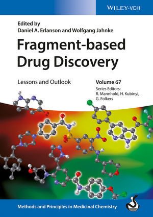
While it may not be possible to judge a book by its cover, the cover of this book does illustrate some of the fruits of the field, with structures of three fragment-derived drugs that have entered the clinic. These are just a small fraction of the 30+ drugs working their way through the pipeline, and of the many more that will spring from the research described and informed by the work presented.