Showing posts with label synergy. Show all posts

09 February 2026

Multivalent fragments in the clinic: Muvalaplin

It’s been a couple years since Practical Fragments last updated our “fragments in the clinic” list. Before doing so it makes sense to highlight some of those we’ve missed. Let’s start with an open-access Nature paper from Laura Michael and collaborators at Lilly and Monash University published in 2024. Truth be told I’ve been waiting for a longer discovery paper, but I’ll go with what’s available now.
 
The researchers were interested in lipoprotein(a), or Lp(a), which has been linked to cardiovascular diseases. Lp(a) forms when low-density lipoprotein (LDL) binds to apolipoprotein(a), or apo(a). This is a two step process, in which the ten subtypes of so-called Kringle IV (KIV) domains in apo(a) bind to lysine residues on LDL, followed by disulfide bond formation between apo(a) and LDL. Blocking the first step in this process should reduce levels of Lp(a).
 
Here's the only description of the initial screen: “Biochemical and biophysical compound screens using purified apo(a) KIV7-8 protein identified interacting small molecules. Optimization of the initial binding molecules led to…LSN3353871.” Whatever the details, LSN3353871 is unequivocally a rule-of-three compliant fragment. It is also a very ligand-efficient binder with high nanomolar affinity for the KIV8 domain. LSN3353871 disrupted the formation of Lp(a) in vitro at low micromolar levels and decreased levels of Lp(a) in cynomolgus monkeys when dosed orally.
 
As noted above, the apo(a) protein contains multiple KIV domains, and a classic method for improving potency is by making dimeric ligands that can bind to two domains simultaneously. The researchers did just this in the form of LSN3441732, which binds to apo(a) and disrupts formation of Lp(a) in vitro at picomolar concentrations.
 
If dimeric ligands are better than monomeric ones, why not go for multimeric ligands? The trimeric molecule LY3473329, or muvalaplin, was synthesized and crystallographically shown to bind to three copies of KIV8. It blocked formation of Lp(a) in vitro and reduced Lp(a) levels in cynomolgus monkeys.
 
Kringle domains are found not just in apo(a) but also in plasminogen, the zymogen form of plasmin, which is responsible for degrading blood clots. Fortunately, subtle differences between the Kringle domains in apo(a) and human plasminogen provide selectivity for the former protein, especially for multivalent ligands such as muvalaplin, and a phase 1 clinical study showed that Lp(a) could be lowered without affecting plasmin activity.
 
This is a nice application of applying fundamental multivalent principles to develop a potent molecule. It is also another example of a molecule that may not look like a drug but works like one: despite containing four basic nitrogen atoms, three carboxylic acid moieties, and sporting a molecular weight above 700 Da, muvalaplin is orally bioavailable. It is currently in a phase 3 trial in up to 10,450 patients. Cardiovascular disease is the leading cause of death in the developed world, and Practical Fragments wishes luck to everyone involved in these studies.
 
In the meantime, watch for more Practical Fragments posts on new entries to our fragments in the clinic list, which will be updated later this year.

06 October 2025

Exploiting avidity for finding fragments

As our poll last year demonstrated, there is no shortage of methods to find fragments. But that doesn’t mean new approaches aren’t welcome, particularly when they also apply to fragment growing. This is the promise of a recent paper in J. Med. Chem by Thomas Kodadek and collaborators at University of Florida Scripps and Deluge Biotechnologies. (Tom and first author Isuru Jayalath also presented this at the DDC meeting earlier this year.)
 
The researchers were inspired by the concept of avidity, the observation that multiple copies of a ligand bound to a multiprotein assembly can form a more stable complex than monomeric ligands bound to monomeric proteins. Could this phenomenon be exploited to find weak fragments?
 
A previous DNA-encoded library screen on streptavidin had identified 28 macrocycles, all of which contained one of two closely related fragments. The affinity of the more potent fragment came in at 706 µM using SPR. The researchers coupled this fragment to TentaGel beads, 10 µm wide polystyrene spheres covered in polyethylene glycol (PEG) chains terminated by amine groups. The PEG makes the beads water soluble. The beads were soaked in a solution of fluorescently labeled streptavidin, washed, and analyzed. Importantly, streptavidin exists as a tetramer, so each tetramer could bind up to four bead-bound fragments.
 
Streptavidin bound avidly to the beads, even when incubated at low (50 nM) concentrations. A control protein did not bind, nor did streptavidin bind to beads modified with a negative control fragment. Moreover, a monomeric version of streptavidin did not bind to the beads, illustrating the importance of avidity. Finally, adding the natural ligand biotin kept streptavidin from binding to the beads.
 
TentaGel beads have long been used in combinatorial synthesis, so the researchers built a small library in which the initial fragment was coupled to 48 carboxylic acids. These were then incubated with labeled streptavidin, and some of the beads showed more intense fluorescence, suggesting more protein binding. SPR analysis revealed that these new molecules had improved affinity, with the best coming in at 90 µM as a monomer. Thus, the primary screen can rank order affinities.
 
This is great for oligomeric proteins, but what about the large number of targets that are monomeric? Many recombinant proteins are expressed as fusions with glutathione S-transferase (GST), which facilitates purification. Importantly, GST exists as a homodimer in solution. The researchers screened a GST fusion of the oncology target Rpn13 against a small library of 94 fragment-coupled beads and found five hits. SPR studies confirmed weak (KD > 2 mM) binding for two hits to pure Rpn13 (ie, without the GST fusion), and this binding could be competed with a known peptide ligand of Rpn13.
 
Screening beads in individual wells is one thing, but to really increase throughput it would be nice to be able to screen mixtures of different beads. To do so, the researchers developed a photocleavable linker between bead and fragment. The linker also contained an alkyne group that could be modified with a brominated imidazopyridinium moiety. This tag is UV active, ionizes well, and the bromine’s unique isotopic signature helps distinguish true hits from noise. Beads containing more than 50 different compounds, including the two fragment hits we mentioned above, were incubated with labeled streptavidin. Beads to which protein bound were separated by fluorescence-activated cell sorting (FACS), clicked with the tag, cleaved from the beads, and analyzed by mass spectrometry. Only the two known binders were identified, demonstrating the specificity of the approach.
 
This is a neat paper well worth reading. I particularly like the fact that the method can be done with minimal equipment. I look forward to seeing how it works against more targets.

02 August 2021

Linking fragments on DNA, revisited

Three years ago we discussed using DNA-encoded libraries to find and link fragments. In a new open-access Bioorg. Med. Chem. article, Nicolas Winssinger and colleagues at University of Geneva report a different version of this approach.
 
Rather than using DNA, the researchers constructed their libraries with peptide nucleic acids (PNAs), which can be assembled using traditional solid-phase peptide synthesis and will also hybridize to DNA. Each PNA is coupled to a different fragment, and the fragment-PNA molecules are then bound to microarrays of DNA such that two fragment-PNA molecules bind to a single DNA strand. In this case the researchers used 250,000 combinations of fragment pairs.
 
Next, a protein of interest (here the anti-apoptotic cancer target BCL-xL) was screened at 50 nM. Binding to specific pairs of fragments was assessed by fluorescent detection of the protein at various spots on the microarray.
 
Trying to figure out which of the fragments are best – and how to link them – is “not trivial,” so the researchers took a combinatorial approach. Based on the first screen, they generated a new library of 10,000 molecules in which 10 sulfonamide-containing fragments were linked to 100 heterocycle-containing fragments using 10 different linkers. These compounds were screened using the same microarray technology, and the best binders were then resynthesized with a biotin tag rather than the PNA.
 
The biotinylated molecules were able to pull down recombinant BCL-xL in solution. Two of them, including compound 80-28, were even able to pull down recombinant protein that was spiked into cell lysate. Importantly, neither fragments 80 nor 28 did this by themselves. The affinity of 80-28 was measured by SPR to be 96 nM. 
 
 
Finally, the researchers tested 80-28 in K562 cells and found that it was cytotoxic with EC50 = 1.7 µM. They compare this favorably to venetoclax, the second fragment-based drug to be approved. However, this is a disingenuous comparison: venetoclax was specifically designed to bind less tightly to BCL-xL than to the related protein BCL-2. A more appropriate comparison would be a molecule such as navitoclax or the specific BCL-xLbinder A-1155463.
 
Like most BCL-family binders, compound 80-28 is also a rather unusual looking molecule, with a high molecular weight of 735. Unlike navitoclax and venetoclax, it also has 7 hydrogen bond donors and many more rotatable bonds. The long floppy linker in particular is something the earlier DNA-based fragment-linking work sought to fix. As we noted then, such linkers may be an inherent liability with the approach. From a technology perspective this is interesting work. But from a drug discovery perspective it still has some way to go to prove itself practical.

28 May 2018

Fragments vs the common cold (via NMT)

Anyone who has spent much time in drug discovery will have been asked what they've done to cure the common cold. In a paper just published in Nature Chemistry, Robert Solari, Edward Tate and collaborators from Imperial College London and institutions throughout the UK have taken a stab at this challenge.

One of the problems with rhinovirus, which causes the common cold, is that there are more than 100 different serotypes, thwarting vaccine development. To make matters worse, the virus replicates rapidly and sloppily, thereby increasing the odds of resistance mutations. To sidestep both problems, the researchers decided to target a host protein rather than a viral protein.

After the rhinovirus genome is translated in cells as a single polyprotein, it is cleaved and processed into component proteins which self-assemble to form the virion. One of the proteins, VP0, has a fatty acid attached to its N-terminus by host proteins called N-myristoyltransferases (NMT1 and NMT2 in humans). Mutagenesis studies had previously suggested that this modification is important for infectivity, so the researchers sought inhibitors against the NMTs.

High-throughput screens had previously identified two unrelated series of compounds, and crystallography revealed that they bind at adjacent but overlapping regions within the enzyme active site. Fragment-sized compound IMP-72 makes multiple interactions with the protein; an inhibitor from the other series makes a key interaction with an active-site serine. This molecule was trimmed back to a fragment (IMP-358) which showed minimal enzyme inhibition on its own but which dramatically increased the potency of IMP-72. Crystallography confirmed that the two fragments could bind NMT1 simultaneously.


A sort of fragment linking was conducted in which the key hydrogen bond acceptor of IMP-358 was attached to the more potent fragment, leading to a low nanomolar inhibitor. Further structure-guided optimization led to IMP-1088, which inhibits both human NMT1 and NMT2 with IC50 < 1 nM and shows picomolar binding by surface plasmon resonance (SPR).

So does it work? IMP-1088 is able to block myristoylation of VP0 in human cells. More importantly, the molecule shows antiviral activity against a range of rhinovirus serotypes and is able to rescue cells from viral cytotoxicity. Further mechanistic work suggests that inhibiting NMT activity blocks virus assembly.

Of course, lots of human proteins are myristoylated – NMT1 and NMT2 are human enzymes after all. Reassuringly, IMP-1088 itself did not reduce viability of uninfected cells. Although SPR had shown very slow off-rates, NMT proteins are constantly being resynthesized, and NMT activity had fully recovered after 24 hours. The researchers suggest that an early diagnosis and short treatment could be both safe and effective.

There is still much to do, notably pharmacokinetic and animal efficacy studies. And of course, the fear of toxicity will hang all the more heavily over antiviral strategies that target host proteins. So the next time someone asks whether scientists have invented a cure for the common cold, you’ll still have to tell them no. But at least we’re working on it.

30 April 2018

Fragment linking vs IMPDH (with a little help from the literature)


Mycobacterium tuberculosis (Mtb), the cause of its eponymous disease, is making an unwelcome comeback. Current treatments are lengthy, and highly resistant strains are emerging – and spreading. Chris Abell’s lab at the University of Cambridge has been working on multiple tuberculosis targets, and his latest results – in collaboration with Tom Blundell, David Ascher, and collaborators at the University of Cape Town and the University of Melbourne – has just published open access in J. Med. Chem.

The researchers were interested in inosine-5’-monophosphate dehydrogenase, or IMPDH, which is important for the synthesis of guanine nucleotides and is essential for every pathogen examined. They started by screening 960 fragments at 1 mM in a biochemical (spectrophotometric) assay. The IMPDH enzyme from a related organism was used due to its better expression and higher diffracting crystals; selected compounds were cross-checked with the Mtb enzyme and showed similar behavior.

This screen identified 18 molecules that gave at least 50% inhibition. IC50 values were determined for the six most active, and these ranged from 325-675 µM. These molecules were soaked into crystals of IMPDH, but only compound 2 produced a structure. As if to compensate, two molecules of compound 2 bound in the active site. Moreover, these two fragments bound in the same region as a previously reported molecule, compound 1.


Initial attempts at fragment growing yielded only modest improvements in potency, so the researchers tried to link the two copies of compound 2. One linking attempt failed outright, a second gave a 58 µM inhibitor, but a breakthrough came when the linker taken from compound 1 was used. The (S) enantiomer of compound 31 is 2500 times more active than the starting fragment, and crystallography revealed that it binds as designed. Unfortunately, and in contrast to compound 1, compound 31 showed no activity against Mtb in culture. The researchers hope to figure out why.

This paper illustrates several points. First, fragment linking can be quite effective. Second, consistent with our poll from a few years ago, this is not necessarily easy. Indeed, given the reliance on the structure of compound 1, this study can be considered an example of fragment-assisted drug discovery as much as fragment linking. And finally, as we’ve said before, biochemical potency all too often does not translate to cell-based activity – let alone good pharmacokinetic properties. Potency is just the first step in the long march to a drug.

17 November 2014

Deconstruction, superadditivity, and selectivity

One of the more exciting phenomena in fragment-based approaches is synergy (or superadditivity), in which the binding energy of linked fragments is greater than the sum of the binding energies of the individual fragments. Extreme cases are relatively rare, and the underlying thermodynamics can be counterintuitive, so it is always fun to see new examples. Cosimo Altomare and collaborators at the University of Bari and Consiglio Nazionale delle Ricerche (Italy) describe one in a recent paper in J. Med. Chem.

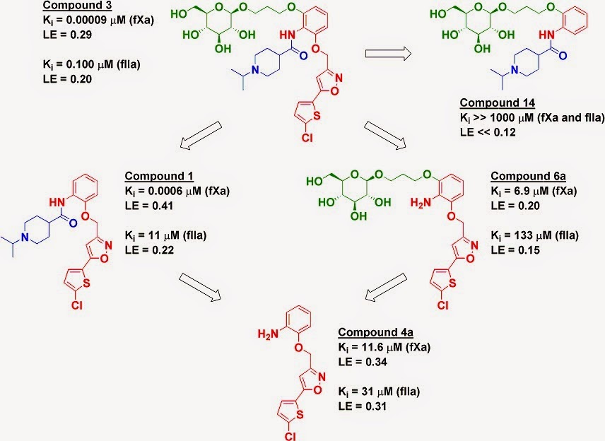
The proteases factor Xa (fXa) and thrombin (fIIa) are two heavily-studied anticoagulant targets. The paper characterizes a previously described molecule (compound 3) that is selective for fXa but still potent against fIIa, leading to good anticoagulant activity in human plasma as well as profibrinolytic activity. The researchers took a fragment deconstruction approach to better understand the binding to both targets.

As seen previously for fXa, the chlorothiophene moiety (red) is essential for binding, and removing it (compound 14) obliterates any detectable activity on both enzymes. However, while removing the glucose moiety (green) to give compound 1 reduced affinity for fXa by less than ten-fold, it reduced affinity for fIIa by more than two orders of magnitude. In contrast, removing the piperidine moiety (blue) to give compound 6a reduced affinity to both enzymes by several orders of magnitude.


However, these results are context-dependent. Removing both the piperidine moiety and the glucose moiety gives compound 4a, which has similar activity against fIIa as compounds 1 and 6a, where only a single moiety has been removed. In fact, compound 4a (without the glucose) is actually slightly more potent than compound 6a (with the glucose) against fIIa. But, as mentioned above, adding the glucose to compound 1 gives an impressive 110-fold boost in affinity for fIIa. In comparison, a famous early example of cooperativity in an NMR by SAR study gave only a 14-fold boost.

The researchers solved the crystal structure of compound 3 bound to fIIa, which reveals several hydrogen bond interactions between the glucose moiety and amino acid residues that have been previously implicated in allosteric activation of the protein. Perhaps compound 3 is exploiting this allosteric mechanism to bind more tightly.

This is a careful, thorough study and serves as a useful reminder that cooperativity can be huge, but it is still difficult to explain, much less predict.

16 October 2012

Fragment linking, enthalpy, and entropy: not quite so simple

The strategy of fragment linking dates to the origins of fragment-based lead discovery. The idea that two low affinity binders can be linked to produce a more potent molecule is based on the theory that the binding energies of linked fragments will at least be additive. Indeed, sometimes superadditivity can be observed; in those cases, the binding energy of the linked molecule is considerably better than the sum of the binding energies of the separate fragments. The most common explanation for this is that linking two fragments “pre-pays” the entropic cost of binding to the protein; rather than two fragments locking into fixed binding modes, only a single linked ligand pays this entropic penalty. This makes sense intuitively, but is it correct?

An early example of fragment linking was reported by Abbott researchers in 1997: two fragments that bound to the matrix metalloproteinase stromelysin were linked together to give a molecule that bound about 14-fold more tightly than the product of the affinities of the two fragments. Thermodynamic analyses were conducted to explore the roles of entropy and enthalpy, but these were complicated by the fact that one of the fragments contained an acidic phenol that was removed in the course of linking. In a new paper published in Bioorg. Med. Chem. Lett., Eric Toone and colleagues at Duke University have re-examined this system.

The researchers dissected several of the originally reported linked molecules into component fragments and examined their thermodynamics of binding using isothermal titration calorimetry. All of the experiments produced similar results; a particularly illustrative example is shown in the figure, in which a single bond in compound 1 was conceptually broken to yield component fragments 5 and 8.


As the researchers note, weirdly, the “favorable additivity in ligand binding – that is a free energy of binding greater than the sum of those for the constituent ligand fragments – is enthalpic in origin,” not entropic. It is not clear why this is the case, but what is clear is that the results are completely different from those obtained by Claudio Luchinat and colleagues on another matrix metalloproteinase. In that report, the enhanced affinity of the linked molecule was entirely entropic in origin, as might be expected. So what’s going on here?

One clue is provided by Fesik and colleagues in their original analysis of their stromelysin inhibitors. They noted that, when fragment 8 (acetohydroxamic acid) was added to the protein, biphenyl ligands similar to fragment 5 bound considerably more tightly than when fragment 8 was not present. In other words, the ligands displayed cooperative binding even when they were not covalently linked, probably due to non-covalent interactions between the two bound ligands or possibly to changes in protein structure and dynamics.

It is easy to assume that two ligands bind independently to two sites on a rigid protein, when in fact proteins are anything but rigid, and the addition of one ligand to a protein can dramatically change its properties. Thermodynamics measures changes in the entire system, not just the ligands, and if the protein changes upon ligand binding things can quickly get complicated. As Fesik and coworkers noted:

The observed cooperativity between the two ligands is a factor that should be considered when optimizing compounds for binding to nearby sites, since a portion of the binding energy is due to the cooperativity rather than interactions between the ligands and the protein.

All of which is to say that we remain woefully ignorant of the forces driving ligand binding, let alone fragment linking. But assessing how much better (or worse) a linked molecule binds than its component fragments can still be a useful exercise to guide optimization, even if the thermodynamic origins of the effects are unclear.

30 January 2012

Fragment linking: flexible rules

Linking two fragments together to achieve a boost in potency has been done a number of times (see here, here, here, and here), though it often doesn’t work as well as might be hoped (see here). To better understand the energetics of fragment-linking, Marc Nazaré, Hans Matter, and colleagues at Sanofi-Aventis Deutschland have analyzed ligands for the blood coagulation enzyme factor Xa (fXa) and published their results in a recent issue of Angew. Chem. Int. Ed.

The researchers “deconstructed” potent fXa inhibitors into component fragments, measured their inhibition constants (and thereby inferred their binding energies), and compared these binding energies with those of the original linked molecules. One of the first observations was that many of the component fragments bound so weakly as to show no measurable activity, a phenomenon that has been observed previously.

In an exemplary case, cleaving a single bond connecting the two component fragments of a 2 nM ligand (1a, below) yielded one fragment (1g) with 58 micromolar activity and another (1d) whose activity was worse than 10 millimolar. Because the second fragment has such low affinity, the binding energy of linking is really just a lower estimate, but it seems to be at least 3.3 kcal/mol, which is greater than the binding energy of fragment 1d itself. In other words, the affinity brought about by linking is greater than the affinity of the weakly binding fragment. The superadditivity provided by the linker in this case is about 300-fold, a similar value to that observed in the unrelated MMP-12 system. This is perhaps all the more remarkable given the fact that the fragments are connected by a linker containing several rotatable bonds, the entropy of which should partially counter the advantages of linking.


In fact, a common strategy to improve the potency of two linked fragments is to rigidify the linker. Often this doesn’t work: in a second case, the Sanofi-Aventis researchers cleaved one bond of a 3 nM ligand (2a, below) to yield two fragments with roughly equal potency. However, even though the linker is more rigid than in the previous example, the binding energy due to linking is less – just 2.0 kcal/mol, representing a boost of about 30-fold.


As the authors note:
The introduction of rigid aromatic moieties as a common approach to increase affinity does not necessarily maximize the benefit from the linker effect as detrimental affinity contributions might originate from suboptimal orientation and accommodation of specific binding elements.
There are many more examples in this paper than can be covered in a blog post; the authors dissect compounds 1a and 2a at a number of different points, and while the component fragments typically bind less tightly than simple additivity would suggest, there are lots of interesting details.

Finally, it is interesting to note that ligands 1a and 2a consist of a relatively hydrophobic fragment (1g or 2g) connected to a more polar fragment (1d or 2h). The fact that these show superadditivity is consistent with Mark Whittaker and colleagues' proposal last year that linking such fragments is likely to maximize additivity, although given the precise interactions made by both parts of the molecules the details get a bit messy. We’re not yet at the point where the universe of molecular interactions can be distilled to rigid rules.

25 July 2011

Fragment linking: oil and water do mix

Fragment linking is one of the most seductive forms of fragment-based lead discovery: take two low-affinity binders, link them together, and get a huge boost in potency. But what’s appealing in theory is difficult in practice: the linked molecule rarely binds more tightly than the product of the fragment affinities, and sometimes there is not even an improvement over the starting fragments. In a recent paper in Molecular Informatics, Mark Whittaker and colleagues at Evotec suggest a strategy to maximize the chance of success.

The researchers start by briefly reviewing nine published examples of fragment linking where affinities for both fragments as well the linked molecule are provided (some of these have been discussed previously here, here, and here). Of these, only three examples showed clear superadditivity (in which the linked molecule has a significantly higher affinity than the product of the affinities of the individual fragments), and two of these examples are rigged systems in which a molecule already known for its potency (such as biotin) is dissected into fragments. The challenges of linking are succinctly summarized:
The keys to achieving superadditivity upon linking are to maintain the binding modes of the parent fragments, not introduce both entropy and solvation penalties while designing the linker, and also make any interactions with the intervening protein surface that need to be made.
Also, of course, the resulting molecule needs to be synthetically accessible. Having a certain amount of flexibility in the linker can be useful, as this will allow the fragments some room to shift around, but too much flexibility introduces an entropic cost that defeats the purpose of linking in the first place. Software tools such as those by BioSolveIT can help design the linker, but what if some fragments themselves are inherently better suited for linking?

All three of the examples that show superadditivity start with one fragment that is highly polar and makes hydrogen bonds or metal-mediated bonds with the protein. The researchers suggest that such fragments are likely to pay a heavy thermodynamic penalty when they are desolvated, and that this cost can be reduced by linking them to a hydrophobic fragment. Thus, to maximize your chances of successful linking, the authors suggest you should choose
a fragment pair that consists of one fragment that binds by strong H-bonds (or non-classical equivalents) and a second fragment that is more tolerant of changes in binding mode (hydrophobic or vdW binders).

This is an interesting proposal, though because there are so few examples it is hard to assess. Indeed, the only other case of clear superadditivity I found involves dimerizing a fragment that is reasonably hydrophobic (ClogP = 2.4), albeit negatively charged. Hopefully we’ll see more examples in the coming years, but in the meantime, linking a water-loving fragment to an oily one is worth a shot.

11 June 2010

Fragment linking: how much is it worth?

Fragment linking is a topic we’ve discussed a few times. One of its great appeals is that, all other things being equal, the entropic cost of binding one linked molecule is less than the cost of binding two separate molecules. Thus, linking two fragments should give more than an additive increase in binding energy. As the late William Jencks noted, for two fragments A and B:
Kd(AB) = Kd(A) * Kd(B) * E
Where
Kd(AB) is the dissociation constant for the linked molecule AB
Kd(A) is the dissociation constant for fragment A
Kd(B) is the dissociation constant for fragment B
E is a “linking coefficient”, reflecting the costs and benefits of linking

The lower the Kd the better, so ideally E < 1, though in practice finding a suitable linker can be tricky and all too often E > 1 (sometimes >> 1). But how low can E go? How much of a boost can you get by linking two fragments? Claudio Luchinat and colleagues at the University of Florence looked at this question experimentally in a recent paper in J. Med. Chem.

The researchers took PMAHA, a known inhibitor of the matrix metalloproteinase MMP-12, and dissected it into two fragments, AHA and PMS, by conceptually “cleaving” the bond connecting them (see figure). This simplifies analysis: since the two fragments are almost identical to the linked molecule, there are no concerns that atoms in the linker interact with the protein.


The crystal structure of PMAHA bound to MMP-12 had been previously reported, but Luchinat and co-workers solved the co-crystal structure of AHA and PMS bound simultaneously to MMP-12. The two fragments overlay fairly well with the parent molecule: AHA binds to the catalytic zinc, while PMS binds in the S1’ pocket. The AHA fragment is rotated with respect to its position in PMAHA, though it makes the same interactions in both structures.

Thermodynamic binding parameters for the three molecules were determined (see figure). As expected, PMAHA binds considerably more tightly than the product of the affinities of the two fragments: E << 1 (in fact, about 0.0021). And in nice accord with theory, this enhanced affinity is entropic: both fragments bind with favorable enthalpy and unfavorable entropy, while the linked molecule has both favorable enthalpy and entropy. In other words, the salutary effect of linking these two fragments does seem to come entirely from entropic effects.

One of the more interesting lessons from this paper is a sense of how much of a boost in potency you can expect if fragment linking goes well: about 500-fold. In theory you could do better, but in practice you should expect much more modest benefits: a prominent success of SAR by NMR on a different metalloproteinase reported a 14-fold boost in affinity. But just like the lottery, the hope of a big payout will continue to attract people to the linking game.

17 December 2008

50% ain’t half-bad

In the world of fragment-based ligand discovery, researchers hope that two fragments, when linked together, will behave at least additively: the free energies of binding for each fragment will sum together, with a multiplicative effect on affinity. In ideal cases, linked fragments will behave synergistically (see for example the post from 18 August, below). But all too often, linking two fragments produces disruptive behavior, and the resulting molecule actually binds less tightly than would be predicted based on the binding energies of the individual fragments. This occurs not just when linking fragments, but in fragment merging and growing as well. Can such phenomena be modeled?

The mathematical groundwork was described more than forty years ago by Spencer Free and James Wilson at the old Smith Kline and French company, and came to be known as a Free-Wilson analysis. In a nice update of this work, Julen Oyarzabal and co-workers have applied this technique to the screening results of eight libraries consisting of several hundred compounds total. The molecules belong to five diverse chemical scaffolds (shown), and were tested against a variety of different targets, including a kinase, GPCRs, ion channels, and P450s.


For each library tested against each target, the authors asked whether the binding contribution due to a substituent Rx was additive, partially additive, or non-additive with the binding contribution of a substituent Ry. The mathematics get pretty intense, and the paper goes far beyond what I can summarize in a blog post, but the main conclusion is surprisingly encouraging: roughly half of all the data sets (10 of 19) show clear additive behavior, while another quarter (5 of 19) show partially additive effects. Only 4 data sets show non-additive behavior.

In many fields, a 50% success rate wouldn’t look too impressive, but in medicinal chemistry (in fact in much of chemistry in general), half-right sounds pretty good. The authors don’t further divide the non-additive data sets into sub-additive versus super-additive categories. In other words, the non-additive effects could well be due to synergy, the quality those of us pursuing FBLD ardently desire. But even if synergy is elusive, the paper suggests that you’ve got a better than even shot of producing a whole that is at least equal to the sum of its parts.