Showing posts with label selectivity. Show all posts

27 July 2026

Ligand affinities for isolated proteins vs in cells and cell lysates

Earlier this year we highlighted a study that compared the affinities of kinase inhibitors for isolated proteins vs those same proteins in cells and found differences – sometimes dramatic. A new open-access paper in J. Am. Chem. Soc. by Blake Peterson and colleagues at Ohio State University addresses the same question, and explores insights from cell lysates as well.
 
To measure affinity in cells, the earlier paper used NanoBRET, which relies on a fusion protein consisting of the kinase and a luminescent protein. It is possible that this artificial protein may behave differently from the native protein, so the Peterson group developed the Fluorescent Probe Cellular Binding Assay (FPCBA). This technique makes use of a fluorophore conjugated to a ligand for the protein of interest. The protein of interest is overexpressed, the binding of fluorescent probe causes cells to become fluorescent, and the level of fluorescence can be quantified using flow cytometry. Adding a (non-fluorescent) ligand displaces the fluorescent probe, which is rapidly effluxed, and the loss of fluorescence can be used to calculate the affinity of the ligand.
 
For the sake of brevity I’m skipping lots of important details, such as detailed characterization of efflux and use of a second fluorescent protein to correlate expression of the target, but these are described extensively in the paper. The researchers focus on a fluorescent probe based on dasatinib, an approved drug that binds to dozens of kinases.
 
The researchers compared the affinity of dasatinib and imatinib, another approved drug, to the kinase ABL1 using both FPCBA and NanoBRET. The results were reassuringly similar, though dasatinib bound 3 or 4-fold more tightly in the PBCBA assay than the NanoBRET assay while imatinib bound up to 2-fold less tightly, which might be due to changes in the regulatory mechanism of the ABL1 fusion protein required for NanoBRET.
 
Whole cell assays tend to be lower throughput, so cell lysates are often used instead. For kinases, a common assay makes use of “kinobeads.” Native kinases are captured from cell lysates on beads and quantified using mass spectrometry. An inhibitor blocks binding of the kinase to the bead, and this reduction in binding can be used to calculate the affinity. The researchers measured the affinity of dasatinib and imatinib against a score of kinases using both FPCBA and kinobeads.
 
Of 21 kinases, 14 showed similar affinities (within four-fold) for dasatinib by both FPCBA and kinobeads. Six of the remaining seven showed up to 13-fold tighter binding in FPCBA than kinobeads, which the researchers attribute to the kinases being in a more active state in cells than in lysates. (A similar phenomenon has been seen for the helicase WRN.)  In contrast, the kinase SRC showed 11-fold weaker activity in cells than lysates, which the researchers attribute to autoinhibitory regulation.
 
The situation becomes even more discordant when comparing affinities in cells to the affinities of isolated recombinant kinase domains using the KINOMEscan assay. Since a picture is worth several hundred words (the length of this blog post), I’ve graphed the results. While possible to see a correlation, it is also possible to see dragons in the shapes of clouds.
 
The affinity of dasatinib was generally higher (often by more than 10-fold) in the KINOMEscan than the FPCBA assay, which is not surprising given the lack of ATP in the biochemical assay. But there were exceptions, most notably EPHA4, which binds slightly more tightly to dasatinib in cells than in the KINOMEscan assay. Interestingly, the affinity in the cell lysate assay was lower; the researchers suggest that in cells the kinase is in a more active form than it is in lysates, perhaps due to membrane localization.
 
In the end, this paper is another useful reminder that assays with isolated proteins do not necessarily translate to cells. And of course, activity in cells does not necessarily translate to activity in vivo. Understanding why is part of what makes drug discovery both frustrating and exciting.

09 March 2026

Selectivity in cells may vary

Last year we celebrated the ten-year anniversary of the Chemical Probes Portal. One of the key requirements for a chemical probe is selectivity, which was set to >30-fold vs related targets when the Portal launched in 2015. For enzymes such as kinases, selectivity is often measured in cell-free assays. A new open-access J. Med. Chem. paper by Matthew Robers, Alison Axtman, and collaborators at Promega and University of North Carolina at Chapel Hill suggests that such data don’t necessarily translate to cellular assays.
 
Kinases are one of the most heavily mined classes of targets this century; five of the eight FBLD-derived approved drugs target kinases. With more than 500 in the human proteome, selectivity has long been a focus. One common method for assessing selectivity in cell-free assays is the Eurofins DiscoverX panel, which currently includes more than 450 kinases. Each kinase has a DNA tag and is paired with a promiscuous high affinity binder attached to a solid support. Test compounds are added, and qPCR is used to assess and quantify which kinases are displaced. The competition assay allows determination of dissociation constants.
 
To measure the binding of compounds to kinases in living cells, the researchers turned to the NanoLuc-bioluminescence resonance energy transfer (NanoBRET) assay. This is also a displacement assay that relies on a bivalent molecule containing a kinase ligand and a fluorophore. Kinases are tagged with NanoLuc, which causes luminescence of the fluorophore when it is in close proximity (ie, bound to the kinase). Ligands that bind to the kinase displace the bivalent molecule, decreasing luminescence.
 
The researchers started with four promiscuous kinase inhibitors, two of which (dasatinib and sorafenib) are approved drugs. They ran these against 240 or 300 kinases in the NanoBRET assay and compared the values with published DiscoverX dissociation constants. Most of the compounds were more potent in the DiscoverX assay than in the cell-based assay, and the researchers suggest several possible reasons. First, the DiscoverX assay is run in the absence of the cofactor ATP, which can compete with ligands that bind to the active site. Second, cell (im)permeability could decrease cellular potency. Finally, most of the DiscoverX kinases are truncated, whereas the NanoBRET kinases are full length.
 
For these and other reasons, it is common for compounds to be less active in cell assays than biochemical or biophysical assays. Surprisingly though, for a few kinases the compounds were actually more potent in the cellular assay than they were in the DiscoverX assay.
 
To extend these findings, the researchers tested additional kinase inhibitors and found that three kinases were particularly susceptible to inhibition in cells. One of these kinases, PIP4K2C, was engaged at mid nanomolar potency by cabozantinib, and the researchers suggest this could be useful for immuno-oncology. More worrisome, several approved drugs bind to the tumor suppressor STK11 in cells, raising the potential that these compounds could inhibit exactly the wrong pathway.
 
On the bright side, the researchers find that some molecules that look moderately selective in the DiscoverX assay are actually quite selective in cell assays, and they propose new chemical probes for the little-studied kinases BRSK1/2 as well the kinases DDR1/2.
 
Kinases are certainly not the only class of targets for which compounds’ performance differs outside vs inside cells; we wrote about covalent WRN inhibitors here. This paper is a good reminder that as useful as cell-free assays are, things can go weird once you go into cells – for better or for worse.

13 January 2025

Berotralstat: an overlooked fragment-derived drug

At the end of 2023 I mentioned that a paper by Dean Brown listed berotralstat as a fragment-derived drug. Readers will notice this molecule does not appear on our “fragments in the clinic” list. Did we miss it? After reading a (2021!) J. Med. Chem. paper by Pravin Kotian and colleagues at BioCryst, I believe the answer is yes.
 
Hereditary angioedema (HAE) is a rare genetic disease caused primarily by deficiencies in a protein that inhibits a serine protease called plasma kallikrein, or PKal. Drugs had already been developed to replace the inhibitor protein, but these need to be injected or infused. Since PKal is an enzyme, the researchers sought to make a small molecule inhibitor that could be taken as a pill.
 
BioCryst had developed an earlier drug called BCX4161, which is potent but has poor oral bioavailability. To find a better molecule, the researchers turned to the rich literature around serine protease inhibitors, which led them to make compound 2, a fragment of previously reported inhibitors of other serine proteases. The protonated benzylamine was expected to bind in the S1 pocket of the enzyme, and indeed the molecule did show weak but measurable activity.
 

Fragment growing led to compound 4, with double-digit micromolar activity. Building off the new phenyl ring led to more potent molecules such as compound 13, with low micromolar activity. Further structure-based design eventually led to BCX7353, or berotralstat. The paper provides good descriptions of the design rationale. For example, the fluorine was added to improve permeability, and the nitrile was added to improve the ADME profile. Modeling was used both to improve potency as well as to gain selectivity over other serine proteases. This proved to be successful: berotralstat is a subnanomolar inhibitor of PKal and at least several thousand-fold selective over trypsin and other serine proteases such as thrombin and FXa.
 
The pharmacokinetic properties of berotralstat in rats and monkeys were also good, and according to clinicaltrials.gov the molecule first entered the clinic in 2015. In December of 2020 the FDA approved berotralstat for prophylactic treatment of HAE attacks.
 
This is a nice story, and I agree with Dean that the discovery of berotralstat was “based on a legacy clinical candidate and fragment approaches.” The earlier molecule BCX4161 contained a benzamidine moiety, which was in part responsible for the poor oral bioavailability. Replacing this with a benzylamine fragment from the literature is a classic fragment strategy, and compound 2 is fully compliant with the rule of three.
 
So how was it missed? The abstract only states that berotralstat was discovered “using a structure-guided drug design strategy.” Indeed, the word “fragment” appears precisely once in the paper, albeit in a very telling sentence: “We evaluated these fragments in our PKalpur inhibitor assay…”
 
From a timeline perspective, the approval of berotralstat makes it the fifth approved fragment-derived drug, after pexidartinib and before sotorasib. I’ll include it in the next update of clinical compounds, along with my standard disclosure that “the list is almost certainly incomplete.” What else are we missing?

22 July 2024

Multiplexing (native) mass spectrometry

Native mass spectrometry (nMS) is one of the less commonly used fragment-finding methods. The approach entails mixing proteins and ligands and gently ionizing them under non-denaturing conditions to look for complexes. As with many other methods, multiple fragments can be screened in a single sample. In a new ACS Med. Chem. paper, Ray Norton and collaborators at Monash University and CSIRO report screening multiple proteins in a single sample.
 
The researchers were interested in fatty acid-binding proteins, or FABPs. As their name suggests, these transporter proteins shuttle lipophilic molecules such as fatty acids around cells. The ten human isoforms are expressed in different tissues and have different functions in metabolic signaling, but their similarity to one another has made finding selective chemical probes difficult. Enter nMS.
 
FABP isoforms 1-5 are the most heavily studied, and these were first assessed individually. They ionized well, though in some cases peaks corresponding to both the native protein and a complex with acetic acid was observed, not surprising given that the buffer contained 50 mM ammonium acetate.
 
Next, all five proteins were mixed together at 10 µM each. All the proteins could still be observed (with or without bound acetate), though some proteins did give stronger signals than others due to differences in ionization efficiency.
 
Adding small molecule WY14643, which the researchers had previously found to bind to FABPs in a fluorescence polarization (FP) assay, led to a more complex spectrum, with peaks corresponding to unbound proteins, proteins bound to WY14643, proteins bound to acetate, and proteins bound to both acetate and WY14643. When WY14643 was added at 10 µM, the selectivity profile was consistent with the FP data. Interestingly though, when ligand was added at the total concentration of all protein isoforms (50 µM), the selectivity profile changed. The researchers suggest this may be due to nonspecific binding at higher ligand concentrations, as has been seen previously for nMS.
 
To explore the generalizability of multiplexing nMS, the researchers turned to more potent (nanomolar) ligands. As with WY14643, these molecules showed good agreement with published selectivity rankings at lower ligand concentrations with some non-specific binding at higher concentrations.
 
When I first wrote about nMS back in 2010, I noted that “the stability of protein-small molecule complexes in native mass spectrometry assays does not necessarily correlate with the (more relevant) solution-phase affinity,” and this fact is investigated in the paper. Careful optimization of the experimental conditions, including ionization voltage and temperature, led to good relative selectivity rankings for a given ligand across the different FABP isoforms but differences in absolute values from those measured by ITC.
 
Another challenge is the fact that the five FABP isoforms tested have similar molecular weights; in one case a ligand complexed with FABP3 was difficult to distinguish from free FABP2. The researchers could solve this by using different protein constructs, such as a hexa-histidine-tagged version of FABP3.
 
Overall this is an interesting approach, and the paper does an excellent job describing the technical details and limitations. Along with protein-observed 19F NMR, mass spectrometry is a rare experimental technique suitable for screening mixtures of proteins in solution. Indeed, this becomes even easier when screening covalent binders, as seen in this paper from 2003, since there is no need to worry about ligand dissociation during ionization. And with the increasing interest in covalent drugs, the use of MS is only likely to increase.

28 August 2023

Affinity measurements in a single NMR tube?

Last week we highlighted a ligand-detected NMR method to measure affinities of protein-ligand interactions. That technique, R2KD, requires preparing multiple NMR samples with the ligand at different concentrations. In a new open-access paper published in J. Am. Chem. Soc., Serena Monaco and collaborators at University of East Anglia and Universidad de Sevilla describe a method that can be done in a single NMR tube.
 
The researchers have actually combined two methods, chemical shift imaging (CSI) and Saturation Transfer Difference (STD) NMR, to create imaging STD NMR. We’ve written previously about STD NMR, which relies on the transfer of magnetization from an irradiated protein to a bound ligand. In CSI, chemical shift information is recorded at multiple slices along the length of an NMR tube. Normally the solution in an NMR tube is homogenous and so the chemical shifts would be identical at the bottom and top of the NMR tube. Here, though, the researchers create concentration gradients by carefully pipetting a solution containing ligand on top of a solution containing protein and allowing the ligand to diffuse the length of the NMR tube.
 
Like all things NMR-related, the mathematics get a bit complicated. One important factor is the rate of diffusion for a given small molecule. This “diffusion coefficient” can be experimentally measured by creating a concentration gradient in the absence of protein and measuring the ligand concentration at various positions in an NMR tube after a given length of time (typically more than 12 hours). Diffusion is dependent on molecular weight, so it is also possible to calculate the diffusion coefficient, and in fact the researchers found that the calculated values matched the experimental values for three different small molecules.
 
Knowing the diffusion coefficient helps establish the maximum ligand concentration to use and the ideal diffusion time. The researchers examined three different protein-ligand pairs, all of which had weak affinities, with KD values from 0.2 to 2 mM. Measuring STD signals at different slices along the NMR tube effectively yields STD signals at different concentrations of ligand, and fitting this to an equation allows calculation of the dissociation constant. For the three model systems the affinities agreed with literature values, which had been determined using ITC or WAC.
 
One nice feature of imaging STD NMR is that it can identify non-specific binding. This is because STD signals vary depending in part on how close a proton on the ligand is to the protein, resulting in different STD signals for different protons for specific binders. If this “epitope pattern” is lost at higher concentrations, this suggests non-specific binding, where the ligand can bind in random orientations to multiple sites on the protein. The researchers demonstrated this for one of their model systems: tryptophan binds specifically to bovine serum albumin with a dissociation constant of 0.2 mM, but above 1 mM or so the epitope disappears, suggesting non-specific binding.
 
Imaging STD NMR does have some limitations. For one thing, it requires a high initial concentration of ligand: 30 mM in the case of tryptophan, and even higher for the other two ligands. Most small molecules are nowhere near this soluble in water. The researchers suggest that ligands could be dissolved in DMSO and placed on the bottom of the NMR tube, with the protein solution gently layered on top. They show that the concentration gradients develop in a similar manner as a fully aqueous system, but acknowledge that high DMSO concentrations may not play well with most proteins.
 
Also not stated is the sensitivity of the method for higher affinity binders. Last week’s R2KD could measure affinities as tight as 10 µM, but it is unclear how much below 200 µM imaging STD NMR can go.
 
Finally, as we noted in 2019, STD effects are remarkably complex and not well-correlated with affinity. In particular, binding kinetics can play a role in the strength of the signal. It would have been nice to see more than three protein-ligand pairs tested.
 
All that said, this is an intriguing approach. Laudably, the researchers provide extensive supporting information, including mathematical derivation of the fitting equations, a spreadsheet, NMR pulse sequences, and macros. I’ll be curious to see how it works for others.

30 January 2023

Fragments in the clinic: MK-8189

Just over seven years ago Practical Fragments highlighted work out of Merck describing the discovery and optimization of potent, selective inhibitors of phosphodiesterase 10A (PDE10A), a potential target for schizophrenia (see here and here). An open-access paper in the latest issue of J. Med. Chem. by Mark Layton and colleagues tells how these were ultimately advanced to a clinical compound.
 
To recap, a biochemical fragment screen identified the highly ligand-efficient compound 1, which was optimized to the potent compound 2. However, this molecule had poor pharmacokinetics and multiple other liabilities. Further optimization led to Pyp-1, which we noted at the time would make a good chemical probe.
 

The new paper continues SAR around the central ring, in particular to try to reduce lipophilicity. Also, the methyl-pyrazole in Pyp-1 was associated with high clearance in rats, so this substituent was replaced with a methyl-1,3,4-thiadiazole moiety. To cut a long story short, this ultimately led to MK-8189.
 
Not only is MK-8189 a picomolar biochemical inhibitor of PDE10A, it is a low nanomolar inhibitor in cells. Moreover, it shows excellent pharmacokinetics in rats and rhesus monkeys as well as selectivity against various off-targets such as CYPs and hERG. Importantly for a drug intended to reach the brain, the molecule is permeable, not effluxed, and achieves relevant concentrations in the rat striatum after oral dosing. Finally, it decreased psychomotor activity and improved episodic memory in rat models of schizophrenia. With all these positives, MK-8189 has been taken into the clinic.
 
Several lessons emerge from this story. First, as experienced drug hunters will recognize, systematic exploration of multiple positions is important to generate a molecule with the right balance of properties to become an investigational drug. Second, as the researchers note, ligand efficiency was roughly maintained throughout the optimization process. Finally, this publication is a reminder of the long lag that can occur between research and publication. We already mentioned that the first papers describing this series appeared in 2015, but according to ClinicalTrials.gov MK-8189 first entered the clinic a year earlier, in 2014. Our list of fragment-derived clinical compounds will forever be incomplete and out of date. But on a positive note, this means that fragments may be having even more of an impact than the list shows.

23 August 2021

Fragments vs DYRK1A and DYRK1B: Part 2

Last week we highlighted work out of Vernalis and Servier in which fragment-based methods were used to identify potent and selective inhibitors of DYRK1A and 1B, potential targets for cancer and neurodegenerative diseases. The NMR screens yielded 166 hits, only one of which was advanced in that paper. A second J. Med. Chem. paper by Andras Kotschy and collaborators describes the optimization of another fragment.
 
Compound 1 is a whoppingly potent fragment with impressive ligand efficiency. If you’ve ever worked on kinases you probably think you know how it binds, as the diaminopyrimidine moiety is a common hinge-binding motif. In fact, crystallography revealed that the molecule binds in a completely different orientation and that the methoxy group makes a single hydrogen bond to the hinge amide NH. Cyclizing the molecule led to compound 10, with a satisfying boost in affinity.

 
Unfortunately, compound 10 was also a potent inhibitor of the kinase CKD9. To gain selectivity, the researchers took advantage of the fact that one of the backbone carbonyl oxygens in the hinge adopts an unusual orientation in DYRK1A, making room for the methyl group in compound 33. Next, the researchers replaced the benzofuran core for reasons of “synthetic tractability, metabolic stability, and freedom to operate.” This exercise ultimately led to compound 40.
 
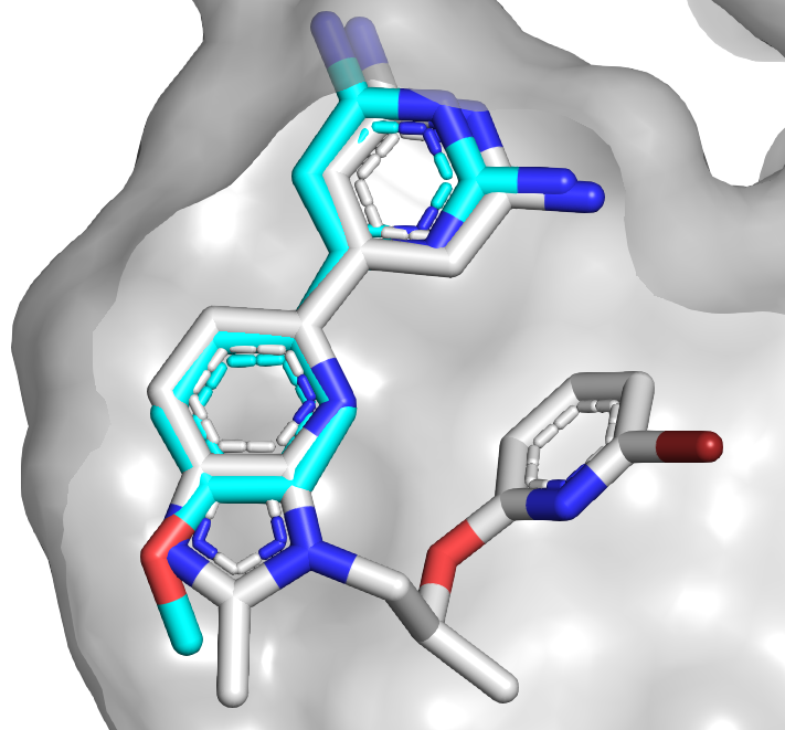
This compound was profiled against 442 kinases and found to be quite selective, with only 8 kinases significantly inhibited at 1 µM. One of these was the related kinase DYRK2, but further growing led to selective compound 58. An overlay of the initial fragment (blue) with compound 58 (gray) reveals how the binding mode has been maintained, in contrast to the series described last week.

Compound 40 had only modest antiproliferative activity against human cancer cell lines that were grown in 2D culture but was more active when the cells were grown in 3D culture. The molecule had good oral bioavailability in mice, and xenograft studies revealed that it inhibited tumor growth, though it was also toxic at higher doses. The researchers do not mention brain penetration, though given the number of hydrogen bond donors I would be surprised if it crosses the blood-brain barrier.
 
This paper is a nice example of how getting high affinity is often only the beginning of a long journey. In combination with the story from last week it is also a useful reminder of how many starting points a single fragment screen can provide: just two fragments led to two completely independent series. Whether molecules from these series advance to the clinic, they provide useful tools to further understand the biology of DYRK1A.

16 August 2021

Fragments vs DYRK1A and DYRK1B: Part 1

The dual-specificity tyrosine-phosphorylation-regulated kinases 1A and 1B (DYRK1A and DYRK1B) belong to a family of five serine/threonine kinases implicated in several cancers as well as Down’s syndrome and other neurodegenerative diseases. For the latter indications in particular, brain penetration would be essential for any inhibitor, just as in the LRRK2 story last week. In a new (open access) J. Med. Chem. paper, Rod Hubbard and collaborators at Vernalis and Servier describe the discovery of a chemical probe.
 
The researchers started by testing their in-house library of 1063 fragments in pools of six, each at 500 µM, in three ligand-detected NMR screens. This resulted in a whopping 166 hits. Crystal structures of the eight most ligand-efficient fragments bound to DYRK1A were obtained, including compound 5. Fragment growing led to compound 16, which bound the kinase 200-fold more tightly. 
 

The crystal structure of compound 16 bound to DYRK1A was compared to structures of other known ligands and suggested the possibility for an alternative binding mode. This led to the synthesis of compound 24, with low nanomolar affinity against both DYRK1A and DYRK1B (only the former is shown in the figure). This compound turned out to be surprisingly unstable in slightly acidic aqueous solution (below pH 5), but replacing the oxygen with a nitrogen fixed this, and further tweaking ultimately led to compound 34.
 
Compound 34 was profiled at 1 µM against a panel of 442 kinases and found to be fairly selective, with only 15 kinases inhibited by at least 50%. It is orally bioavailable in mice, brain penetrant, and inhibited the proliferation of glioblastoma cells, although the potency was significantly attenuated by serum. In a xenograft study the compound caused tumor growth delays and was well-tolerated.
 
This is a nice example of fragment-based lead discovery heavily dependent on structural information. Comparing the binding mode of compound 34 (gray) with that of compound 5 (light blue) reveals the significant shift in binding mode of the initial fragment.

The paper is also a useful reminder of how long it can take for industry research to be published. Work began in 2009, and Rod presented some of it at the CHI FBDD conference in 2019. But this is not the end of the DYRK1A story: stay tuned for next week!

09 August 2021

Fragments vs LRRK2 with the help of a surrogate and a magic methyl

Leucine-rich repeat kinase 2 (LRRK2) has been implicated in Parkinson’s disease and has thus long been targeted by drug hunters. Dozens of inhibitors have been approved for other kinases, making the protein class appear “easy”, but LRRK2 is particularly challenging. First, it is a large multidomain protein that has resisted crystallography. Second, an inhibitor for a chronic disease such as Parkinson’s will need to be highly selective. Finally, the fact that LRRK2 is in the brain means that inhibitors will need to cross the treacherous blood-brain barrier (BBB), whose function is to exclude anything unusual. A recent J. Med. Chem. paper by Douglas Williamson and collaborators at Vernalis and Lundbeck addresses the first two of these issues.
 
The researchers started by screening 1313 fragments (at 200 µM each) against the disease-relevant G2019S mutant of LRRK2. The screen was run using the DiscoveRx KINOMEscan, which relies on displacement of a kinase from an immobilized ligand. Some 80 hits were then triaged in a kinase activity assay.
 
Rather than banging their heads against the wall that has blocked X-ray structures of LRRK2, the researchers turned to a crystallographic surrogate. The readily crystallizable kinase CHK1 has some similarity to LRRK2, and some inhibitors bind to both kinases. Introducing ten mutations into CHK1 around the ATP-binding site led to a LRRK2 surrogate – an approach we’ve previously mentioned.
 
Among the fragment hits were adenine (compound 7) and two closely related molecules. Crystallization of compound 7 with wild-type CHK1 and the LRRK2 surrogate revealed two different binding modes. Similarity-based searches of in-house and literature compounds led to molecules with nanomolar potency, and subsequent optimization led to compound 17 (all molecules were tested against both wild-type LRRK2 as well as the G2019S and had similar affinities).
 

Interestingly, crystal structures of these molecules revealed them to bind more similarly to the complex of compound 7 bound to wild-type CHK1 rather than the surrogate. Adding a methyl group to compound 17 led to compound 18 with a satisfying 100-fold boost in potency: a true magic methyl, as the other enantiomer had slightly worse affinity than compound 17. Crystallography with the surrogate revealed hydrophobic interactions between the methyl and an alanine side chain. It is worth noting that compound 18 is still fragment-sized and yet has high picomolar affinity for LRRK2.
 
Extensive medicinal chemistry followed, ultimately leading to compound 45. Profiling against 468 kinases in the KINOMEscan assay demonstrated it was quite selective, binding only three other targets with dissociation constants less than 1 µM. The compound was active in cells containing either wild-type or G2019S LRRK2. Unfortunately, while the compound showed good oral bioavailability in dogs, it was not orally bioavailable in rats. Moreover, brain to plasma ratios were low in mice, and the molecule was a substrate for the human protein BCRP, a transporter that pumps small molecules across the BBB.
 
Although these liabilities halted further work on the series, this is nonetheless a nice fragment to lead story that highlights the utility of crystallographic surrogates. But the different binding modes for the initial fragment are a reminder that multiple binding modes are not uncommon, and it is best to employ crystallography not just early but often, when you can.

05 October 2020

Fragments vs (lots of) RNA

RNA is hot. Hundreds of millions of dollars have gone to startups focused on finding small molecules that bind RNA, and plenty of large pharmaceutical companies are pursuing this class. As with all difficult targets, fragments have a role to play, as demonstrated by a paper just published (open access!) in ChemBioChem by Harald Schwalbe and collaborators at Johann Wolfgang Goethe-University Frankfurt and Saverna Therapeutics.
 
The researchers assembled a collection of 101 fluorine-containing fragments and pooled these into five sets of 20-21 compounds each. These were then screened (at 50 µM) against 14 different RNA targets, ranging from 14-nucleotide hairpins to a 127-nucleotide riboswitch. The primary screen was ligand-detected 19F NMR using CPMG, in which ligand binding causes a change in relaxation which is detected as a decreased signal. All hits from the mixtures were confirmed as single molecules. To assess selectivity, all the compounds were also screened against 5 DNA targets and 5 proteins.
 
The results are not entirely unexpected. Some of the fragments did not hit any targets, while others were rather promiscuous: a couple showed strong binding to 8 of the 14 RNA targets. That said, strong is a relative term; the highest affinity of any fragment measured was 375 µM.
 
The RNA targets spanned a variety of structures, but the most hit-rich were aptamers and riboswitches, which bind specific small molecules or ions. These had 7 to 26 hits each. In contrast, the other RNA targets tested all had six or fewer fragment hits. For the aptamers, competition experiments with the natural ligands suggested binding at the orthosteric sites in some cases but not others.
 
Hit rates against four of the five proteins were also high, with 16 to 55 hits each. The fifth protein, a challenging phosphatase, had only four hits. This was still better than the 24-nucleotide DNA duplex, with a single hit. The four G-quadruplex DNA targets had between 12 and 20 hits, consistent with prior research. Taken together, the results suggest that RNA riboswitches and aptamers may be reasonably ligandable, while RNA targets that do not normally bind small molecules may be more challenging.
 
The researchers also conducted cheminformatic analyses. Not surprisingly given the relatively small library, there were no strong correlations between molecular features and targets bound, though fragments that hit had a slight tendency towards more aromatic atoms and fewer sp3-hybridized carbons. This is consistent with a vigorously debated paper we highlighted in July.
 
Finding weak hits is one thing, but advancing them has been challenging for RNA targets. The researchers provide an example in which they link a fragment to an intercalator to generate a low micromolar binder, but intercalators are often nonspecific, and affinity would still need to be further improved. Practical Fragments first highlighted a fragment screen against RNA more than a decade ago, and earlier this year we noted a high nanomolar binder, but I have yet to see an attractive low nanomolar lead emerge.
 
Nonetheless, this paper provides a solid launching point for such an effort to succeed. In particular, the researchers laudably include structures of all the library members as well as the raw screening data of all the hits on all the targets in the Supporting Information. If you are feeling adventurous, you now have plenty of starting points to choose from.

20 July 2020

Fragments vs JAK1, a sequel from LEO

Four years ago we highlighted a paper from LEO Pharma describing inhibitors of the kinase JAK1, which is implicated in a host of inflammatory conditions. Although they developed low nanomolar inhibitors, these showed phototoxicity, which was unacceptable for the topical applications the researchers had in mind. The molecules were also not selective against closely related JAK2, whose inhibition can cause neutropenia and anemia. A recent paper in J. Med. Chem from Andrea Ritźen and collaborators at LEO and GVK Biosciences describes a more selective series.

As mentioned previously, hits came from about 500 fragments screened against JAK2 using SPR and validated in a biochemical assay against JAK1. Most fragments had similar activities against both proteins, but compound 1 was moderately selective for the latter. Initial SAR around the fragment revealed that the methyl group was essential to activity and that methylating the pyrazole nitrogen atoms also obliterated binding. The molecule looks like a hinge-binder, but because it can assume four different tautomers docking was difficult. Fortunately, replacing the difluoromethyl substituent with a phenyl ring in compound 6 improved affinity and led to a crystal structure, which showed the methyl group making lipophilic interactions with the protein.


The crystal structure also revealed that the phenyl ring didn’t quite fill the lipophilic ribose-binding pocket, so the researchers replaced this with the more three-dimensional cyclohexyl substituent in compound 7, which yielded a ten-fold improvement in biochemical potency as well as the first cellular activity. The philosophy behind further optimization is described eloquently: “in the spirit of fragment-based drug design – start small and make every added atom count – small substituents with balanced polarity were added to the cyclohexyl analogue 7.” Unfortunately, although several polar substituents introduced onto the cyclohexyl ring improved biochemical potency, they did not do much for cell activity.

Replacing the cyclohexane moiety of compound 7 with a norbornane led to compound 11, which was not only more potent against JAK1 but also less lipophilic and more soluble. The researchers then borrowed a nitrile from an approved pan-JAK inhibitor, leading to compound 40. This molecule has low nanomolar activity against JAK1 and is somewhat selective against closely related JAK2, JAK3, and TYK2. It is quite selective against a panel of 50 other kinases and does not inhibit several cytochrome P450 enzymes or bind to hERG. Oral bioavailability in rats is fairly low, but this should not be a problem for topical indications.

As noted in the paper, the researchers were able to benefit from published work from multiple other companies that has led to five approved JAK inhibitors plus several more in clinical development. While another JAK inhibitor may not be the most pressing medical need, this paper is still a nice example of structure-based design that illustrates several points. First, ligand efficiency was improved during the optimization process, in contrast to common perceptions. Second, fragment selectivity was also improved during optimization. And finally, although this sounds banal, it matters what you have in your fragment library: had the des-methyl version of compound 1 been the only representative of this core in the LEO library the researchers would not have discovered it. In other words, while simplifying your fragments will decrease molecular complexity, sometimes a single methyl group can make all the difference.

01 June 2020

BETting on fast follower fragments

A common approach in drug discovery is to improve a previously reported molecule. An example of such a “fast follower” approach has just been published in J. Med. Chem. by Cheng Luo, Bing Zhou, and colleagues at Shanghai Institute of Materia Medica.

The researchers were specifically interested in bromodomains, which recognize acetylated lysine residues in proteins and play major roles in gene expression. In 2018 we described AbbVie’s fragment-based discovery of ABBV-075, which had entered phase 1 clinical trials. Although reasonably selective for BET-family bromodomains, it also strongly inhibits EP300, which could lead to toxicity. Thus, more selective molecules have been sought.

The new paper starts with a thermal shift assay of 1000 fragments against the two separate bromodomains of BRD4, BD1 and BD2. Hits were validated using an AlphaScreen assay. Compound 47 was found to be active against both BD1 and BD2, with high ligand efficiency (all IC50 values shown are for BD1; values for BD2 are similar). Modeling suggested this fragment could be merged with ABBV-075, and indeed the resulting compound 26 was quite potent. (Note: structures of compounds 26 and 38 were originally drawn incorrectly - now fixed.)


Compound 26 was metabolically unstable, but further optimization, aided by crystallography and modeling, ultimately led to compound 38. This molecule has good oral bioavailability in mice and promising pharmacokinetics in both mice and rats. It inhibits the expression of cancer-driving genes such as c-Myc and BCL-2, inhibits the growth of several cancer cell lines, and demonstrated good tumor growth inhibition in a mouse xenograft study. Compound 26 does not inhibit five cytochrome P450 enzymes or hERG. Finally, it is much more selective than ABBV-075 against EP300 and indeed most other bromodomains aside from BET family members. The researchers conclude that “compound 38 is a highly promising preclinical candidate.”

Unfortunately, selectivity for BET-family bromodomains may not be sufficient to avoid toxicity. Indeed, as we described earlier this year, AbbVie has dropped clinical development of ABBV-075 in favor of ABBV-744, which is selective for BD2 over BD1. Whether or not the same could be done for this series, the paper is still another nice example of appending a fragment onto a previously discovered molecule.

02 March 2020

FBLD meets DEL

FBLD, of course, starts with small libraries of small fragments. DNA-encoded chemical libraries (DEL) usually start from the opposite extreme. Massive numbers of molecules are combinatorially synthesized attached to DNA, screened against a target using affinity selection, and hits identified by sequencing the DNA. A recent paper in J. Med. Chem. by Christopher Wellaway and colleagues at GlaxoSmithKline uses information from both approaches to generate a high-quality candidate.

The researchers were interested in bromodomain and extraterminal (BET) family proteins – the same targets we discussed last week. GlaxoSmithKline had already put molecules into the clinic, but they were looking for structurally different backup candidates, so they performed a DEL screen on the BD1 domain of BRD4. A library of 117 million compounds yielded potent compound 10, and crystallography revealed that the 2,6-dimethylphenol moiety bound in the acetyl-lysine-binding pocket.


Phenols are often metabolic liabilities, and indeed compound 10 was rapidly cleared in mice. However, GlaxoSmithKline has a long and successful history of fragment screening against bromodomains; Teddy first described some of their seminal work back in 2012, when the world didn’t end. Compound 16 had been found in a previous screen as a hit against BRD4, and crystallography revealed that the pyridone binds in a similar fashion to the phenol moiety. (Similar pyridones had been reported by others, for example this one.) Merging the molecules led – after a bit of tweaking – to compound 20a. In addition to BRD4, this molecule binds another bromodomain, BAZ2A, which the researchers wanted to avoid. Structure-based design led them to the more selective compound 20i.

Although compound 20i is potent in cells, it still has moderate clearance in rats. Unsubstituted benzimidazole rings have been reported to be unstable, so the researchers systematically explored a series of substitutions, ultimately arriving at compound 24 (I-BET469). Not only is this compound potent and soluble, it is remarkably stable, with “no detectable turnover in rat, dog, and human microsomal and hepatocyte preparations.” Oral bioavailabilities approach 100%, and the compound proved to be effective in acute and chronic mouse inflammation models. Although selectivity against non-BET family bromodomains members is good, compound 24 does strongly bind to both BD1 and BD2 domains of all four BET family members, and as we saw last week this may lead to toxicity.

Nonetheless, this is a lovely example of using a fragment to replace a problematic moiety in a larger molecule, as we’ve seen previously for chymase, Factor VIIa, and Factor XIa. Throughout the optimization the researchers paid close attention to molecular properties such as lipohilicity and molecular weight, and this resulted in a molecule with excellent pharmacokinetics despite the presence of potentially unstable moieties such as the morpholine. If nothing else, this will be a useful in vivo chemical probe.